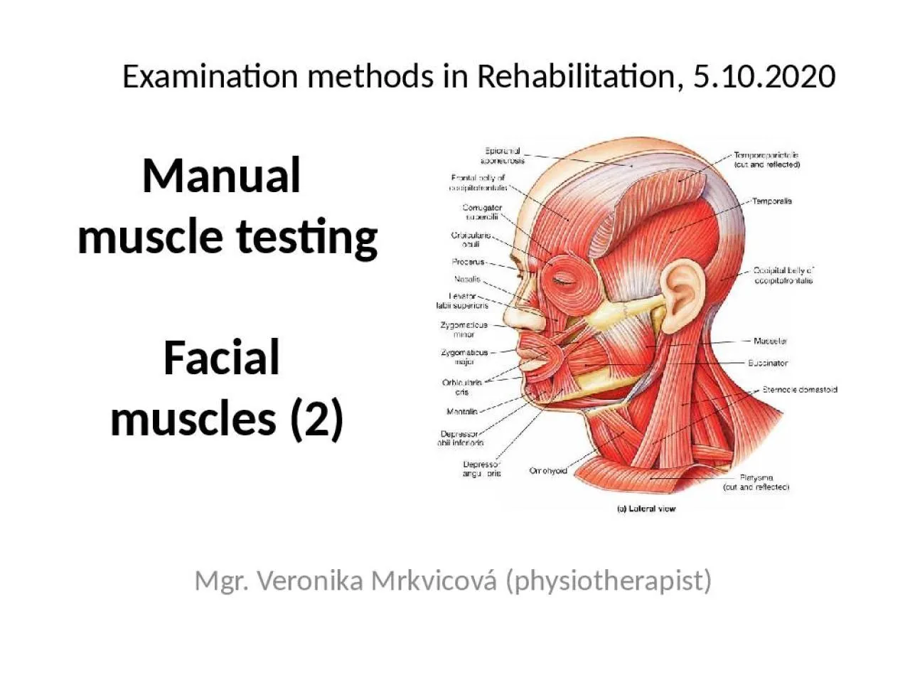
Facial muscles 2 Mgr Veronika Mrkvicová physiotherapist Examination methods in Rehabilitation 5102020 Introduction Facial muscles MMT grading Facial nerve ID: 912430
Download Presentation The PPT/PDF document "Manual muscle testing" is the property of its rightful owner. Permission is granted to download and print the materials on this web site for personal, non-commercial use only, and to display it on your personal computer provided you do not modify the materials and that you retain all copyright notices contained in the materials. By downloading content from our website, you accept the terms of this agreement.
Slide1
Manual muscle testingFacial muscles (2)
Mgr. Veronika Mrkvicová (physiotherapist)
Examination methods in Rehabilitation, 5.10.2020
Slide2IntroductionFacial musclesMMT, gradingFacial nerveFacial paralysis
Slide3Slide4Facial NerveMost of the facial nerve is comprised of motor innervation of the muscles
of facial expressionIn addition, it subserves several other functions including: taste perception from the anterior two-thirds of the tongue
perception of cutaneous stimuli in the external auditory canal and over part of the pinna and mastoid region innervation of the stapedius muscle in the middle ear innervation of the lacrimal gland and two of the salivary glands (the submaxillary and submandibular)
Slide5Facial expressionThe most prominent deficit noted by patients with facial nerve damage is weakness of muscles of facial expressionCareful observation of the patient's face during conversation and at rest almost always reveals facial weakness
Additionally, the face may "droop" on the side of damage due to the effects of gravity
Slide6Facial nerve: testThe nerve can be further tested by functional activities: having the patient
close their eyes and lips tightly having the patient grimace (show their teeth) having the patient look up (elevating the eyebrows and creasing the forehead)
having the patient fill their cheeks with air with their lips tightly pursed. If one or both sides of the face are weak, s/he will have difficulty holding the air in
Slide7Bell´s palsyThe most common cause of facial weakness, an idiopathic condition that may result from viral infection-induced inflammatory swelling of the facial nerve in its canalSince the canal is very long and tight, swelling can put
pressure on the nerve, resulting in damage either by direct effects or by impairing blood flow in the nerveIn some cases, facial palsy is produced by a very clear viral infection with Herpes Zoster, often associated with ear pain and vesicles on the tympanic membrane
Lyme disease also has a predisposition to produce facial palsy, sometimes bilateral
Slide8Bell´s palsyThe characteristic of peripheral facial palsy is that it involves the entire side of the face, including weakness of the forehead muscles as well as those around the eye and mouth
This is because fibers to all of these regions of the face are packed together in the facial canalMost cases of uncomplicated Bell's palsy recover quite well In its most severe form, infarction of the nerve may occur with a prolonged and not infrequently incomplete process of regeneration
This is more common when a longer course of the nerve is affected, accompanied by ageusia (loss of taste) and hyperacusis
Slide9Bell´s palsy
Slide10Central paralysisCorticobulbar (pyramidal) projections from the motor cortex (precentral gyrus) through the genu
of the internal capsule are the major pathway for voluntary facial movementThe cerebral cortical projections to the facial motor neurons innervating the upper face are essentially bilateral (i.e., each cortical hemisphere provides innervation to both sides)
Therefore, unilateral lesions (such as a stroke affecting one hemisphere or the internal capsule) will not produce weakness of the upper face muscles
Slide11Central paralysisOn the other hand, facial motor neurons that innervate the muscles of the lower face receive input largely from the contralateral hemisphere (i.e., the right hemisphere activates motor neurons of the left facial nucleus, and vice-versa)
Therefore, a lesion involving the right motor cortex (e.g., carotid-middle cerebral arterial system occlusion and hemispheric infarction) causes a weakness of voluntary left lower facial movement that is especially noticeable while the patient is talking, grimacing (usually elicited by asking the patient to bare their teeth or gums), or resting
Slide12Central paralysisThe corner of the mouth droops and there may be some widening of the palpebral fissure (eye)On the other hand, the forehead is normally creased when a person raises their eyebrows or looks toward the ceiling
This distinguishes the "supranuclear" weakness of the face from the weakness of the whole side of the face due to damage of the peripheral facial nerve, as seen with Bell's palsy.
Slide13Peripheral vs. central lesion
Slide14Manual muscle test - grading5 Normal contraction of
the muscle, no assymetry compared to healthy
side4 Nearly normal contraction, little assymetry compared to healthy side3 Contraction of the muscle in one half compared to healthy side2 Contraction of the muscle in one quarter compared to healthy side
1
Trace
of
muscle contraction can be
seen
/
palpated
0
The
muscle
demonstrates no palpable muscle
contraction
Slide15Muscles of facial expression
Slide16Occiptofrontalis groupThe occiptofrontalis, or scalp, group consists of the frontalis and occipitalis
These muscles provide movement of the eyebrows, forehead, and scalp
Slide17M. occipitofrontalisRaises forehead, pulls scalp backward
Slide18Orbital groupThe orbital group consists of:orbicularis oculicorrugator
superciliiThese muscles provide movement of the eyelid and periorbital skin
Slide19M. corrugator supercilii
Draws eyebrow medially
Slide20M. orbicularis oculi
Closes eyelid
Slide21Nasal groupThe nasal group consists of:ProcerusNasalisThese muscles provide movement of the nose and
perinasal skin
Slide22M. procerusM. nasalis
M.
nasalisM. procerus
Slide23Oral GroupThe oral group consists of:obicularis
orisdepressor anguli oris, levator anguli
oriszygomaticus major et minorlevator labii sup., levator labii sup. alaeque nasi risoriusdepressor labii inferioris mentalis buccinatorThese muscles provide movement of the lips
Slide24M. levator labii superioris alaeque nasi
Raises upper lip and widens nostril
Slide25M. buccinator
Moves “bolus” of food
Slide26M. zygomaticus major et minor
Raises angle of mouth
Slide27M. risorius
Smile widely
Slide28M. risorius, m. depressor labii inferioris
Slide29M. levator labii superiorisM. depressor labii inferioris
Lowers lower lip
Slide30M. orbicularis oris
Closes or purses lips
Slide31M. depressor anguli oris
Slide32M. mentalis
Raises chin, protrudes lower lip, and decreases
depth of lower vestibule
Slide33Neck groupThe neck group consists of the platysmaIt provides movement of the skin of the neck
Slide34M. platysma
Raises skin of neck and lowers corner of mouth
Slide35Thank you for attention
The
Seven Universal Facial Expressions of Emotion