/
Article published online 20210802 Article published online 20210802

Article published online 20210802 - PDF document

stella
stella . @stella
Follow
343 views
Uploaded On 2022-09-09

Article published online 20210802 - PPT Presentation

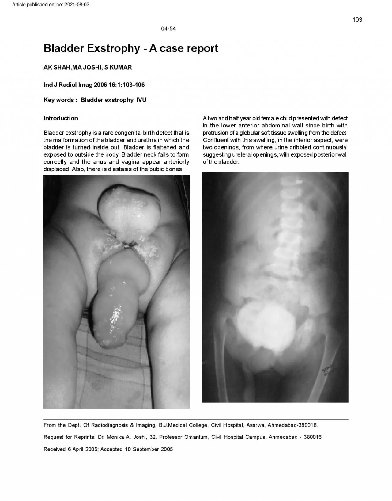
103 0454 Bladder Exstrophy A case report AK SHAHMA JOSHI S KUMAR Ind J Radiol Imag 2006 161103106 Key words Bladder exstrophy IVU Introduction A two and half year old female chil ID: 953872

exstrophy bladder anterior wall bladder exstrophy wall anterior abdominal cloacal urinary reconstruction defect urethra diagnosis openings short exposed distal

Share:

Link:

Embed:

Download Presentation from below link

Download Pdf The PPT/PDF document "Article published online 20210802" is the property of its rightful owner. Permission is granted to download and print the materials on this web site for personal, non-commercial use only, and to display it on your personal computer provided you do not modify the materials and that you retain all copyright notices contained in the materials. By downloading content from our website, you accept the terms of this agreement.


Presentation Transcript

Article published online: 2021-08-02 103 04-54 Bladder Exstrophy - A case report AK SHAH,MA JOSHI, S KUMAR Ind J Radiol Imag 2006 16:1:103-106 Key words : Bladder exstrophy , IVU Introduction A two and half year old female child presented with defect in the lower anterior abdominal wall since birth with Bladder exstrophy is a rare congenital birth defect that is protrusion of a globular soft tissue swelling from the defect. the malformation of the bladder and urethra in which the Confluent with this swelling, in the inferior aspect, were bladder is turned inside out. Bladder is flattened and two openings, from where urine dribbled continuously , exposed to out side the body . Bladder neck fails to form suggesting ureteral openings, with exposed posterior wall correctly and the anus and vagina appear anteriorly of the bladder . displaced. Also, there is diast asis of the pubic bones. From the Dept. Of Radiodiagnosis & Imaging, B.J.Medical College, Civil Hospit al, Asarwa, Ahmedabad-380016. Request for Reprint s: Dr . Monika A. Joshi, 32, Professor Omantum, Civil Hospit al Campus, Ahmedabad - 380016 Received 6 April 2005; Accepted 10 September 2005 104 104 AK Shah et al Umbilicus was not seen sep arately . Patient had another prolapsing elongated soft tissue inferior to the globular swelling, with an opening through which patient used to defecate, suggesting prolapsed rectum, which was somewhat anteriorly placed than normal individuals. Labia were widely separated and clitoris was bifid [Fig 1]. Plain KUB Radiographs were taken in antero-posterior and lateral projections [Fig 2,3] which revealed a globular soft tissue shadow arising from the anterior aspect of lower abdomen and pelvis. Another elongated sof t tissue opacity was seen below it arising from the pelvis and directed caudally.There was evidence of diastasis of pubic bones.Coccyx was absent. IJRI, 16:1, February 2006 Intravenous Urography IVP [Fig 4] was done by injecting 15 cc of 76% Urografin intravenously . There was prompt excretion of contrast from both kidneys. There was e/o fulln

ess of right renal pelvis with gross dilatation and tortuosity of both ureters, that of the right side being affected more than left, with abrupt tapering at their distal ends. Non filling of bladder was noted with leakage of contrast through ectopic ureteral openings s/o partial obstruction at distal ends of both ureters. Diagnosis was put as exstrophy of bladder with bilateral ectopic ureteral openings with distal partial strictures with back pressure changes in the form of bilateral hydroureter , more on the right side, with rectal prolapse and omphalocoele. 105 IJRI, 16:1, February 2006 Bladder Exstrophy 1 0 5 Discussion Exstrophy of bladder is a rare condition with incidence of 1 per 30,000 - 50, 000 live births with male to female ratio ranging from 1.5 : 1 to 5 :1 [1,2,4]. The risk of having sibling with bladder exstrophy is 1% [7]. The condition is intermediate in severity between epispadias and cloacal exstrophy . The condition is thought to be caused by incomplete development of the infra- umbilical part of the anterior abdominal wall, associated with incomplete development of the anterior wall of the bladder owing to delayed rupture of the cloacal membrane. Persistence of the cloacal membrane prevents medial mesenchymal ingrowth, causing the abdominal wall to remain lateral and the posterior bladder wall to be exposed to the external surface [2, 7]. Trigone of the bladder and ureteral openings are exposed and sometimes there is mild prolapse. The anterior abdominal wall defect involves the entire urethra and bladder neck [4].The pubic symphysis is always widened [3] with diastasis of rectus abdominis. Umbilicus is low set [4, 7] . Frequently there is omphalocoele [4,7] which is confluent with exstrophic bladder . [4] In males the penis is short, stubby , curved upwards and is drawn into the exstrophic area [2,4]. Unilateral or bilateral cryptorchidism may be present [2,4]. Inguinal hernia may be associated [2,4]. In females, the urethra is short, often buried in the exstrophied bladder . The clitoris tends to be bifid. The labia are also wi

dely separated. The vagina is short and orifice may be stenotic. Uterine prolapse or unicornuate uterus may be present [2, 4]. Anus is anteriorly placed and may be patulous, and this is more commonly seen in girls [4]. Distal ends of ureters are slightly dilated, and curve laterally , then medially and slightly upwards in the shape of a hook before entering the bladder . [4]. In untreated patients, due to continuous dribbling of urine, there can be mucosal abrasion, infection, squamous metaplasia resulting in acquired VUJ obstruction. The detrusor muscle may become fibrotic and scarred. Instances of adenocarcinoma of bladder have been reported in untreated adult patients. [4]. Diagnosis of exstrophy - epispadias complex can be made antenat ally . Antenat al USG findings include [5,6].­ Repeated failure to visualize the bladder . Lower anterior abdominal wall mass. Low set umbilicus with omphalocoele. Abnormal genitalia. Increased pelvic diameter . Associated renal anomalies, myelomeningocoele and limb anomalies, which are more common in cloacal exstrophy . In neonates, exstrophy of bladder is diagnosed on clinical examination; workup includes baseline RFT before complex reconstruction of the urinary tract. Renal USG is done to rule out renal agenesis, hydronephrosis and ectopic kidney . Af ter bladder reconstruction is done, USG is done to look for upper urinary tract deterioration which may result from increased bladder pressure or repeated infection. S pinal USG , radiography and MRI is done to exclude myelodysplasias or vertebral anomalies. [7]. An early assessment by examination under anaesthesia should be carried out in a center experienced with the condition. The goals of surgery are to close the bony pelvic ring, close the bladder , posterior urethra and close the anterior abdominal wall defect and reconstruct the genitalia. In the first year of life, the bladder is closed following osteotomy of both iliac bones just lateral to SI joints. Later reconstruction of bladder neck and sphincter is done [2]. Complications of closure is the failure to

reach adequate capacity and thus augmentation or reconstruction may be necessary [2]. Another option is urinary diversion if continence is poor following bladder reconstruction. This can be done by ureterosigmoid anastomosis or formation of ileal conduit, colonic conduit or continent urinary diversion [1,2,3].Complications include stricture at the site of anastomosis, increased chances of adenomas and adenocarcinomas at the site of ureterocolic anastomosis [3], and hyperchloraemic acidosis. References 1. J Ben- Chain, CG Docimo, RD Jeffs and JP Gearhart, Dep artment of Urology , John Hopkins Hospit al and University , School of Medicine, Baltimore, USA, Journal of Royal Society of Medicine, VOL. 89 Issue 1: 39-46. Copyright 1996 by Royal Society of Medicine. 2. RCG Russell, Norman S Williams, Christopher JK Bulstrode. The urinary bladder - congenital defects of bladder , in Bailey and Love Short Practice of Surgery , 23rd edition. Arnold Publishers : Pages 1202-1203,. 3. R. de Bruyn, I. Gordon and K. McHugh: Imaging of the kidneys and urinary tract in children, in Grainger and Allison's diagnostic radiology- A textbook of medical 106 106 AK Shah et al IJRI, 16:1, February 2006 imaging , 4th edition., V ol 2 , 2001:1733- 1734. : 2123-2124. 4 . Frederic N Silverman, Jerald P . Kuhn. Chap 34 ­ abnormalities of pelvocaliceal system, Ureter , Bladder 6 . Pinnette MG , Pan YQ, Pinnette SG , et al.: Prenat a l diagnosis of fetal bladder and cloacal exstrophy by and Urethra, Section 3 , Epispadias - Exstrophy complex, in Caf fey's Pediatric X ray diagnosis - An integrated ultrasound - A report. Reproduction Medicine, 1996, Feb; 41(2): 132-134. 5 . approach, Mosby , 1993: 1298 - 1301. Hamada H T akanok, Shiina H et al.: New 7 . Gearhart JP , Jef fs RD : Exstrophy - Episp adias complex and bladder anomalies, In Campbell MF , Relik AB, Ultrasonographic criteria for the prenatal diagnosis of Cloacal Exstrophy , Journal of Urology , Dec 1999, 162(6) V aughan B, Campbell's Urology , 7th edition, Philadelphia;Pa: W . B. Saunders,1998:1939 - 1990.