/
Int. J. Electrochem. Sci., Int. J. Electrochem. Sci.,

Int. J. Electrochem. Sci., - PDF document

yoshiko-marsland
yoshiko-marsland . @yoshiko-marsland
Follow
363 views
Uploaded On 2016-05-01

Int. J. Electrochem. Sci., - PPT Presentation

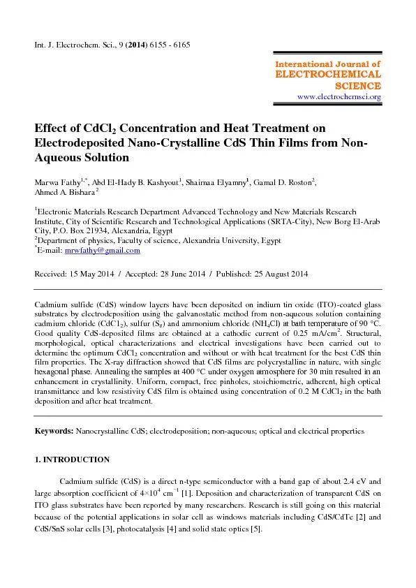
9 2014 6155 6165 International Journal of ELECTROCHEMICAL SCIENCE wwwelectrochemsciorg Effect of CdCl 2 Concentration and Heat Treatment on Electrodeposited Nano Crystalline CdS Thin Films ID: 301991

( 2014) 6155 - 6165 International Journal of ELECTROCHEMICAL SCIENCE www.electrochemsci.org Effect

Share:

Link:

Embed:

Download Presentation from below link

Download Pdf The PPT/PDF document "Int. J. Electrochem. Sci.," is the property of its rightful owner. Permission is granted to download and print the materials on this web site for personal, non-commercial use only, and to display it on your personal computer provided you do not modify the materials and that you retain all copyright notices contained in the materials. By downloading content from our website, you accept the terms of this agreement.


Presentation Transcript

Int. J. Electrochem. Sci., 9 ( 2014) 6155 - 6165 International Journal of ELECTROCHEMICAL SCIENCE www.electrochemsci.org Effect of CdCl 2 Concentration and Heat Treatment on Electrodeposited Nano - Crystalline CdS Thin Films from Non - Aqueous Solution Marwa Fathy 1,* , Abd El - Hady B. Kashyout 1 , Shaimaa Elyamny 1 , Gamal D. Roston 2 , Ahmed A. Bishara 2 1 Electronic Materials Research Department Advanced Technology and New Materials Research Institute, City of Scientific R esearch and Technological Applications (SRTA - City), New Borg El - Arab City, P.O. Box 21934, Alexandria, Egypt 2 Department of physics, Faculty of science, Alexandria University, Egypt * E - mail: mrwfathy@gmail.com Received: 1 5 May 201 4 / Accepted: 28 June 201 4 / Published: 25 August 201 4 Cadmium sulfide (CdS) window layers have been deposited on indium tin oxide (ITO) - coated glass substrates by electrodeposition using the galvanostatic method from non - aqueous solution containing cadmium chloride (CdC1 2 ), sulfur (S 8 ) and ammonium chloride (NH 4 Cl) t bth temperture of 90 °C. Good quality CdS - deposited films are obtained at a cathodic current of 0.25 mA/cm 2 . Struc tural, morphological, optical characterizations and electrical investigations have been carried out to determine the optimum CdCl 2 concentration and without or with heat treatment for the best CdS thin film properties. The X - ray diffraction showed that CdS films are polycrystalline in nature, with single hexgonl phse. Anneling the smples t 400 °C under oxygen tmosphere for 30 min resulted in n enhancement in crystallinity. Uniform, compact, free pinholes, stoichiometric, adherent, high optical trans mittance and low resistivity CdS film is obtained using concentration of 0.2 M CdCl 2 in the bath deposition and after heat treatment. Keywords: Nanocrystalline CdS; electrodeposition; non - aqueous; optical and electrical properties 1. INTRODUCTION Cadmi um sulfide (CdS) is a direct n - type semiconductor with a band gap of about 2.4 eV and large absorption coefficient of 4 × 10 4 cm − 1 [1]. Deposition and characterization of transparent CdS on ITO glass substrates have been reported by many researchers. Research is still going on this material because of the potential applications in solar cell as windows materials including CdS/CdTe [2] and CdS/SnS solar cells [3], photocatalysis [4] and so lid state optics [5]. Int. J. Electrochem. Sci., Vol. 9 , 201 4 6156 The complete devices and individual layers of the devices are the subjects of processing/performance studies, which are sometimes empirical and sometimes present back tracking in order to establish the means by which they are effecti ve [6]. A variety of deposition techniques have been reported in the literature for the preparation of CdS thin films; including sputtering [7], MOCVD [8], anodic oxidation, cathodic reduction, chemical vacuum deposition [9], spray pyrolysis [10,11], chemi cal bath deposition [12,13], successive ionic layer adsorption and reaction (SILAR) [14], electrochemical atomic layer epitaxy (ECALE) [15,16] or electrodeposition [17 – 19] from aqueous and non - aqueous solutions containing a soluble metal salt and sulfur co mpounds that has been developed. The latter method has drawn a special attention because of low cost, easy control of growth parameters and good adhesion of the obtained films to the substrate. Morphology and quality of the CdS films and, in consequence, e fficiency of resultant photovoltaic cells, are strongly dependent on several parameters which should be very carefully adjusted: composition of the solution, especially sources and concentration of sulfur and cadmium ions [20 - 23], pH [20,24,25] and temper ature [22,25 - 27] of electrodeposition bath as well as value of current density [28]. In this work, transparent CdS thin films on ITO glass substrates are electrodeposited using galvanostatic technique from non - aqueous solution bath consisting of Cadmium ch loride, sulfur and ammonium chloride in ethylene glycol solution and the deposited films are annealed under oxygen atmosphere. Structural, morphological, optical and electronic properties are investigated to optimize the fabrication parameters to produce u niform, stable, adherent, stoichiometric, and conductive CdS thin films on ITO glass substrate. 2. EXPERIMENTAL 2.1. Methodology The substrates are soda - lime glasses coated with Indium - Tin oxide thin film (ITO) layer using sputtering technique [29]. The overall thickness of ITO is about 650 nm. The substrate has high transparency and sheet resistance of about 12 Ω / □ . The substrates are cleaned in an ultrasonic bath with acetone, methanol and distilled water respectively. The substrates are taken out from the bath, and then dried under a stream of nitrogen [30]. ITO glass substrates are then etched for 30 sec in 0.1 M HCl at room temperature in order to remove impurities and increase the roughness of ITO surface, and finally w ashed in de - ionized water. 2.1.1. Preparation of CdS non - aqueous solution Different concentrations of CdCl 2 ranged from 0.1 M to 0.3 M are added to 0.02 M of S 8 in ethylene glycol (non - aqueous solvent) due to its relative high boiling point (about 197.4 °C) s well s its stability at high temperatures [31]. Ammonium chloride is added with a concentration of 0.1 M, which plays an important factor as a supporting electrolyte and performs several functions in the Int. J. Electrochem. Sci., Vol. 9 , 201 4 6157 electrochemical process [ 32 ]. The solution t emperture is djusted t bout 120 °C to dissolve S 8 and then is kept t bout 90 °C with stirring rte of 300 rpm. 2.1.2. Electrodeposition of CdS thin films A potentio - galvano scan wenking pgs 95 is used for the electrodeposition process. Good quality CdS films have been deposited using different concentration of CdCl 2 (as shown in Table 1) at current density of 0.25 mA/cm 2 for 1x1 cm 2 ITO glass substrate [29]. Table 1 . Preparation parameters of CdS thin films Samples CdCl 2 Concentration (M) Sulfur Concentration (M) NH 4 Cl Concentration(M) Annealing t 400 ˚C for 30 min S1 0.1 0.02 0.1 - S2 0.2 0.02 0.1 - S3 0.3 0.02 0.1 - S4 0.1 0.02 0.1 Annealed S5 0.2 0.02 0.1 Annealed S6 0.3 0.02 0.1 Annealed Platinum sheet of size 2x2 cm 2 is used s the node. The deposition bth temperture is 90 °C and a deposition time of 30 min to produce thin film thickness of 100 nm according to equation (1) [33]. Where d is the film thickness, J is the current density (mA/cm 2 ), M is the molecular weight (g), t is the deposition time (sec), n is the number of electrons, F is Frdy constnt (96500 C) nd ρ is the CdS densit y (4.84 g/cm 3 ). Figure 1. Optical microscope image of CdS thin film electrodeposited on ITO glass substrate at bath temperature of 100˚C . Int. J. Electrochem. Sci., Vol. 9 , 201 4 6158 The effect of bath deposition temperature is studied in the literature, where as the deposition bath temperature increases, the grain size increases. Also the rise in temperature of the bath enhances the rate of diffusion, increases the ionic mobility and hence the conductivity of the bath. So the best deposition bth temperture is 90 °C becuse more homogenous crystlliztion is formed t this temperture [23]. So, below 90 °C, non uniform in nture, nd bove it, very thin visible crcks is produced [22] , as shown in Figure 1. The source used for sulfur ions is pure S 8 and the overall reaction is assumed to be [ 34 ] . Cd ++ + S + 2e - ↔ CdS This method has the advantage of being simple and needs only a power supply and a temperature controller to control the bath temperature during the deposition process. It has been demonstrated to be useful to avoid concentration gradients and precipitation. The obtained CdS films are homogeneous, smooth with a bright yellow color and good adhesion to the substrate. After deposition, the samples are rinsed in DI water and blown dry with filtered compressed air. The produced smples re nneled under oxygen tmosphere t 400˚ C for 30 min. 2.2. Characterization methods X - ray powder diffraction measurements performed using Shimadzu 7000 XRD, with CuK α radiation (λ= 1.5418 Å) generted t 30 kV nd 30 mA with scnning rte of 4º min - 1 for 2 θ values between 20 and 70 degrees. The surfaces of the CdS thin films are investigated with high - resolution scanning electron microscopy (JEOL, JSM - 6360 LA) to examine the morphology and the homogeneity of the surface. Thin films of gold are sputtered onto the samples to get charge free surfac es and an accelerated voltage of 30 kV is used . Composition of solid phases is estimated by energy dispersive spectrometry (EDS). Optical transmittance measurements are performed with double beam UV - Vis spectrophotometer under normal incidence in the rang e of 190 - 900 nm. The band gap energy (Eg) of CdS films is calculated using transmittance spectra. Electricl properties of the films with thickness (t) of 0.8 μm re studied by Hll Effect measurement system (MMR Technologies, Inc.1400 North Shoreline Bl vd., Unit A5, CA 94043). Resistivity, Sheet resistance Rs, mobility, Hall coefficient and type of carriers are measured by four probs. All measurements are made at room temperature. 3. RESULTS & DISCUSSION 3.1. Structural analysis X - ray diffraction is used to analyze the formation of the crystalline phases. Generally, the hexagonal phase and the cubic phase are two crystalline modifications of CdS. X - ray diffraction (XRD) spectra of cadmium sulfide Samples; S1, S2, S3, S4, S5 and S6 are presented in Fi gure 2 as a function of the CdCl 2 concentration and heat treatment, respectively. Int. J. Electrochem. Sci., Vol. 9 , 201 4 6159 Fig ure 2. XRD spectra of electrodeposited CdS samples using different CdCl 2 concentrations, and before and after heat treatment. For S1; the peks t 2θ = 26.45 ˚ , 34.6˚ re ssocited with  mixture of hexgonl (002)/cubic (111) planes [35] and the hexagonal (110)/cubic (220) planes of CdS, respectively. The peak at 2 θ= 24.8˚, 36.6˚, 47.8˚ nd 51.8˚ correspond to the hexgonl ( 102), (110), (103) and (112) planes , respectively (according to JCPDS card no. 41 - 1049) [36]. Beside these, no other peaks of cubic phase CdS appeared. Indicating that, the structure of the films is predominantly hexagonal which consider as the stable phase of CdS at room temperature [7] . F or S2 and S3; the intensity of the peaks is increased which indicate that the crystallinity increased as the CdCl 2 concentration increase. Table 2 . Crystallite size, EDS analysis, and energy bandgap of CdS samples Samples Crystallite size (L) (nm) Atomi c ratio Eg (eV) S Cd S/Cd S1 24 68.04 31.96 2.1 2.6 S2 30 56.88 43.12 1.3 2.43 S3 25 43.4 56.5 0.7 2.58 S4 34 65.11 34.89 1.8 2.497 S5 28 51.45 48.55 1.05 2.375 S6 29 45.13 54.87 0.8 2.543 For S4, S5, and S6, it is seen that no new peaks appeared, indicating that there are no new crystalline phases. However, the preferred orientation of heat treated CdS samples seems to be slightly Int. J. Electrochem. Sci., Vol. 9 , 201 4 6160 affected. The intensity of the peak increases and very sligh t shift towards lower scattering angles is observed. Increasing the peak intensity indicates improved crystallinity due to heat treatment process. The shift towards a lower and the consequent increase in the lattice parameter resulting in enlarging of the CdS lattice is due to the filling of the vacancies and the appearance of sulfur interstitial. Both defects contribute to the enlarging of the CdS cell [25]. Also, the position shift of the peaks may correspond to the relaxation of tensile stress [6] and recrystallization process [37]. The individul crystllite size (L) is determined using Scherrer’s formul [38] (eqution where k is Scherrer’s constnt, which is  reference vlue corresponding to the qulity fctor of the apparatus measured with a reference single crystal and dependent on the crystallite shape (0.89 – 0.9). λ is the X - ray wave length, B is the FWHM (full widt h at half maximum) or integral breadth of the diffrction pek nd θ is the Brgg ngle [39]. The calculated data of the crystallite size are summarized in Table 2. For CdS thin films, the crystalli te size increased from 24 nm for S1 to 34 nm for S4 and i ncreased from 25 nm for S3 to 29 nm for S6 as a result of heat treatment [39]. But for sample S2, there is a slight decrease in the crystallite size by heat treatment from 30 nm (S2) to 28 nm (S5). 3.1.2. Morphological analysis Figure 3 Shows the SEM mic rographs of CdS thin films grown on ITO/glass substrate using different CdCl 2 concentrations before and after heat treatment (S1, S2, and S3) and (S4, S5, and S6), respectively. Generally; CdS thin films grown on the ITO/glass substrates had a compact and regular structure having an average grain size of about 35 nm with very well define grain boundaries [30, 40, 41] as shown for S1, and S2. But for S3, with the increasing CdCl 2 concentration, CdS particles are aggregates and the grain sizes increased obvio usly. It is necessary to optimize the CdCl 2 concentration to obtain pore - free and compact CdS films. After heat treatment under oxygen atmosphere, there is no much difference, no pinholes and cracks among grain boundaries are observed, any oxide barriers can found on crystals are removed and the crystals are grown together [37] for S4 and S5 sampl es but S6 some particles are found on the surface of the CdS film and grains coalesce between them producing larger sizes and holes which is also observed in the other reports [8]. EDS analysis of all CdS samples is summarized in Table 2. It confirms that the composition of cadmium and sulfur in CdS films on ITO/glass substrate is related to the CdCl 2 concentration. S1, the S/Cd atomic ratio is 2.1, which indicates free sulfur content. Increasing the Cd co ncentration to 0.2 M and maintaining S ions as 0.02 M, S/Cd ratio becomes 1.3 , which approximated to the stoichiometric ratio [7]. By further increasing the Cd concentration to 0.3 M, S/Cd ratio is reduced to 0.76 due to produce Cd rich sample. After heat treatment under oxygen atmosphere, sulphur deficiency is observed in all the films. This may be due to the fact that sulphur has great affinity towards oxygen, so it might have converted to SO 2 and then evaporated. EDS result of S5 reveals that the deposit ed films are very close to the nominal composition [42]. Int. J. Electrochem. Sci., Vol. 9 , 201 4 6161 Fig ure 3. SEM images of electrodeposited CdS thin films samples. 3.1.3. Optical and electrical analysis The Transmittance spectra of as - deposited and heat treated CdS samples are studied at normal incidence in the wave length range from 300 to 900 nm, as shown in Figure 4. Fig ure 4. Optical transmittance spectra of CdS thin film samples. The spectra exhibited interference fringes and the value of refractive index is estimated by the envelop method as follows [43, 44]: Int. J. Electrochem. Sci., Vol. 9 , 201 4 6162 [ n= [N+( - N = - Where n and n s are the refractive index of the sample and the substrate, respectively, T max , T min are the maximum and minimum transmittances at the same wavelength in the fitted envelope curve on  trnsmittnce spectrum, α is the bsorption coefficient, d is the film thickness, λ 1 nd λ 2 are the wavelengths at the two adjacent maxima or minima. The absorption coefficient for the direct allowed transition can be described as a function of photon energy [45] as equation (8).  ≈ A (hν – E g ) 2 Where A is the constnt which is relted to the effective msses ssocited with the bnds, ν is the frequency of the incident photons nd hν is the photon energy [46]. For as deposited samples (S1, S2 and S3), the average transmittance of the films on ITO/glass substrates is about 76, 77, and 74%, and the Eg is 2.6, 2.43, and 2.58 eV (as shown in Table 2), respectively. The higher transmittance (S1 and S2) indicates lower defect density and better electrical properties of the CdS films because the absorption of light in the wavelength longer region is usually caused by crystalline defects such as grain bound aries and dislocations [7] but S3 exhibits lower transmission because it is cadmium rich, also no fringes observed which indicate low film adhesion on ITO/glass substrate. After heat treatment, the optical transmission for S4, S5, and S6 increases and the absorption edge becomes much sharper. This is due to improve the crystallinity of the samples. Also, a decrease of the band gap of S4, S5 and S6 to 2.49, 2.37, and 2.54eV, respectively is observed as reported previously by other authors [25]. This may be d ue to electron - electron an d electron impurity interaction. The electrical properties of the CdS films are examined at room temperature by resistivity and Hall measurements using Van der Pauw method and summarized in Table 3. Table 3 . The electr ical properties of CdS thin films. Samples Resistivity (Ω.cm) Mobility (cm 2 /N s ) Carriers Density (cm - 3 ) Hall coefficient (cm 3 /coul.) Sheet resistance (Ω/cm 2 ) S1 2.3 6.79  10 - 5 3.97  10 12 1.57  10 6 4.6  10 4 S2 6.6  10 - 1 2  10 2 4.67  10 16 1.335  10 2 8.29  10 3 S3 2.2  10 - 1 5.45  10 3 5.1  10 15 1.2  10 3 2.77  10 3 S4 3.67 2.01  10 3 8.46  10 13 7.38  10 3 7.33  10 4 S5 2.31 4.5  10 3 6  10 14 1.04  10 4 2.89  10 4 S6 2.2  10 1.18  10 3 2.37  10 13 2.63  10 5 2.28  10 5 Int. J. Electrochem. Sci., Vol. 9 , 201 4 6163 It is observed that, for S1, S2, and S3 samples, the resistivity decreases and Hall mobility increases with CdCl 2 concentration increases . This may be due to the change in film stoichiometry (excess cadmium or sulphur vacancies, which are electron donor sites that provide the additional carriers and decrease the resistivity) [47]. The conduction of CdS films at room temperature can also be explained considering the micro - structural aspects. S2 has higher carrier concentration than another two samples. Consequently; S2 has low er sheet resistance than S1 which strongly depends on the variations in charge - carrier density rather than the mobility. The density of the major carriers of the CdS film can be relatively calculated from the following relation [48, 49]. Where N is the density of the majority carriers, e is the Charge of the electron, and R H is the hall coefficient [50]. Also, the grain boundaries between the crystallites dominate the ele ctrical properties of the polycrystalline thin film semiconductors [51 ]. Carrier traps at the grain boundaries are responsible for the potential barrier that limits carrier mobility. The carrier mobility is inversely proportional to the film roughness and the carrier concentration is proportional to the grain size [52]. Experimentally; the type of CdS film is found as n – type, depending on the value of Hall Factor (R H .) which is calculated by using the equation: R H is measured for the film at B = 0.1 Tesla. Where; I is the current flow through the film, V H is the generated voltage on both sides of the film, and B is the magnetic field. The influence of heat treatment process on the resistivity of the films is depicted in Table 3. The magnitude of the resistivity increased with heat treatment for all samples. It is attributed to the change in film stoichiometry (excess of sulfur vacancies which are electron donner sites that provide the additional carriers and decrease the resistivity). In addition, during the heat treatment process, oxygen fills the S vacancies so the donor sites are eliminated, the free carrier concentration is reduced a s shown in table. The absorbed oxygen offsets the decrease in resistivity due to the excess carriers provided by the excess cadmium obtained on heat treatment resulting in a net increase of resistivity [47]. The carrier mobility increases for S4 and S5 sam ples from 6.79  10 - 5 to 2.01  10 3 (cm 2 /N s ), and from 2  10 2 to 4.5  10 3 (cm 2 /N s ), respectively. It is attributed to, after heat treatment, the crystals are grown together and the electrical contacts between them are improved [37]. But for S6, The grain bo undaries between crystals dominate the electrical properties of the sample. Traps at grain boundaries are responsible for the potential barrier that limits carrier mobility [47]. 4. CONCLUSIONS The structural, morphological, optical and electrical pro perties of CdS film electrodeposited on ITO glass substrate from non - aqueous solution as a function of different CdCl 2 concentrations and heat treatment in oxygen atmosphere are investigated. XRD patterns indicated that the structure of CdS film Int. J. Electrochem. Sci., Vol. 9 , 201 4 6164 is hexagon al and the crystallinity increased after heat treatment under oxygen atmosphere. The as prepared CdS film using 0.2 M CdCl 2 , 0.02 M S 8 , and 0.01M NH 4 Cl is uniform, compact and free of holes and after heat treatment, the grain sizes of the film are almost the same as the deposited film and no pinholes and cracks among the grain boundaries are observed. Increasing CdCl 2 concentration in the deposition bath and heat treatment also make some effect on the optical transmittance of the CdS films and the optical bandgap of the film. The film stoichiometry plays an important role in the resistivity of the CdS film. During the heat treatment under oxygen atmosphere, the oxygen fills the S vacancies in the CdS and the doner sites are eliminated which effect the reduc tion in the carrier concentration and consequently the resistivity is increased. ACKNOWLEDGEMENT A part of this work has been supported by the Ministry of Science Research (ASRT), Egypt, under the Science and Technology Development Fund Program (STDF). P roject ID: 1396 with title “Synthesis of thin films to produce photovoltic solr cells”. References 1. K. Senthil, D. Mangalaraj, Sa. K. Narayandass, B. Hong, Y. Roh, C.S. Park, J. Yi, Semicond. Sci. Technol. 17 (2002) 97 - 103. 2. M. Soliman, A. B. Kashyout , M. El - Gamal, M. Shabana, 17 th European Solar Energy Conference, (2001). 3. J. Xu, Y. Yang , Energy Conversion and Management , 78 (2014) 260 - 265 . 4. H. Zhang, G. Chen, D.W. Bahnemann, J. Mater. Chem . 19 (2009) 5089 - 5121. 5. Y. Wang, Z. Tang, M.A. Correa - Duarte, L.M. Liz - Marzan, N.A. Kotov , J. Am. Chem. Soc . 125 (2003) 2830 - 2831. 6. M.K. Al Turkestani, K. Durose, Sol. Energy Mater. Sol. Cells , 95 (2011) 491 - 496. 7. M. Cao, L. Li, B.L. Zhang, J. Huang, K. Tang, H. Cao, Y. Sun, Y. Shen, J. Alloys and Compounds 530 (2012) 81 - 84. 8. J. Han, C. Liao, T. Jiang, G. Fu, V. Krishnakumar, C. Spanheimer, G. Haindl, Kui Zhao, A. Klein, W. Jaegermann, Mater. Res. Bull . 46 (2011) 194 - 198. 9. N.I. Fainer, M.L. Kosinova, Yu.M. Rumyantsev, E.G. Salman, F.A. Kuznetsov, Thin Solid Films 280 (1 996) 16 - 19. 10. S. Mathew, P.S. Mukerjee, K.P. Vijayakumar, Thin Solid Films 254 (1995) 278 - 284. 11. J. Hiie, T. Dedova, V. Valdna, K. Muska, Thin Solid Films 511 – 512 (2006) 443 - 447. 12. J.P. Enriquez, X. Mathew, Sol. Energy Mater. Sol. Cells 76 (2003) 313 - 322. 13. R.S. Mane, C.D. Lokhande, Mater. Chem. Phys . 65 (2000) 1 - 31. 14. B.R. Sankapal, R.S. Mane, C.D. Lokhande, Mater. Res. Bull . 35 (2000) 177 - 184. 15. B.W. Gregory, J.L. Stickney, J. Electroanal. Chem . 300 (1991) 543 - 561. 16. M. Innocenti, S. Ca ttarin, M. Cavallini, F. Loglio, M.L. Foresti, J. Electroanal. Chem . 532 (2002) 219 - 225. 17. I. Sisman, M. Alanyahoglu, U. Demir, J. Phys. Chem . C 111 (2007) 2670 - 2674. 18. D. Lincot, Thin Solid Films , 487 (2005) 40 - 48. 19. M. Bouroushian, Ed. F. Scholz , Electrochemistry of Metal Chalcogenides, Springer, Verlag Berlin Heidelberg (2010) 91 . Int. J. Electrochem. Sci., Vol. 9 , 201 4 6165 20. J. Nishino, S. Chatani, Y. Uotani, Y. Nosaka, J. Electroanal. Chem . 473 (1999) 217 - 222. 21. M. Takahashi, S. Hasegawa, M. Watanabe, T. Miyuki, S. Ikeda, K. Iida, J. Appl. Electrochem . 32 (2002) 359 - 367. 22. S.J. Lade, C.D. Lokhande, Mater. Chem. Phys . 49 (1997) 160. 23. F. Kadirgan, D. Mao, W. Song, T. Ohno, B. McCandless, Turk. J. Chem . 24 (2000) 21 - 33. 24. G.P. Power, D.R. Peggs, A.J. Parker, Electrochim. Acta 26 (1981) 6 81 - 682. 25. G. Sasikala, R. Dhanasekaran, C. Subramanian, Thin Solid Films 302 (1997) 71 - 76. 26. S. Dennison, Electrochim. Acta 38 (1993) 2395 - 2403. 27. S. J. Lade, M.D. Uplane, C.D. Lokhande, Mater. Chem. Phys . 53 (1998) 239 - 242. 28. B.E. McCandless, A. Mondal, R.W . Birkmire, Sol. Energy Mater. Sol. Cells 36 (1995) 369 - 379. 29. A.B. Kashyout, M. Fathy, M. B. Soliman, International J. Photoenergy, A rticle ID 139374 (2011) 1 - 6. 30. M. Rami, E. Benamar, M. Fahoume, A. Ennaoui, Physica Status Solidi 172 (1999) 137 - 147. 31. R. K. Pandy, S. N. Sahu, S. Chandra, Hand book of semiconductor electrodeposition, Marcel Dekker Inc., New York (1996) 95. 32. A. B. Kashyout, M.M. Shabana, M.A. El - Gamal , M. B. Soliman, The effect of preparation parameters on the properties of CdTe films thesis, Alexandria university (2001) 66. 33. O.A .Ileperuma, C. Vithana, K. Premaratne, S.N. Akuranthilaka, S.M. MCGregor, I.M. Dharmadasa, J. Materials Science: Materials in electronics , 9 (1998) 367 - 372. 34. W.J. Danaher, L.E. Lyons, G.C. Morris, Sol. Ene rgy Mater. Sol. Cells , 12 (1985)137 - 148. 35. S. Butt, N. A. Shah, A, Nazir, Z. Ali, A. Maqsood , J. Alloys and Compounds , 587 (2014) 582 – 587. 36. H.Kim, D. Kim, Sol. Energy Mater. Sol. Cells , 67 (2001) 297 - 304. 37. J. Hiie, K. Muska, V. Valdna, V. Mikli, A. Taklaja , A. Gavrilov, Thin solid films , 516 (2008) 7008 - 7012. 38. B. D. Cullity, Elements of X - Ray Diffraction, 2nd ed. Addison Wesley, U.S.A. (1978) Chap. 3, 102. 39. S. Chun, K.S. Han, J.S. Lee, H.J. Lim, H. Lee, D. Kim, Current Applied Physics , 10 (2010) S196 – S200. 40. J. Hernândez - Borj, Y.V. Vorobiev , R. Rmrez - Bon, Sol. Energy Mater. Sol. Cells , 95 (2011) 1882 – 1888. 41. J. - H. Lee, Thin Solid Films , 515 (2007) 6089 – 6093. 42. A. Hasnat, J. Podder, J. Bangladesh Academy of Sciences , 37 (2013) 33 - 41. 43. M. Miyake, K. Murase, T. Hirato, Y.Awakura, J. Electroanal.Chem , 562 (2004) 247 - 253. 44. M. Miyake, K. Murase, T. Hirato, Y.Awakura, J. Electrochem. Soc , 150 (2003) C413. 45. J. Herrero and C. Guillen, J. Appl. Phys ., 69 (1991) 429 - 434. 46. E. Fatas, P. Herrasti, T. Garcia, Materials Ch emistry and Physics , 13 (1985) 497 - 502. 47. K.R. Murali, M. Matheline, R. John, Chalcogenide Letters 6 (2009) 483 – 490. 48. R. Hill, Ed. T. J. Coutts, Active and passive thin film devices, Academic press, New York (1978). 49. J. I. B. Wilson, J. Woods, J. Physics and Chemistry of Solids , 34 (1973) 171 - 181. 50. A. M. Suhail, W. A. A. Twej , S. S. Abdulah, N. M. Saeed, IBN AL - HAITHAM J. FOR PURE & APPL. SCI, 22 (2009) 2 - 10. 51. L.L. Kazmerski, Polycrystalline and amorphous thin film devices, Academic press, New York (19 80). 52. W. C. Shin and S. G. Yoon, J. Electrochem. Soc . 144 (1997) 1055 - 1060. © 2014 The Authors. Published by ESG ( www.electrochemsci.org ). This article is an open access article distributed under the terms and conditions of the Creative Commons Attribution license (http://creativecommons.org/licenses/by/4.0/).