
Normal parathyroid Incidental cyst not important capsule The neoplasm Normal parathyroid Parathyroid adenoma Tumor behavior is much more important than morphology Tumor invasion in the capsule ID: 930464
Download Presentation The PPT/PDF document "Fat Oxyphil cells Chief cells" is the property of its rightful owner. Permission is granted to download and print the materials on this web site for personal, non-commercial use only, and to display it on your personal computer provided you do not modify the materials and that you retain all copyright notices contained in the materials. By downloading content from our website, you accept the terms of this agreement.


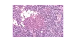
Slide1
Fat
Oxyphil
cells
Chief cells
Normal parathyroid
Slide2Incidental cyst
(not important)
capsule
The neoplasm
Normal parathyroid
Parathyroid adenoma
Tumor behavior is
much more important
than morphology
Slide3Tumor invasion in the capsule
Parathyroid carcinoma
Slide4Parathyroid hyperplasia
Same as normal tissue but expanded
…and somehow, adipose tissue is
diminished
Slide5Exocrine pancreas
(acini with zymogenic color of the cells)
Islet of Langerhans
Normal pancreas
Slide6Lymphocytes
Inflamed islet
Insulitis
DM type 1
Slide7Amyloid
Advanced stage
o
f
DM2
Islet of Langerhans
Slide8Islet
Normal pancreas
Islet cell tumor
(= pancreatic
neuroendocrine
tumor)
Trabeculae of tumor cells
…they can grow in many patterns
…here: trabecular pattern
May be benign
May be malignant
The tumor behavior
i
s the most important
Slide9Islet cell tumor
a
lso a trabecular pattern
is seen here
…resembling trabecular
bone
This is a neuroendocrine tumor (like carcinoid)
So when you look at the nuclei:
Slide10Zona glomerulosa
Zona
fasciculata
Zona reticularis
Medulla
Cells with
g
ood amount of basophilic
cytoplasm
(chromaffin cells)
Normal adrenal gland
Cortex
Slide11Atrophic adrenals
Normal adrenals
Hypertrophic glands
e.g., exogenous steroids
(low ACTH) or
Addison
e.g., ACTH
oversecretion
Golden cortex
Dark medulla
Slide12Neuroblastoma that have grown to the degree of displacing
the liver to the left
Liver
Neuroblastoma
Slide13An adrenal medullary tumor
…mainly in children
Slide14*It is a small round blue cell tumor
*High mitotic and apoptotic
activity and necrosis
mitoses
Slide15A well-circumscribed adrenocortical tumor
Adrenocortical adenoma
Slide16Adrenocortical adenoma
Resembling
adrenocortical
cells
Slide17Adrenocortical carcinoma
Discovered at an advanced stage because the retroperitoneal
s
pace permits this degree of enlargement
Slide18After staining with
d
ichromate...due to
t
he oxidation of
catecholamines
Pheochromocytoma
An adrenal medullary tumor
Slide19Part of
pheochromocytoma
tumor
…the cells have good
amount of basophilic cytoplasm
Pheochromocytoma
May be benign
May be malignant
The most important is behavior
Cortex
Slide20Capillary network
Nests
of polygonal to
spindle-shaped
chromaffin
cells
(
zellballen
pattern)
that are supplied by a rich vascular network
Pheochromocytoma
Slide21Capillary network
Pheochromocytoma
Sustentacular
cells
Nest
Nest
Nest
This nested
p
attern is called:
Zellballen
pattern
Slide22Thank You