/
Stomach   Presented by  Msc Stomach   Presented by  Msc

Stomach Presented by Msc - PowerPoint Presentation

bery
bery . @bery
Follow
65 views
Uploaded On 2024-01-03

Stomach Presented by Msc - PPT Presentation

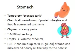
Dr Reham Saad Kadhum Gastrointestinal Tract Esophagus Abdominal Portion The esophagus is a muscular collapsible tube about 10 in 25 cm long that joins the pharynx to the stomach ID: 1037732

left stomach artery gastric stomach left gastric artery part greater curvature lesser fibers pyloric omentum orifice extends pylorus vein

Share:

Link:

Embed:

Download Presentation from below link

Download Presentation The PPT/PDF document "Stomach Presented by Msc" is the property of its rightful owner. Permission is granted to download and print the materials on this web site for personal, non-commercial use only, and to display it on your personal computer provided you do not modify the materials and that you retain all copyright notices contained in the materials. By downloading content from our website, you accept the terms of this agreement.


Presentation Transcript

1. Stomach Presented by Msc. Dr. Reham Saad Kadhum

2. Gastrointestinal TractEsophagus (Abdominal Portion)The esophagus is a muscular, collapsible tube about 10 in. (25 cm) long that joins the pharynx to the stomach. The greater part of the esophagus lies within the thorax (see page100). The esophagus enters the abdomen through an opening in the right crus of the diaphragm at level T 10 . After a course of about 0.5 in. (1.25 cm), it enters the stomach on its right side.

3. Blood SupplyArteriesThe arteries are branches from the left gastric arteryVeinsThe veins drain into the left gastric vein, a tributary of the portal vein Lymph DrainageThe lymph vessels follow the arteries into the left gastric nodes.Nerve SupplyThe nerve supply is the anterior and posterior gastric nerves (vagi) and sympathetic branches of the thoracic part of the sympathetic trunk.

4. stomachLocation and DescriptionThe stomach is the dilated portion of the alimentary canal .The stomach is situated in the upper part of the abdomen, extending from beneath the left costal margin region into the epigastric and umbilical regions.The stomach is relatively fixed at both ends but is very mobile in between .It is roughly J-shaped Has two opening the cardiac and pyloric orifices; two curvatures, the greater and lesser curvatures; and two surfaces, an anterior and a posterior surface , two borders right ( lesser curvature) and left border ( lesser curvature ) .It tends to be high and transversely arranged in the short, obese person (steer-horn stomach) and elongated vertically in the tall, thin person (J-shaped stomach).

5. Parts of stomach 1. Fundus: This is dome-shaped and projects upward and to the left of the cardiac orifice. It is usually full of gas.2. Body: This extends from the level of the cardiac orifice to the level of the incisura angularis, 3. Pyloric antrum: This extends from the incisura angularis to the pylorus .4. Pylorus: This is the most tubular part of the stomach.The thick muscular wall is called the pyloric sphincter, and the cavity of the pylorus is the pyloric canal .

6. The lesser curvature forms the right border of the stomach and extends from the cardiac orifice to the pylorus . It is suspended from the liver by the lesser omentum.The greater curvature is much longer than the lesser curvature and extends from the left of the cardiac orifice, over the dome of the fundus, and along the left border of the stomach to the pylorus The gastrosplenic omentum (ligament) extends from the upper part of the greater curvature to the spleen, and the greater omentum extends from the lower part of the greater curvature to the transverse colon .The cardiac orifice is where the esophagus enters the stomach . Although no anatomic sphincter can be demonstrated here, a physiologic mechanism exists that prevents regurgitation of stomach contents into the esophagus .The pyloric orifice is formed by the pyloric canal, which is about 1 in. (2.5 cm) long. The circular muscle coat of the stomach is much thicker here and forms the anatomic and physiologic pyloric sphincter. The pylorus lies on the transpyloric plane ( L 1 ) ,

7.

8.

9. Layers of Stomach The mucous membrane of the stomach is thick and vascular and is thrown into numerous folds, or rugae, that are mainly longitudinal in direction. The folds flatten out when the stomach is distended, So its responsible for increase surface area of stomach . The muscular wall of the stomach contains longitudinal fibers, circular fibers, and oblique fibers The peritoneum (visceral peritoneum) completely surrounds the stomach. It leaves the lesser curvature as the lesser omentum and the greater curvature as the gastrosplenic omentum and the greater omentum.

10.

11.

12. Relations■■ Anteriorly: The anterior abdominal wall, the left costal margin, the left pleura and lung, the diaphragm, and the left lobe of the liver■■ Posteriorly: The lesser sac, the diaphragm, the spleen, the left suprarenal gland, the upper part of the left kidney, the splenic artery, the pancreas, the transverse mesocolon, and the transverse colon

13. Blood SupplyArteriesThe arteries are derived from the branches of the celiac artery The left gastric artery arises from the celiac artery. It supplies the lower third of the esophagus and the upper right part of the stomach. The right gastric artery arises from the hepatic artery. It supplies the lower right part of the stomach.The short gastric arteries arise from the splenic artery at the hilum of the spleen and pass forward in the gastrosplenic omentum (ligament) to supply the fundus.The left gastroepiploic artery arises from the splenic artery at the hilum of the spleen ,supply the stomach along the upper part of the greater curvature.The right gastroepiploic artery arises from the gastroduodenal branch of the hepatic artery. supplies the stomach along the lower part of the greater curvatureFig ( 5. 20)

14.

15. VeinsThe veins drain into the portal circulation The left and right gastric veins drain directly into the portal vein. The short gastric veins and the left gastroepiploic veins join the splenic vein. The right gastroepiploic vein joins the superior mesenteric vein.Lymph DrainageThe lymph vessels follow the arteries into the left and right gastric nodes, the left and right gastroepiploic nodes, and the short gastric nodes. All lymph from the stomach eventually passes to the celiac nodes located around the root of the celiac artery on the posterior abdominal wall.

16. Nerve SupplyThe nerve supply includes sympathetic fibers derived from the celiac plexus and parasympathetic fibers from the right and left vagus nerves .The sympathetic innervation of the stomach carries a proportion of pain-transmitting nerve fibers, whereas the parasympathetic vagal fibers are secretomotor to the gastric glands and motor to the muscular wall of the stomach. The pyloric sphincter receives motor fibers from the sympathetic system and inhibitory fibers from the vagi.

17.

18.