PPT-13 Plasma lipids and lipoproteins
Author : lauren | Published Date : 2022-06-20
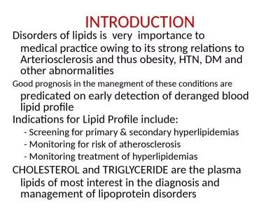
Plasma lipids 200 Lipoproteins 201 Fatty acids 200 Disorders of lipid metabolism 207 Cholesterol 201 Investigation of hyperlipidaemias 214 FATTY ACIDS
Presentation Embed Code
Download Presentation
Download Presentation The PPT/PDF document "13 Plasma lipids and lipoproteins" is the property of its rightful owner. Permission is granted to download and print the materials on this website for personal, non-commercial use only, and to display it on your personal computer provided you do not modify the materials and that you retain all copyright notices contained in the materials. By downloading content from our website, you accept the terms of this agreement.
13 Plasma lipids and lipoproteins: Transcript
Download Rules Of Document
"13 Plasma lipids and lipoproteins"The content belongs to its owner. You may download and print it for personal use, without modification, and keep all copyright notices. By downloading, you agree to these terms.
Related Documents