
MaruYama Takashi Gifu Univ School of Med Introduction Nuclear I κ B family proteins form a complex with NF κ B and control NF κ B target gene regulation I κ B ζ a member of this protein family can be induced in T cells in response to TGF ID: 908909
Download Presentation The PPT/PDF document "I k B NS controls Th17 differentiation ..." is the property of its rightful owner. Permission is granted to download and print the materials on this web site for personal, non-commercial use only, and to display it on your personal computer provided you do not modify the materials and that you retain all copyright notices contained in the materials. By downloading content from our website, you accept the terms of this agreement.


Slide1
I
k
BNS controls Th17 differentiation and Experimental Autoimmune Encephalomyelitis
MaruYama
, Takashi (Gifu Univ., School of Med.)
Introduction
Nuclear I
κ
B family proteins form a complex with NF-
κB and control NF-κB target gene regulation. IκB-ζ, a member of this protein family, can be induced in T cells in response to TGF-β+IL-6 and positively regulates Th17 differentiation. Thus, IκB-ζ-deficient mice are able to resist some Th17-dependent autoimmune diseases, such as experimental autoimmune encephalomyelitis (EAE). However, these mice develop different types of autoimmune disorders such as Sjogren syndrome with advanced age, limiting the utility of IκB-ζ as a therapeutic target. In this study, we focus on another nuclear IκB family protein, IκBNS (IκBNS is a most similar homolog of IκB-ζ) and asked whether IκBNS have a potential therapeutic target of EAE without any immune dysregulations.
Ankyrin
-repeats
Ankyrin
-repeats
Transactivation
domain
Nuclear
Localize
S
ignal
I
k
B
-
z
I
k
B
NS
43% match
・
Less
Th17 differentiation
Nature
. 2010
29;464:1381-5
・
Sjogren
syndrome like diseases
Immunity
.
2013 21;38:450-60
.
・
Not observe immune dysregulation
with ages
Deficient mice
Materials & Methods
Plasmids:
Expression
vectors encoding FLAG-tagged mouse
I
B-
were constructed as described previously (
JBC
2005 280
:
1678-1687
)
.
Mouse I
B
NS
was inserted into a pcDNA3-FLAG vector at the
EcoR
I
and
BamH
I
sites.
M
ouse
IL-17A promoter (-6647 to +1) was inserted into a pGL4.12 vector at the
Nhe
I
&
Hin
dIII
sites.
EAE induction
:
M
ice
were injected subcutaneously (on the lower back) on day 0 with emulsions containing complete Freund’s adjuvant (
CFA),
100
μg
MOG
peptide,
and 0.5 mg
Mycobacterium tuberculosis
H37RA.
In addition, these mice received 500 ng pertussis
toxin
by
i.p
. injection to boost immunological responses on day 0 and
2.
In vitro
T cell
culture:
Purified
CD4
+
CD25
-
T cells were cultured in RPMI 1640 medium containing 10% heat-inactivated fetal calf
serum
at
37°C
in 5%
CO
2
.
Th0 condition:
anti-CD3 (1
μg
/mL) + anti-CD28 (1
μg
/mL)
stimulation.
Th17 condition:
anti-CD3 (1
μg
/mL) + anti-CD28 (1
μg
/mL) with TGF-
1 (2 ng/mL) and IL-6 (50 ng/mL)
stimulation.
Data Analysis
: Data (Fig.1-3)
shown represent mean ± S.E. Paired data were evaluated using the Student’s t test. *
p
< 0.05, **
p
< 0.01.
Results
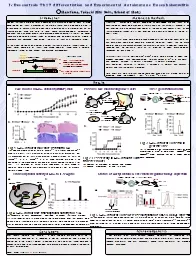
Fig.2 Less severity in
I
k
B
NS
deficient T cells
transfer model
(
A
) Diagram of methods of Passive EAE models.
(
B
) Disease progression of EAE
(
n =
3-4)
EAE
model in
I
k
B
NS
deficient (
Nfkbid
-/-
) mice
Fig.1
I
k
B
NS
deficient mice have resistant to EAE
(
A
) Disease progression of EAE in
Nfkbid
+/+
(n = 11–13) and
Nfkbid
-/-
mice (n = 9–11). (
B-C
)
Analysis of mice 12 days after immunization. (
B
) Measurement
of IL-17A
supernatant
concentrations by ELISAs (
Nfkbid
+/+
: n = 5;
Nfkbid
-/-
: n = 6), using cultured draining LNs incubated in the presence or absence of MOG peptide (10 ng/ml) for 72h.
(
C
)
Histology of spinal cord specimens in EAE models.
S
ections
were stained with
hematoxylin
and eosin (HE
) or
Klüver
-Barrera staining (
KB)
A
B
C
Passive EAE model using CD4
+
T cells
MOG+CFA
12 days
after immunization
Inguinal
draining LNs
Cultured
in the presence of
MOG
Purified CD4+ T cells
& transfer to WT mice
A
B
Th17 generation
In vitro
S
pleen
Naïve CD4
+
T
Cultured
With/without
TGF-
b
+IL-6
ELISA (A)
Supernatant
RT-PCR (B)
Cells
A
B
Fig.3
I
k
B
NS
deficient T cells fail to
generate Th17
(
A, B
)
Expression of IL-17A
protein (
A
)
or
Il-17a
mRNA
(
B
) in
CD4
+
T cells from
Nfkbid
+/+
and
Nfkbid
-/-
mice, cultured for 48 h under Th0 or Th17 conditions.
Transcriptional activity of
I
k
B
NS
to IL-17A gene
TCR
ROR
g
t
I
k
B
-
z
TGF-
b
IL-17 A
IL-6
I
k
B
NS
?
A
B
Fig.4
I
k
B
NS
did not have transcriptional activity to IL-17A
(
A
) Schematic of Th17 development controlled by nuclear
I
k
B
family
proteins.
(
B
)
Il-17a
reporter
activity presented as the fold-increase over that observed in HEK293 cells transfected with the empty vector.
Data shown are the mean ± S.D. of duplicate samples and are representative of 3 independent experiments.
Status of Acetyl Histone & Th17 master regulator ‘
ROR
g
t
’ expression
Fig.5
I
k
B
NS
deficient T cells were less acetylation of CNS1 &
ROR
g
t
expression
(
A
) Scheme of IL-17A gene conservation. (
B
)
ChIP
analysis of H3K27Ac. Cells were cultured under
Th17
conditions for 48 h.
(
C
)
RORγt
expression in CD4
+
T cells from
Nfkbid
+/+
and
Nfkbid
-/-
mice, cultured for 72 h under
Th17
conditions. Data are representative of three independent experiments.
S
pleen
Naïve CD4
+
T
Cultured (48h)
With
TGF-
b
+IL-6
ChIP
(B)
Cells
FACS (C)
ROR
g
t
&
I
k
B
-
z
binding element
A
B
C
Conclusions
O
ur
results indicate that
Nfkbid
-/-
T cells showed impaired Th17 cells differentiation because of a reduction in
RORγt
expression and histone H3 acetylation in the CNS 1 region. In conclusion, we show that I
BNS deficiency causes resistance to Th17-dependent autoimmune disease through the control of Th17 differentiation.PLoS One 2014 Oct 27;9(10):e110838.
Acknowledgements
I am
grateful to Dr.
Muta
,
Tatsushi
(Tohoku University) for supervising this research and for helpful discussions. We are also grateful to Mr.
Goto
,
Yasuyuki
(Tohoku University) and Ms. Takahashi, Kyoko (Gifu University) for providing technical assistance.