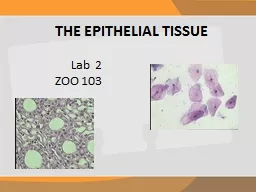
/
Epithelial Tissue Lab *Draw the diagram and answer the questions for each slide Epithelial Tissue Lab *Draw the diagram and answer the questions for each slide

Epithelial Tissue Lab *Draw the diagram and answer the questions for each slide - PowerPoint Presentation

mia
mia . @mia
Follow
343 views
Uploaded On 2022-06-11

Epithelial Tissue Lab *Draw the diagram and answer the questions for each slide - PPT Presentation

1 Simple Columnar   A Diagram cells are the layer of pink elongated cells the purple cells within them are goblet cells   B Name 2 areas where it is located in the body   C What is the function of a goblet cell ID: 917349

diagram cells stratified pink cells diagram pink stratified observe layer outer dark squamous purple body ciliated simple epithelial elongated

Share:

Link:

Embed:

Download Presentation from below link

Download Presentation The PPT/PDF document "Epithelial Tissue Lab *Draw the diagram ..." is the property of its rightful owner. Permission is granted to download and print the materials on this web site for personal, non-commercial use only, and to display it on your personal computer provided you do not modify the materials and that you retain all copyright notices contained in the materials. By downloading content from our website, you accept the terms of this agreement.


Presentation Transcript

Slide1

Epithelial Tissue Lab

*Draw the diagram and answer the questions for each slide

Slide2

1) Simple Columnar


A. Diagram (cells are the layer of pink, elongated cells, the purple cells within them are goblet cells)


B. Name 2 areas where it is located in the body.  C. What is the function of a “goblet” cell?

Slide3

2) Simple Squamous


A. Diagram (you are observing many of these purple cells clumped together; the darker purple dot in the middle of each cell is the nucleus)


B. Why do these cells need to be thin and smooth?

Slide4

3) Simple cuboidal


A. Diagram (these cells are lining each circle in the field of view)


B. Name 2 places it is located in the body.

Slide5

4) Fallopian Tube


A. Diagram (cells are outer layer of pink, elongated cells; notice the “fuzzy” cilia on the top)


B. Why are ciliated cells needed in the fallopian tubes?

Slide6

5) Stratified Squamous


A. Diagram (these cells are the lighter pink cells underneath the dark pink line)


B. Why do these cells need to be in layers? C. What material fills some of them?

Slide7

6) Ciliated Epithelium


A. Diagram (observe the outer layer of cells; note the cilia on top)


B. What is the area under the ciliated cells called?

Slide8

7) Stratified Columnar


A. Diagram (observe the outer layer of elongated cells)


B. Why is this called stratified?

Slide9

8) Pseudostratified


A. Diagram (observe the outer layer of dark pink cells)


B. Why is it referred to as “pseudo” stratified?

Slide10

9) Human Thyroid Gland


A. Diagram (observe the dark pink cells lining the circles)


B. Since this is a gland what type of cells are these?

Slide11

10) Human Stratified Squamous Smear


A. Diagram (observe the dark pink cells in the center of the field of vision)


B. This is a smear scraped off the surface of the body. Why would these cells be lost constantly? C. What cellular process replaces these cells?

Slide12

11)

A. Why do we not usually get infections in stratified squamous epithelium?

B. Why do epithelial cells not have blood vessels?

C. What are the 3 arrangements of cells in epithelial tissue?