
Essential idea Eukaryotes have a much more complex cell structure than prokaryotes T he background image above is an electron micrograph of pancreatic exocrine cells It clearly shows the complex structures present in eukaryote cells ID: 597484
Download Presentation The PPT/PDF document "1 .2 Ultrastructure of cells" is the property of its rightful owner. Permission is granted to download and print the materials on this web site for personal, non-commercial use only, and to display it on your personal computer provided you do not modify the materials and that you retain all copyright notices contained in the materials. By downloading content from our website, you accept the terms of this agreement.


Slide1
1
.2 Ultrastructure of cells
Essential idea: Eukaryotes have a much more complex cell structure than prokaryotes.
The background image above is an electron micrograph of pancreatic exocrine cells. It clearly shows the complex structures present in eukaryote cells.
By Chris Painehttps://bioknowledgy.weebly.com/
http://
medcell.med.yale.edu
/
systems_cell_biology_old
/
liver_and_pancreas
/images/
exocrine_pancreas_em.jpgSlide2
Understandings, Applications and Skills
Statement
Guidance
1.2.U1
Prokaryotes have a simple cell structure without compartmentalization.
1.2.U2
Eukaryotes have a compartmentalized cell structure.
1.2.U3
Electron microscopes have a much higher resolution than light microscopes.
1.2.A1
Structure and function of organelles within exocrine gland cells of the pancreas and within palisade mesophyll cells of the leaf.
1.2.A2
Prokaryotes divide by binary fission.
1.2.S1
Drawing of the ultrastructure of prokaryotic cells based on electron micrographs.
Drawings of prokaryotic cells should show the cell wall,
pili
and flagella, and plasma membrane enclosing cytoplasm that contains 70S ribosomes and a nucleoid with naked DNA.
1.2.S2
Drawing of the ultrastructure of eukaryotic cells based on electron micrographs.
Drawings of eukaryotic cells should show a plasma membrane enclosing cytoplasm that contains 80S ribosomes and a nucleus, mitochondria and other membrane-bound organelles are present in the cytoplasm. Some eukaryotic cells have a cell wall.
1.2.S3
Interpretation of electron micrographs to identify organelles and deduce the function of specialized cells.Slide3
1
.2.U3
Electron microscopes have a much higher resolution than light microscopes.Light microscopes are limited in resolution by the wavelengths of visible light (400–700 nm).
Electrons have a much shorter wavelength (2 – 12 pm) therefore electron microscopes have a much higher resolutionLight microscopes are usually limited to 1000x because, due to the resolution, nothing is gained by increasing the magnification – try zooming in on an image on your laptop or phone after a certain point there is no benefit to zooming in as the image becomes pixelatedResolution
is defined as the shortest distance between two points that can be distinguished
resolution
Millimetres
(mm)
Micrometres
(
μm
)
Nanometres(nm)Human eye0.1100100,000Light microscopes0.00020.2200Electron microscopes0.0000010.0011
http://
upload.wikimedia.org
/
wikipedia
/commons/c/c5/
Electron_Microscope.jpgSlide4
1
.2.U3 Electron microscopes have a much higher resolution than light microscopes.
Light microscopes allow us to see the structure of cells
Electron microscopes allow us to see the
ultrastructure
of
cells, such as these
pancreatic exocrine cells
Electron microscopes can see viruses (0.1μm diameter) , but light microscopes cannot
Ultrastructure
is all the structures
of a biological specimen that are at least 0.1nm in their smallest dimensionhttp://medcell.med.yale.edu/systems_cell_biology_old/liver_and_pancreas/images/exocrine_pancreas_em.jpgSlide5
1
.2.U1
Prokaryotes have a simple cell structure without compartmentalization.Slide6
1
.2.U1 Prokaryotes have a simple cell structure without compartmentalization.
Good web-links for learning about prokaryote ultrastructure:
http://
www.wiley.com/college/boyer/0470003790/animations/
cell_structure
/
cell_structure.htm
http://
www.sheppardsoftware.com
/health/anatomy/cell/
bacteria_cell_tutorial.htmSlide7
1
.2.U1 Prokaryotes have a simple cell structure without compartmentalization.
http://www.tokresource.org/tok_classes/biobiobio/biomenu/metathink/required_drawings/index.htm
Ultrastructure of
E. coli as an example of a prokaryote
E. Coli
is a model organism used in research and teaching. Some strains are toxic to humans and can cause food poisoning.
We refer to the cell parts/ultrastructure of prokaryotes rather than use the term organelle as very few structures in prokaryotes are regarded as organelles.Slide8
Prokaryotes reproduce asexually using the process of
b
inary fissionThe DNA is replicated semi conservatively [2.7.U1]The two DNA loops attach to the membrane
The membrane elongates and pinches off (cytokinesis) forming two separate cellsThe two daughter cells are genetically identically (clones)Binary fission is also used by some organelles in eukaryotes [links to 1.5.U3]
1.2.A2 Prokaryotes divide by binary fission.
http://
cronodon.com
/images/Bacteria_dividing_3b.jpg
https://
commons.wikimedia.org
/wiki/
File:Binary_Fission.png
http://youtu.be/vTzH1P3aQjgSlide9
1.2
.
S1 Drawing of the ultrastructure of prokaryotic cells based on electron micrographs.Slide10
1.2
.
S1 Drawing of the ultrastructure of prokaryotic cells based on electron micrographs.
Warning: only draw and label features you can clearly see – don’t put structures in because you think they should be there.Slide11
1.2
.S1 Drawing of the ultrastructure of prokaryotic cells based on electron micrographs
.Slide12
1.2
.U2 Eukaryotes
have a compartmentalized cell structure.
There are several advantages in being compartmentalized:Effeciency of metabolism - enzymes and substrates can localized and much more concentratedLocalised conditions - pH and other such factors can be kept at optimal levels. The optimal pH level for one process in one part of the cell
Toxic / damaging substances can be isolated, e.g. digestive enzymes (that could digest the cell itself) are stored in lysosomesNumbers and locations of organelles can be changed dependent on the cell’s requirements.
http://
medcell.med.yale.edu
/
systems_cell_biology_old
/
liver_and_pancreas
/images/
exocrine_pancreas_em.jpgSlide13
1.2.U2 Eukaryotes have a compartmentalized cell structure.
This is a good drawing, but the nucleus should show a double membrane and small pores (gaps) in the membrane.Slide14
1.2.U2 Eukaryotes have a compartmentalized cell structure.
Vesicle
m
embrane sac containing proteins ready for secretion
http://www.tokresource.org/tok_classes/biobiobio/biomenu/eukaryotic_cells
/liver_cell_500.jpg
This drawing is a better diagram, but lack a scale bar.Slide15
1.2
.A1 Structure and function of organelles within exocrine gland cells of the pancreas and within palisade mesophyll cells of the leaf.
http://
upload.wikimedia.org/wikipedia/commons/5/57/Micrograph_of_a_cell_nucleus.png
http://courses.lumenlearning.net/biology/wp-content/uploads/sites/5/2014/02/Figure_03_03_05.jpgNucleusGenerally spherical with a
double membrane
Pores (holes) are present in the membrane
Contains genetic information in the form of chromosomes (DNA and associated histone proteins)
Uncoiled chromosomes are referred to as chromatin – they stain a dark colour and are concentrated at the edges of the nucleus
mRNA is transcribed in the nucleus (prior to use in protein synthesis in the cytoplasm)
mRNA leaves the nucleus via the pores
(DNA is too large to move through the pores)Slide16
1.2
.A1 Structure and function of organelles within exocrine gland cells of the pancreas and within palisade mesophyll cells of the leaf
.
http://commons.wikimedia.org/wiki/File:Mitochondria,_mammalian_lung_-_TEM.jpg
Mitochondria in mammalian lung cellsThe Mitochondrion (pl. Mitochondria)Has a double membrane
A smooth outer membrane and a folded inner membrane
The folds are referred to as cristae
Variable in shape
Site of ATP production by aerobic respiration (if fat is
used as a source of
energy it is digested here)
http://
ibguides.com/images/biology_figure_8.1.2_mitochondrion.pngSlide17
1.2
.A1 Structure and function of organelles within exocrine gland cells of the pancreas and within palisade mesophyll cells of the leaf.
Free ribosomes80S Ribosomes (approx. 20nm diameter) -
larger than the ribosomes found in prokaryotesNo membraneThese appear as dark granules in the cytoplasmSynthesizes proteins to function in the cytoplasm, for use within the cell, e.g. enzymes
http://www.cc.kochi-u.ac.jp/~tatataa/genetics/Q2013/ribosome2.jpgSlide18
1.2
.A1 Structure and function of organelles within exocrine gland cells of the pancreas and within palisade mesophyll cells of the leaf.
The Rough Endoplasmic Reticulum (rER)
The consists of flattened membrane sacs, called cisternaeOften located near to the nucleus80S Ribosomes are attached to the outside of the cisternae are ribosomesrER synthesizes proteins which are transported, by vesicles, to the golgi
apparatus for modification before secretion outside the cellNeed a simple diagram
Smooth Endoplasmic Reticulum
no ribosomes present
we are not studying this structure it this courseSlide19
1.2
.A1 Structure and function of organelles within exocrine gland cells of the pancreas and within palisade mesophyll cells of the leaf.
The Golgi apparatusThis organelle
also consists of flattened membrane sacs called cisternae, like rER.Different to rER:No attached ribosomesOften sited close to the plasma membraneT
he cisternae are shorter and more curved that those of the rERThe Golgi apparatus processes (modifies) proteins from from the rER. The proteins are then repackaged in vesicles
for secretion outside the cell.
Need a simple diagram
http://
commons.wikimedia.org/wiki/File:C_Golgi.jpg
http
://commons.wikimedia.org/wiki/File:Golgi_in_the_cytoplasm_of_a_macrophage_in_the_alveolus_(lung)_-_TEM.jpgSlide20
1.2
.A1 Structure and function of organelles within exocrine gland cells of the pancreas and within palisade mesophyll cells of the leaf.
VesiclesA single membrane with fluid inside
Very small in sizeUsed to transport materials inside of a cellNeed a simple diagram
http://
commons.wikimedia.org/wiki/File:C_Golgi.jpg
http
://commons.wikimedia.org/wiki/File:Golgi_in_the_cytoplasm_of_a_macrophage_in_the_alveolus_(lung)_-_TEM.jpgSlide21
1.2
.A1 Structure and function of organelles within exocrine gland cells of the pancreas and within palisade mesophyll cells of the leaf.
LysosomesGenerally spherical with a single membrane
Formed from Golgi vesicles.They contain digestive enzymes for breakdown of:ingested food in vesiclesunwanted/damaged organellesThe cell itselfHigh concentration of enzymes (a type of protein) cause this organelle to stain heavily and hence appear dark on micrographs
http://1.bp.blogspot.com/-WGspbEtlkls/TgCUbtp7_-I/
AAAAAAAAAJw
/SoIp2vXzH4E/s320/Figure+2-26.bmpSlide22
1.2
.A1 Structure and function of organelles within exocrine gland cells of the pancreas and within palisade mesophyll cells of the leaf.
VacuolesSingle membrane with fluid inside
In Plant cells vacuoles are large and permanent, often occupying the majority of the cell volumeIn animals vacuoles are smaller and temporary and used for various reasons, e.g. to absorb food and digest ithttp://middletownhighschool.wikispaces.com
/file/view/ch13f20.jpg/173915407/ch13f20.jpgSlide23
1.2
.A1 Structure and function of organelles within exocrine gland cells of the pancreas and within palisade mesophyll cells of the leaf.
Flagellum (Flagella pl.)Thin projection (usually singular) from the cell surface.
Contain microtubulesUsed to move the cellhttp://medicine.utah.edu/surgery/andrology/images/P29SERIES2-4.JPG
ANIMALS ONLY*
*Mature plant cells do not possess
possess cilia and
flagella, but some plant gametes are motile and do have them. This is not a common occurrence and you would not be expected to know about this.Slide24
1.2
.A1 Structure and function of organelles within exocrine gland cells of the pancreas and within palisade mesophyll cells of the leaf.
CiliaThin projections from the cell surface.Contain microtubules
Used to either move the cell or to move the fluids next to the cellhttp://cache1.asset-cache.net/gc/139809475-shows-cilia-pseudostratifed-structure-goblet-gettyimages.jpg?v=1&c=IWSAsset&k
=2&d=ut2a821i%2BSbReC5nXMMSneS7x9HksglF3dCo8uKSNT1EglzTjjv58OvCiQSlcyy0ANIMALS ONLY*
*Mature plant cells do not possess
possess cilia and
flagella, but some plant gametes are motile and do have them. This is not a common occurrence and you would not be expected to know about this.Slide25
1.2
.A1 Structure and function of organelles within exocrine gland cells of the pancreas and within palisade mesophyll cells of the leaf.
MicrotubulesSmall cylindrical fibres called microtubules
Have a variety of functions, e.g. part of the structure of flagella and they play a role in cell divisionCentriolesConsist of two groups of nine triple microtubulesAre mainly found in animal cells, not present in vascular plants or fungi.
https://encrypted-tbn1.gstatic.com/images?q=tbn:ANd9GcSrrSOHTtgbp2fiBrTjBtL1sBOhi0i5z0pf4oWpQ9EdTqr_-IN9OQSlide26
1.2
.A1 Structure and function of organelles within exocrine gland cells of the pancreas and within palisade mesophyll cells of the leaf.
ChloroplastMany, but not all, plant cells contain chloroplasts
A double membrane surrounds the chloroplastInside are stacks of thylakoidsEach thylakoid is a disc composed of a flattened membraneThe shape of chloroplasts is variable but is usually ovoidThe site of photosynthesis and hence where glucose is produced.
Starch grains maybe present if photosynthesis is happening quicklyPLANTS ONLY
https://
benchprep.com
/blog/
wp
-content/uploads/2012/08/chloroplast2.jpg
http://
www.biology.arizona.edu
/biochemistry/problem_sets/photosynthesis_1/graphics/chloroplast.GIFSlide27
1.2
.A1 Structure and function of organelles within exocrine gland cells of the pancreas and within palisade mesophyll cells of the leaf.
Cell wallan extracellular component not an organelle.
secreted by all plant cells (fungi and some protists also secrete cell walls).Plant cell walls consist mainly of cellulose which is:Permeable - does not affect transport in and out of the cellStrong – gives support to the cell and prevent the plasma membrane bursting when under pressure
Hard to digest –resistant to being broken down, therefore lasts along time without the need for replacement/maintenancePLANTS ONLY
http://
www.oncoursesystems.com
/images/user/9341/10845583/01-03_PlantCell(L-Large).jpgSlide28
1.2
.S2 Drawing of the ultrastructure of eukaryotic cells based on electron micrographs
.
Draw a pancreas exocrine cellfrom the image below. You should be able to label the following structures:plasma membranemitochondriarERNucleusSecretory granules (dark spheres)
http://medcell.med.yale.edu/systems_cell_biology_old/liver_and_pancreas/images/exocrine_pancreas_em.jpgSlide29
1.2
.S2 Drawing of the ultrastructure of eukaryotic cells based on electron micrographs
.Draw a single palisade mesophyll cell from the image below. You should be able to label the following structures:
cell wallplasma membranechloroplastsvacuolenucleuscytoplasm
mitochondriahttp://www.lifesci.sussex.ac.uk/home/Julian_Thorpe/TEM19.htmSlide30
1.2
.S3 Interpretation of electron micrographs to identify organelles and deduce the function of specialized cells.Slide31
1.2
.S3 Interpretation of electron micrographs to identify organelles and deduce the function of specialized cells.Slide32
1.2
.S3 Interpretation of electron micrographs to identify organelles and deduce the function of specialized cells
.
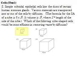
What organelles can you identify? Think about the role of the organelles that occur most common and deduce the function of the cell.Slide33
1.2
.S3 Interpretation of electron micrographs to identify organelles and deduce the function of specialized cells
.
What organelles can you identify? Think about the role of the organelles that occur most common and deduce the function of the cell.Evidence & conclusions:
Nucleus presentNo cell wall – this is an animal cellrER is present and dominates the cell – lots of protein product is made for secretion
Lots of mitochondria – the synthesis of protein requires
enegy
– this a metabolically active cell
Lots of secretory granules/vesicles near the inside border
Likely to be a cell that specializes in secreting a protein product, possibly a hormone or enzyme
Is in fact: a mammalian exocrine secretory cell from the pancreasSlide34
1.2
.S3 Interpretation of electron micrographs to identify organelles and deduce the function of specialized cells
.
What organelles can you identify in the top most layer of cells? Think about the role of the organelles that occur most common and deduce the function of the cell.http://bcrc.bio.umass.edu/histology/files/images/Pseudostratified%20Columnar%20Ciliated%20Epithelium1.jpgSlide35
1.2
.S3 Interpretation of electron micrographs to identify organelles and deduce the function of specialized cells
.
What organelles can you identify in the top most layer of cells? Think about the role of the organelles that occur most common and deduce the function of the cell.http://bcrc.bio.umass.edu/histology/files/images/Pseudostratified%20Columnar%20Ciliated%20Epithelium1.jpg
Evidence & conclusions:Nucleus presentNo cell wall – this is an animal cellCells closely packed – does not allow infiltration of substances from the lumen
Has a cilia dominated ‘brush border’ adjacent to a lumen – cilia are moving fluids in the lumen
Likely to be a protective layer of cells that actively sweep fluids away from the surface
Is in fact: ciliated epithelial cell from a mammalian lungSlide36
1.2
.S3 Interpretation of electron micrographs to identify organelles and deduce the function of specialized cells
.
What organelles can you identify? Think about the role of the organelles that occur most common and deduce the function of the cell.http://www.vcbio.science.ru.nl/images/tem-plant-cell.jpgSlide37
1.2
.S3 Interpretation of electron micrographs to identify organelles and deduce the function of specialized cells
.
What organelles can you identify? Think about the role of the organelles that occur most common and deduce the function of the cell.http://www.vcbio.science.ru.nl/images/tem-plant-cell.jpg
Evidence & conclusions:Cell wall present – must be a plant cellNo chloroplasts – must be found inside the stem or in the rootsVacuoles relatively small – the cell does not have a storage, transport or support function
Nucleus relatively large / cell size small – likely to be a new cell recently undergone mitosis
Possibly recently divided cell tissue from a plant root
Is in fact: a plant cell found at the root-tip