/
Skin  Malignancies   Part Skin  Malignancies   Part

Skin Malignancies Part - PowerPoint Presentation

olivia
olivia . @olivia
Follow
27 views
Uploaded On 2024-02-02

Skin Malignancies Part - PPT Presentation

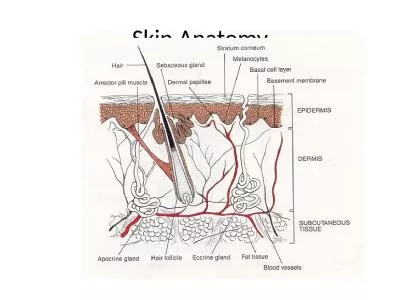
2 By Dr Ahmed Ali A bdelrahim Lecturer of Plastic Surgery Malignant Melanoma Outlines Epidemiology Incidence Risk factors Precancerous lesions Diagnosis of melanoma Growth phases ID: 1043732

skin melanoma node growth melanoma skin growth node common risk malignant phase fig tumor melanomas lymph diagnosis prognosis primary

Share:

Link:

Embed:

Download Presentation from below link

Download Presentation The PPT/PDF document "Skin Malignancies Part" is the property of its rightful owner. Permission is granted to download and print the materials on this web site for personal, non-commercial use only, and to display it on your personal computer provided you do not modify the materials and that you retain all copyright notices contained in the materials. By downloading content from our website, you accept the terms of this agreement.


Presentation Transcript

1. Skin Malignancies Part (2)By Dr. Ahmed Ali AbdelrahimLecturer of Plastic Surgery

2. Malignant Melanoma

3. OutlinesEpidemiologyIncidenceRisk factorsPrecancerous lesionsDiagnosis of melanomaGrowth phasesClinical types of cutaneous malignant melanoma Non-cutaneous malignant melanomaDifferential diagnosisBiopsyStagingPrognostic factorsTreatmentFollow up

4. It develops as a result of the malignant transformation of melanocytes. These cells are derived from the neural crest. Melanocytes are located in the deepest layer of the epidermis. Melanomas usually occur on the skin but can arise in other locations where neural crest cells migrate, such as in the gastrointestinal tract, the retina or the brain.

5. EpidemiologyIncidence Less than 3% of all skin cancers, but cause of 75% of skin cancer-related deaths.Risk factorsUV radiation exposure (both UVA and UVB): A history of blistering sunburns, particularly in early life, correlates to increased risk of some melanoma types.Age: 50% occur in patients older than 50 years.Phenotypic include fair skin (Fitzpatrick I and II) freckling, light eye color, and light hair color. Darker skin is protective against melanoma.

6. Geographic: High altitudes, lower latitudes have increased UV exposure, and therefore increased risk.Gender: Females have lower risk and better prognosis. Lower extremity is the most common site in females; males more commonly have lesions on the head and trunk.Race: Risk is 10 to 20 times higher for whites than blacks. Prognosis in darker skin is worse because of delayed diagnosis.Affluence: Unlike most cancer types, higher socioeconomic status correlates with higher risk.

7. Previous melanoma is a strong predictive factor and confers a 3% to 5% chance of developing a second melanoma.Immunosupression Family history: Vast majority of melanomas are sporadic; however, some hereditary forms exist. Familial melanoma (aka hereditary melanoma): Two or more cases of melanoma in first-degree relatives may indicate familial melanoma, autosomal dominant transference with variable penetrance.

8. Precursor lesionsCongenital nevi: Giant hairy nevi: 10% lifetime risk of melanoma Acquired melanocytic nevi: (< 5 mm) The greater the number of nevi, the greater the chance of melanoma.Dysplastic or atypical nevi: Often appear in puberty, Larger than common nevi (5 to 12 mm). Commonly found in covered areas, May represent a precursor lesion and/or marker for increased risk for melanoma development.

9. Precursor lesionsAtypical junctional melanocytic hyperplasia (AJMH) Also termed “lentigo maligna”; Hutchinson freckleMelanoma in situ: Melanoma precursor lesion; no penetration of atypical cells beyond epidermalDysplastic nevus syndrome (also known as familial atypical multiple mole andmelanoma [FAMMM] syndrome): Patients have a first- or second-degree relative with malignant melanoma and typically have at least 50 melanocytic nevi.

10. Xeroderma pigmentosum (XP)Transmitted through an incomplete sex-linked recessive gene (XR)Onset occurs during early childhood and is characterized by extreme sensitivity to sunlight (photophobia)Initially, diffuse lentigos are noted, with progressive drying and thinning of the skin (xeroderma). (Fig.16).The primary deficiency is that of the enzyme endonuclease, which is needed to repair sunlight-damaged DNA nucleotidesMalignant degeneration into basal cell carcinoma, squamous cell carcinoma, or melanoma is noted during early adult life (with some developing in the 1st few years of life), with death due to metastatic diseaseOther features include corneal opacities, eventual blindness, and neurologic deficits (xerodermic idiocy)Figure (16): Xeroderma pigmentosum with malignant ulcer

11. Diagnosis of melanomaCommon clinical features of melanoma lesions: (ABCDE)A: AsymmetryB: Border irregularityC: Color variationD: Diameter >6 mmE: Evolution: Extra features: Enlarging/evolving lesion, bleeding, itching

12. Growth phasesThe growth of a melanoma is described as having two phases.Radial growth phase: this is the proliferation of neoplastic melanocytes within the epidermis with only focal/single cell invasion of papillary dermis. This is typical of SSM, LMM, acral lentiginous MM.Vertical growth phase: this is the invasion of malignant melanocytes into papillary and reticular dermis and is typical of nodular MM and late LMM. Invasive melanocytes are spindle-shaped and infiltrate along neurovascular structures.

13. Clinical Types of Cutaneous Malignant Melanoma Superficial spreading melanoma Nodular melanomaLentigo maligna melanoma (LMM) Acral lentiginous melanoma Desmoplastic melanomaAmelanotic melanoma

14. Superficial Spreading MelanomaMost common type, 70% casesIntermediate in malignant potencyMost likely to arise from a preexisting nevusAffects both genders equallyMedian age at diagnosis is 50 yearsUpper back in men and lower legs in women are most common sitesIrregular, asymmetric borders with color variegation (Fig. 17)Radial growth phase early, vertical growth phase lateFigure (17): Superficial spreading melanoma

15. Nodular melanomaSecond most common: 15% to 30% casesMost aggressive typeTypically do not arise from preexisting neviMen are affected twice as frequently as womenMedian age at diagnosis is 50 yearsNo clear association with sunlight exposureTypically bluish-black, with uniform, smooth borders(Fig. 18)5% are amelanotic—associated with a poorer prognosis because of delayed diagnosisVertical growth phase is a hallmark feature; no radial growthFigure (18): Nodular melanoma

16. Lentigo maligna melanoma (LMM)10% to 15% of cutaneous melanomasLeast aggressive type, The median age at diagnosis is 70 yearsMost clearly associated with sunlight/UV exposureHead, neck, and arms of elderly (sun-exposed areas) typically affected (Fig. 19)Women are affected more frequently than menUsually greater than 3 cm in diameter; irregular, asymmetric with color variegation, areas of regression may appear hypopigmented.Precursor lesion is lentigo maligna or Hutchinson freckle (histologically equivalent to melanoma in situ, or AJMH): radial growth phase only. Transition to vertical growth phase marks development of LMM.Malignant degeneration is characterized by nodular development.Figure (19): Lentigo maligna melanoma

17. Acral lentiginous melanoma2% to 8% of melanomas in Caucasians, 35% to 60% of melanomas in African-Americans, Hispanics, and AsiansPresents in palms, soles (Fig.20) and beneath nail plate (subungual) (Fig. 21). Must be distinguished from melanonychia, a benign, linear, pigmented streak in the nail, common in African and Asian populations. Due to the risk of melanoma, biopsy of suspect lesions should be performed.Median age at diagnosis is 60 yearsIrregular pigmentation, large size (>3 cm) commonMost common site is great toe or thumbLong radial growth phase, transition to vertical growth phase occurs with high risk of metastasis.Figure (20): Acral lentiginous melanomaFigure (21): Subungual melanoma

18. Desmoplastic melanoma1% of all casesPropensity for perineural invasionImmunohistochemical stain reactive to S-100 proteinHigh rate of regional lymph node spreadAmelanotic melanoma(Fig. 22,23)No pigment by light microscopyDiagnosis by immunohistochemical stainingUsually diagnosed in vertical growth phaseFigure (22): Amelanotic melanomaFigure (23): Metastatic melanoma

19. Non-cutaneous melanomaMucosal melanomaIt represent <2% of melanomas, most commonly presenting within the genital tract, anorectal region, and head and neck mucosal surfaces.(Fig. 24)Difficult to detect; typically advanced at the time of diagnosis with poor prognosis.Figure (24): Mucosal melanoma

20. Ocular melanomaRepresent 2% to 5% of melanomas (Fig.25).Interference with vision leads to earlier diagnosis.The eye has no lymphatic drainage; therefore, no nodal metastasis is seenThe liver is the main site of metastatic diseaseTreatment is by enucleationFigure (25): Ocular melanoma

21. Melanoma with an unknown primaryRepresent 3% of melanomasDiagnosis is by exclusionNodal metastases are the most common presentationPrognosis is similar to metastatic melanomas with a known primary.

22. Differential diagnosisLesions confused with melanoma:Campbell De Morgan spotJunctional or compound naeviKaposi sarcomaPigmented BCCPyogenic granulomaSeborrheic keratosis.

23. Biopsy:Excisional biopsy is preferred for lesions <1.5 cm in diameter. If possible, excise lesion with 1- to 2-mm margins.Incisional biopsy is appropriate when suspicion is low, the lesion is large (>1.5 cm) or is located in a potentially disfiguring area (face, hands, and feet), or when it is impractical to perform complete excision.

24. StagingTNM staging (tab.1):

25. Clark level is based on invasion through the histologic layers of the skin.Breslow thickness is reported in millimeters.

26. Prognostic factors: TNM staging: Tumor thickness, Nodal status, and Metastases(TNM )Breslow thickness is reported in millimeters; thus, it is more accurate and reproducible than Clark level and is a better prognostic indicator.Clark level is based on invasion through the histologic layers of the skin; more subjective.Anatomic location: Trunk lesions generally carry worse prognosis than those on the extremities.Sex: For a given melanoma, women generally have a better prognosis; women are also more likely to have extremity melanomas which carry a better prognosis.Ulceration is a poor prognostic signLymph node involvement or in-transit metastases are more significant than any other prognostic factors.

27. Melanoma treatmentDefinitive management of melanomaWide local excision is the treatment of choice.Recommended surgical margins depend on tumor thickness In situ: 0.5 cm margin1 mm: 1 cm margin1-4 mm: 2 cm margin4 mm: 2 cm marginSubungual melanoma requires amputation proximal to the DIPJ for fingers and proximal to IP joint for the thumb.

28. Management of regional lymph nodesElective lymph node dissection (ELND) involves removal of clinically negative lymph nodes from the nodal basin. Sentinel lymph node biopsy (SLNB)In the sentinel node theory, a sentinel node will be the first lymph node seeded by tumor cells, and therefore, excision of sentinel node(s) alone is adequate to determine nodal status. Sentinel node(s) can be detected in >90% to 95% of patients. SLNB is performed in conjunction with wide local excision of the primary tumor.Lymphatic mapping is performed to determine the first lymph node that drains the primary tumor site (sentinel node).SLNB-positive patients undergo staged regional lymphadenectomy and may be candidates for adjuvant therapy.

29. Surveillance and treatment of melanoma recurrenceAsymptomatic patients should be seen every 3 to 4 months for 2 years, then every 6 months for 3 years, and then annually. Chest X-ray and liver function tests (LDH and alkaline phosphatase) are usually sufficient; more extensive work-ups including CT scans have not altered outcomes.Local recurrences typically occur within 5 cm of the original lesion, usually within 3 to 5 years after primary excision; most often this represents incomplete excision of the primary tumor.

30. The most common sites of recurrence are the skin, subcutaneous tissues, distant lymph nodes, then other sites (lung, liver, brain, bone, GI tract). Re-excision is the primary treatment for local, small, isolated lesions.Surgery is effective for palliation in patients with isolated recurrences in skin, CNS, lung, or GI tract.Chemotherapy: Complete remission is rare. Decarbazine (DTIC), carmustine, cisplatin, and tamoxifen in combination are most frequently used. Isolated hyperthermic limb perfusion for extensive extremity cutaneous disease (melphalan and tumor necrosis factor) is used at some centers.

31. Cytokine therapy has been demonstrated to produce relatively high levels of tumor response. FDA-approved regimens include interferon-α (IFN-α) for stage III disease and interleukin-2 (IL-2) for stage IV disease; however, these therapies demonstrate little or no improvement in overall survival.Immunotherapies with monoclonal antibodies, tumor vaccines, and modified immune cells have been the subject of active investigation for several decades. Despite a number of dramatic successes, these modalities have yet to prove applicable.Mean survival with disseminated disease is 6 months. Respiratory failure and CNS complications are the most common causes of death.

32. Thank You