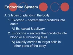
II Adrenal gland Paired organs that lie on top of the kidneys embedded in adipose tissue General structure Covered by capsule of dense collagenous connective tissue Thin septa or ID: 931169
Download Presentation The PPT/PDF document "Histology of Endocrine system" is the property of its rightful owner. Permission is granted to download and print the materials on this web site for personal, non-commercial use only, and to display it on your personal computer provided you do not modify the materials and that you retain all copyright notices contained in the materials. By downloading content from our website, you accept the terms of this agreement.
Slide1
Histology of Endocrine systemII
Slide2Adrenal gland
Paired organs that lie on top of the kidneys, embedded in adipose tissue.
General structure
Covered by capsule of dense
collagenous
connective tissue.
Thin septa or
trabeculae
extend from the connective tissue capsule into the interior of the gland.
Internally there are two major layers called the adrenal cortex and adrenal medulla.
The supporting framework of cortex and the medulla are collectively called the
stroma
. This framework contains many reticular fibers.
Slide3Adrenal gland- Embryological development
In
a sense, the adrenal cortex and medulla may be considered as 2 morphologically distinct endocrine organs.
Similar
to what we saw in the
hypophysis
, the cortical and medullary layers of the adrenal glands have different embryonic origins. That is to say, they are derived from different basic tissue types in the embryo.
As
was the case for the
hypophysis
, we have a situation where neural and non-neural tissues become associated to form an organ.
As
an embryo develops, the cortex of the adrenal gland is derived from
mesodermal
cells in the region of the kidney.
The
cells of the adrenal medulla are derived from a specialized group of neural ectoderm cells that are called the neural crest. These cells are initially part of the larger group of
ectodermal
cells that will form the central nervous system (CNS).
The neural crest cells separate from the developing CNS and migrate through the body's tissues giving rise to many different cell types, tissues, and organs.
Some of the neural crest cells migrate to the developing adrenal cortex, penetrate this tissue and lodge themselves centrally within it to form the adrenal medulla.
So, the cells of the adrenal medulla are sometimes referred to as postganglionic neurons that have lost their axons and dendrites and have become secretory cells.
Slide4Cardiovascular circulation to the adrenal gland
Blood
is supplied to the adrenal glands by a number of arteries.
These
vessels enter through the capsule tissue and then branch out into a sub-capsular plexus of arterioles that give rise to capillaries that extend throughout the cortex.
The
capillaries supply blood to a network of sinusoids in the cortex.
Some
of the arterial branches do not form capillaries in the cortex, but rather run through it to the medulla.
These
are the medullary arteries.
These
arteries form a dense capillary network around the cells of the medulla.
If
we look at the fenestrated endothelium of the capillaries, we find pores that are occluded by a thin membrane. In the cortex, the basal lamina is not continuous. So again, as we might suspect, the circulatory components of this gland are set-up for the exchange of materials between the blood and surrounding cells or visa versa.
Capillaries
of medulla and cortex coalesce to form the adrenal veins that exit the adrenal glands.
Slide5Adrenal cortex
Composed of 3 layers
zona
glomerulosa
zona
fasciculata
zona
reticularis
Slide6Slide7Zona glomerulosa - structure
Columnar
to pyramidal cells that are arranged in folded cords surrounded by capillaries and sinusoids.
Cells
have spherical nucleus and basophilic granules in cytoplasm.
Adjacent
to the endothelium of the capillaries and sinusoids there is a space between the endothelium and surrounding cells.
The
cell surface of the columnar or pyramidal secretory cells adjacent to this space are thrown into folds in this area forming microvilli.
Ultrastructure
is typical for cells involved in lipid
synthesis
Lots
of smooth endoplasmic reticulum with a few short segments of RER.
Well
developed Golgi body.
Mitochondria
are spherical or oval and have tubular cristae.
Small
lipid droplets may be present in cytoplasm associated with smooth endoplasmic reticulum; however, these are not particularly evident in histological sections for light microscopy.
Slide8Zona glomerulosa - function
The
zona glomerulosa cells secrete
mineralcorticoids
, mainly
aldosterone
which is important in maintaining water balance.
These
are steroid hormones that are lipid molecules derived from cholesterol.
Slide9Zona fasciculata - structure
Cells
are polyhedral and are arranged in straight cords (columns) that are 1-2 cells thick with capillaries running between.
Cells
have central nucleus with basophilic cytoplasm. Microvilli are present within the sub-endothelial space (next to the capillaries).
Many
lipid droplets present in cytoplasm. These are extracted during most fixation and embedding procedures, so the cells often appear highly vacuolated in histological sections.
Ultrastructure
is typical for cells involved in lipid synthesis and
secretion
However
, these cells have more RER than those of the zona glomerulosa
This
is why the cytoplasm has an overall basophilic affinity.
Slide10Zona fasciculata - function
Zona
fasciculata cells secrete
glucocorticoids
(important in lipid, protein and carbohydrate metabolism) that are another type of steroid.
Slide11Zona reticularis
Cells
are polyhedral and are arranged in irregular cords with capillaries and sinusoids between.
Lipofuscin
pigment granules in cells.
Cytoplasm
acidophilic. Few lipid droplets.
Cells
secrete
glucocorticoids
.
Slide12Secretion of steroids by adrenal cortex is good example of a feedback system between target organ and pituitary gland.
A
psychological stimulus such as stress or exercise causes
neurosecretory
neurons in the hypothalamus to secrete
adrenocorticotropic
hormone releasing factor into the
capilaries
in the median eminence.
the
releasing factor is carried by the
hypophyseal
portal system from the median eminence to the pars
distalis
.
This
causes secretion of
adrenocorticotropic
hormone (ACTH) by certain cells in the pars
distalis
.
ACTH
causes an increase in corticosteroid secretion (e.g.
cortisol
) by adrenal cortex cells.
As
levels of corticosteroids increase in blood, these inhibit the secretion of releasing factors by the hypothalamus and secretion of ACTH by pituitary.
Slide13Adrenal medulla
Composed of one layer of cords of
polyhedral, epithelioid, secretory cells
that form a compact irregular network surrounded by capillaries, venules and a few sympathetic ganglion cells.
These
epithelioid secretory cells are considered to be
modified postganglionic neurons
.
Nerve
fibers (axons) contact
the
epithelioid cells on the part of their surface that is adjacent to a capillary.
When
stimulated , these nerve fibers release
acetylcholine
that causes the release of the
catecholamine
secretory product accumulated in these cells.
The
secretory product consists of
epinephrine
or
norepinephrine
.
Two
different types of cells, one for each catecholamine.
The
catecholamine secretory granules stain in a specific way when reacted with oxidizing agents. This reaction is called the
chromafin reaction
. As a result, these cells are called
chromafin cells
.
Adrenal
medulla secretes continuously into blood stream.
The
adrenal medulla cells secrete only small amounts of epinephrine and norepinephrine unless stimulated by nervous activity related to emotional reactions.
Increased
secretion of these substances prepares body to react to stressful situations. Blood vessels constrict, blood pressure rises, etc.
Slide14Thyroid gland
Located
below the larynx, partially encircling the esophagus.
Thyroid
is covered by thin layer of loose connective tissue.
Associated
with this loose connective tissue is a dense network of blood and lymphatic capillaries.
Capillaries
are fenestrated.
Septa
extend into organ from the connective tissue capsule.
These
septa separate follicles
of
thyroid tissue from each other.
Septa
composed mainly of reticular fibers.
Slide15Slide16The thyroid tissue within the connective tissue capsule is composed of
follicles
, each containing a lumen filled with a gelatinous substance called colloid.
Each
follicle consists of a
simple
cuboidal
epithelium
surrounding the lumen
The
epithelium changes to simple
squamous
if the follicle is inactive.
Cells
of follicles are responsible for synthesis of thyroid hormones, the most abundant of which is thyroxin.
These
hormones are small molecules consisting of iodinated amino acids that are all formed from the
tyrosyl
radicals of
thyroglobulin
. (i.e. side chains of
thyroglobulin
containing tyrosine are iodinated and separated from the
thyroglobulin
molecule and then processed to form the thyroid hormones.)
Thyroxine
stimulates mitochondrial respiration and
oxadative
phosphorylation
. So, more ATP produced faster.
In
addition to follicle cells,
parafollicular
cells
(
C cells
) are found between the follicles.
These
cells secrete the polypeptide
calcitonin
that causes a reduction of calcium in the blood by inhibiting the activities of
osteoclasts
.
Slide17Control of thyroid hormone secretion
Neurogenic
stimuli provided by axons from parasympathetic and sympathetic ganglia can influence metabolism of thyroid cells.
However
,
thyrotropin
(thyroid stimulating hormone, TSH) is the major controlling factor.
Thyrotropin
is synthesized and secreted by cells in the
adenohypophysis
(pars
distalis
)
.
Thyroid
has feedback system with pituitary similar to adrenal cortex.
Releasing
factors from the median eminence cause the
adenohypophysis
to secrete
thyrotropin
.
This
causes thyroid hormone production by the follicle cells of the thyroid.
As
thyroxin level rises in the blood, it causes an inhibition of the secretion of releasing factors in the median eminence.
Slide18Thyroid hormones are synthesized from the colloid.
Colloid is composed of iodinated
thyroglobulin
.
When thyroid hormones are required, iodinated
thyroglobulin
in colloid is
endocytosed
.
Endocytotic
vesicles merge with
lysosomes
.
Thyroid hormones are synthesized as a result.
This liberates the 4 thyroid hormones into the cytoplasm.
These diffuse across the cell membrane into capillaries where blood carries them to target organs.
Slide19Parathyroid glands
These
are small, but important, organs that are embedded in the wall of the thyroid.
History
- complete removal of the thyroid gland was noted to cause death because of spasms of the laryngeal and thoracic muscles that prevented breathing - called
tetany
In
1892, the French physiologist
Gley
showed that it was actually the removal of the parathyroid glands that "rode" along with the thyroid that was the cause of these titanic seizures. (Due to the lack
of
parathormone
)
Thus
, when the thyroid is removed, it is critical that the parathyroid glands be separated from it and left in the body.
Slide20There are three cell types in the parathryroid glands
Chief
cells
polygonal with vesicular nucleus
slightly acidiphilic, pale staining cytoplasm
secrete parathyroid hormone (parathormone).
Oxyphil
cells
Not
present in thyroid at birth
start
appearing in parathyroid tissues at about age 7 in humans
no
known function
Similar
to, but larger than chief cells.
Adipose
cells
- increase in number as one grows older.
Slide21Major function of parathyroid glands
Secretion
of parathyroid hormone
that causes an increase in calcium in blood by promoting the activities of
osteoclasts
in the breakdown of calcified bone matrix.
So
the
calcitonin
of thyroid
parafollicular
cells and parathyroid hormone of chief cells balance and regulate calcium levels in the body
.