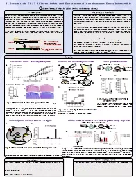
/
S1P effect on cIAP2 knockout mice microglia during EAE, an animal model of multiple sclerosis S1P effect on cIAP2 knockout mice microglia during EAE, an animal model of multiple sclerosis

S1P effect on cIAP2 knockout mice microglia during EAE, an animal model of multiple sclerosis - PowerPoint Presentation

BraveBlackbird
BraveBlackbird . @BraveBlackbird
Follow
343 views
Uploaded On 2022-08-02

S1P effect on cIAP2 knockout mice microglia during EAE, an animal model of multiple sclerosis - PPT Presentation

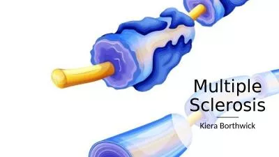
Srikethan Mahavadi Multiple Sclerosis Autoimmune disease of the CNS Demyelination axonal degeneration and cell death More common in women than men Women are 23 times more likely to be diagnosed with ID: 932411

microglia s1p tnf cells s1p microglia cells tnf cell doi experimental ciap2 mice cytotoxicity amp survival ldh signaling inflammation

Share:

Link:

Embed:

Download Presentation from below link

Download Presentation The PPT/PDF document "S1P effect on cIAP2 knockout mice microg..." is the property of its rightful owner. Permission is granted to download and print the materials on this web site for personal, non-commercial use only, and to display it on your personal computer provided you do not modify the materials and that you retain all copyright notices contained in the materials. By downloading content from our website, you accept the terms of this agreement.


Presentation Transcript

Slide1

S1P effect on cIAP2 knockout mice microglia during EAE, an animal model of multiple sclerosis

Srikethan Mahavadi

Slide2

Multiple Sclerosis

Autoimmune disease of the

CNS

Demyelination, axonal degeneration, and cell deathMore common in women than men Women are 2-3 times more likely to be diagnosed with MSLoss of motor skills, vision loss Experimental Autoimmune Encephalomyelitis (EAE) is the mouse experimental model of MS

Slide3

Microglia

Glial

cells which function as immune cells of the CNS

Secrete cytokines such as TNF-  and IL-1These are both signaling proteins involved in inflammation and regulation of immune cellsCytokines are small proteins that have an effect on other cells in this case immune cells

Slide4

TNF Signaling Pathway

Slide5

cIAP2

E3

ubiquitin ligase necessary to activate NF-kB pathway

When cIAP is bound, the cell is thought to be saved from apoptosisLippopolysaccharide (LPS) treated cIAP2-/- mice survived better than wildtype mice cIAP2-/- mice exhibited high inflammation and exaggerated symptoms of EAE

, TNF was highly expressed

Slide6

Previous Experiments

Slide7

Experimental Questions

Why is every cell but microglia dying

?

Is S1P being released, which is saving microglia?

Slide8

Sphingosine-1-phosphate (S1P)

S1P is a lipid mediator involved in extracellular signaling

A study done by Cuviller et al. shows that S1P prevents the features of apoptosis.

It is believed that S1P is secreted by dead cells S1P is present at elevated levels in the CNS during inflammation

Slide9

Experiment Overview

Cell Line: Microglia from mice

Control Group: cells treated with no inflammatory cytokines or S1P

Experimental Group: cells treated with TNF-,, TNF- and S1P, S1PInflammatory cytokines added 30 min prior to S1P and incubated for 24 hoursCytotoxicity measured in 490 nm plate reader

Slide10

LDH-Cytotoxicity

LDH is an enzyme found in all living cells which catalyzes the conversion of

lactate

to pyruvate as well as NAD+ to NADH. An enzyme transfers NADH to NAD+ and reduce WST to formazan(a salt) which shows up orange

Slide11

LDH Cytotoxicity

Slide12

Possible Outcomes

Cytotoxicity levels go down as the S1P is increasing the survival of microglia

Cytotoxicity levels go up as the S1P is not changing the survival of microglia

Possible Further Experiments would include identifying if the S1P is working on the DISC complex or elsewhere or even doing the same experiment and inhibiting the R4 receptor Limitations

Slide13

Discussion

How exactly is S1P increasing the survival of microglia when

inflammatory

cytokines are released?Can S1P be further used as drug therapy for patients with MS?

Slide14

References

Yester, J. W., Bryan, L., Waters, M. R.,

Mierzenski

, B., Biswas, D. D., Gupta, A. S., . . . Kordula, T. (2015). Sphingosine-1-phosphate inhibits IL-1–induced expression of C-C motif ligand 5 via c-Fos–dependent suppression of IFN-β amplification loop. The FASEB Journal, 29(12), 4853-4865. doi:10.1096/fj.15-275180What Is MS? (n.d.). Retrieved from https://www.nationalmssociety.org/What-is-MS Constantinescu, C. S., Farooqi, N., Obrien, K., & Gran, B. (2011). Experimental autoimmune encephalomyelitis (EAE) as a model for multiple sclerosis (MS). British Journal of Pharmacology, 164(4), 1079-1106. doi:10.1111/j.1476-5381.2011.01302.xSilke, J., & Meier, P. (2013). Inhibitor of Apoptosis (IAP) Proteins-Modulators of Cell Death and Inflammation. Cold Spring Harbor Perspectives in Biology, 5(2). doi:10.1101/cshperspect.a008730Conte, D., Holcik, M., Lefebvre, C. A., Lacasse, E., Picketts, D. J., Wright, K. E., & Korneluk, R. G. (2005). Inhibitor of Apoptosis Protein cIAP2 Is Essential for Lipopolysaccharide-Induced Macrophage Survival. Molecular and Cellular Biology, 26(2), 699-708. doi:10.1128/mcb.26.2.699-708.2006Chen, G. (2002). TNF-R1 Signaling: A Beautiful Pathway. Science,296(5573), 1634-1635. doi:10.1126/science.1071924Cuvillier, O., Pirianov, G., Kleuser, B., Vanek, P. G., Coso, O. A., Gutkind, J. S., & Spiegel, S. (1996). Suppression of ceramide-mediated programmed cell death by sphingosine-1-phosphate. Nature, 381(6585), 800-803. doi:10.1038/381800a0Dojindo

Molecular Technologies (2012). LDH Cytotoxicity Assay