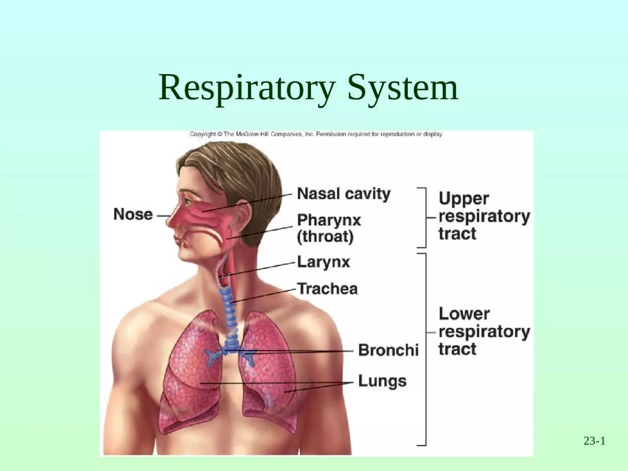
23 2 Respiration Ventilation Movement of air into and out of lungs External respiration Gas exchange between air in lungs and blood Transport of oxygen and carbon dioxide in the blood Internal respiration ID: 933100
Download Presentation The PPT/PDF document "23- 1 Respiratory System" is the property of its rightful owner. Permission is granted to download and print the materials on this web site for personal, non-commercial use only, and to display it on your personal computer provided you do not modify the materials and that you retain all copyright notices contained in the materials. By downloading content from our website, you accept the terms of this agreement.
Slide1
23-1
Respiratory System
Slide223-2
Respiration
Ventilation: Movement of air into and out of lungs
External respiration
: Gas exchange between air in lungs and blood
Transport of oxygen and carbon dioxide in the blood
Internal respiration
: Gas exchange between the blood and tissues
Slide323-3
Respiratory System Functions
Gas exchange: Oxygen enters blood and carbon dioxide leaves
Regulation of blood pH
: Altered by changing blood carbon dioxide levels
Voice production
: Movement of air past vocal folds makes sound and speech
Olfaction
: Smell occurs when airborne molecules drawn into nasal cavity
Protection
: Against microorganisms by preventing entry and removing them
Slide423-4
Respiratory System Divisions
Upper tract
Nose, pharynx and associated structures
Lower tract
Larynx, trachea, bronchi, lungs
Slide523-5
Nose and Pharynx
Nose
External nose
Nasal cavity
Functions
Passageway for air
Cleans the air
Humidifies, warms air
Smell
Along with paranasal sinuses are resonating chambers for speech
Pharynx
Common opening for digestive and respiratory systems
Three regions
Nasopharynx
Oropharynx
Laryngopharynx
Slide623-6
Larynx
Functions
Maintain an open passageway for air movement
Epiglottis and vestibular folds prevent swallowed material from moving into larynx
Vocal folds are primary source of sound production
Slide723-7
Vocal Folds
Slide823-8
Trachea
WindpipeDivides to form
Primary bronchi
Insert Fig 23.5 all but b
Slide923-9
Tracheobronchial Tree
Conducting zone
Trachea to terminal bronchioles which is ciliated for removal of debris
Passageway for air movement
Cartilage holds tube system open and smooth muscle controls tube diameter
Respiratory zone
Respiratory bronchioles to alveoli
Site for gas exchange
Slide1023-10
Tracheobronchial Tree
Slide1123-11
Bronchioles and Alveoli
Slide1223-12
Lungs
Two lungs: Principal organs of respiration
Right lung
: Three lobes
Left lung
: Two lobes
Divisions
Lobes, bronchopulmonary segments, lobules
Slide1323-13
Ventilation
Movement of air into and out of lungsAir moves from area of higher pressure to area of lower pressure
Pressure is inversely related to volume
Slide1423-14
Alveolar Pressure Changes
Slide15Basic Chest X-Ray Interpretation
Deb Updegraff, C.N.S., PICU
Slide16X-rays- describe radiation which is part of the
spectrum which includes visible light, gamma rays and cosmic radiation.Unlike visible light, radiation passes through stuff.
When you shine a beam of X-Ray at a person and put a film on the other side of them a shadow is produced of the inside of their body.
Slide17Different tissues in our body absorb X-rays at different extents:
Bone- high absorption (white)
Tissue- somewhere in the middle absorption (grey)
Air- low absorption (black)
Slide18Film Quality
First determine is the film a PA or AP view.
PA- the x-rays penetrate through the back of the patient on to the film
AP
-
the x-rays penetrate through the front of the patient on to the film.
All x-rays in the PICU are portable and are AP view
Slide19Slide20Quality (cont.)
Is the film over or under penetrated if under penetrated you will not be able to see the thoracic vertebrae.
Slide21Quality (cont)
Check for rotationDoes the thoracic spine align in the center of the sternum and between the clavicles?
Are the clavicles level?
Slide22Slide23Slide24LUNG VOLUMES
The total volume contained in the lung at the end of a maximal inspiration is subdivided into volumes and subdivided into capacities.
There are four volume subdivisions which:
do not overlap.
can not be further divided.
when added together equal total lung capacity.
Slide25Slide26Capacities
Lung capacities are subdivisions of total volume that include two or more of the 4 basic lung volumes.
Slide27Basic lung volumes (memorize)
Tidal Volume (TV).
The amount of gas inspired or expired with each breath.
Inspiratory
Reserve Volume (IRV).
Maximum amount of additional air that can be inspired from the end of a normal inspiration
.
Slide28Basic lung volumes (memorize)
Expiratory Reserve Volume (ERV
). The maximum volume of additional air that can be expired from the end of a normal expiration.
Residual Volume (RV).
The volume of air remaining in the lung after a maximal expiration. This is the only lung volume which cannot be measured with a
spirometer
.
Slide29Basic lung capacities (memorize)
Total Lung Capacity (TLC).
The volume of air contained in the lungs at the end of a maximal inspiration. Called a capacity because it is the sum of the 4 basic lung volumes.
TLC=RV+IRV+TV+ERV
Slide30Basic lung capacities (memorize)
Vital Capacity (VC).
The maximum volume of air that can be forcefully expelled from the lungs following a maximal inspiration. Called a capacity because it is the sum of
inspiratory
reserve volume, tidal volume, and expiratory reserve volume. VC=IRV+TV+ERV=TLC-RV
Slide31Basic lung capacities (memorize)
Functional Residual Capacity (FRC)
. The volume of air remaining in the lung at the end of a normal expiration. Called a capacity because it equals residual volume plus expiratory reserve volume. FRC=RV+ERV
Slide32Basic lung capacities (memorize)
Inspiratory
Capacity (IC)
. Maximum volume of air that can be inspired from end expiratory position. Called a capacity because it is the sum of tidal volume and
inspiratory
reserve volume. This capacity is of less clinical significance than the other three. IC=TV+IRV
Slide33Now you are ready
Look at the diaphram: for tenting
free air abnormal elevationMargins should be sharp (
the right hemidiaphram is usually slightly higher than
the left
)
Slide34Check the Heart
SizeShapeSilhouette-margins should be sharp
Diameter (>1/2 thoracic diameter is enlarged heart)Remember: AP views make heart appear larger than it actually is.
Slide35Cardiac Silhouette
R Atrium
R Ventricle
3. Apex of L Ventricle
Superior Vena Cava
Inferior Vena Cava
6. Tricuspid Valve
Pulmonary Valve
Pulmonary Trunk
9. R PA 10. L PA
Slide36Slide37Slide38Check the costophrenic angles
Margins should
be sharp
Slide39Loss of Sharp Costophrenic Angles
Slide40Check the hilar region
The hilar – the large blood vessels going to and from the lung at the root of each lung where it meets the heart.Check for size and shape of aorta, nodes,enlarged vessels
Slide41Slide42Finally, Check the Lung Fields
InfiltratesIncreased interstitial markingsMasses
Absence of normal marginsAir bronchogramsIncreased vascularity
Slide43Slide44Slide45Slide46Slide47Slide48Slide49Slide50Slide51Slide52Slide53Slide54Slide55Hemothorax
Slide56Slide57Slide58Slide5923-59
Changing Alveolar Volume
Lung recoil
Causes alveoli to collapse resulting from
Elastic recoil and surface tension
Surfactant: Reduces tendency of lungs to collapse
Pleural pressure
Negative pressure can cause alveoli to expand
Pneumothorax is an opening between pleural cavity and air that causes a loss of pleural pressure
Slide6023-60
Pulmonary Volumes
Tidal volume
Volume of air inspired or expired during a normal inspiration or expiration
Inspiratory reserve volume
Amount of air inspired forcefully after inspiration of normal tidal volume
Expiratory reserve volume
Amount of air forcefully expired after expiration of normal tidal volume
Residual volume
Volume of air remaining in respiratory passages and lungs after the most forceful expiration
Slide6123-61
Pulmonary Capacities
Inspiratory capacity
Tidal volume plus inspiratory reserve volume
Functional residual capacity
Expiratory reserve volume plus the residual volume
Vital capacity
Sum of inspiratory reserve volume, tidal volume, and expiratory reserve volume
Total lung capacity
Sum of inspiratory and expiratory reserve volumes plus the tidal volume and residual volume
Slide6223-62
Spirometer and Lung Volumes/Capacities
Slide6323-63
Minute and Alveolar Ventilation
Minute ventilation
: Total amount of air moved into and out of respiratory system per minute
Respiratory rate or frequency
: Number of breaths taken per minute
Anatomic dead space
: Part of respiratory system where gas exchange does not take place
Alveolar ventilation
: How much air per minute enters the parts of the respiratory system in which gas exchange takes place
Slide6423-64
Physical Principles of Gas Exchange
Partial pressure
The pressure exerted by each type of gas in a mixture
Dalton’s law
Water vapor pressure
Diffusion of gases through liquids
Concentration of a gas in a liquid is determined by its partial pressure and its solubility coefficient
Henry’s law
Slide6523-65
Physical Principles of Gas Exchange
Diffusion of gases through the respiratory membrane
Depends on membrane’s thickness, the diffusion coefficient of gas, surface areas of membrane, partial pressure of gases in alveoli and blood
Relationship between ventilation and pulmonary capillary flow
Increased ventilation or increased pulmonary capillary blood flow increases gas exchange
Physiologic shunt is deoxygenated blood returning from lungs
Slide6623-66
Oxygen and Carbon Dioxide
Diffusion Gradients
Oxygen
Moves from alveoli into blood. Blood is almost completely saturated with oxygen when it leaves the capillary
P0
2
in blood decreases because of mixing with deoxygenated blood
Oxygen moves from tissue capillaries into the tissues
Carbon dioxide
Moves from tissues into tissue capillaries
Moves from pulmonary capillaries into the alveoli
Slide6723-67
Changes in Partial Pressures
Slide6823-68
Hemoglobin and Oxygen Transport
Oxygen is transported by hemoglobin (98.5%) and is dissolved in plasma (1.5%)Oxygen-hemoglobin dissociation curve shows that hemoglobin is almost completely saturated when P0
2
is 80 mm Hg or above. At lower partial pressures, the hemoglobin releases oxygen.
A shift of the curve to the left because of an increase in pH, a decrease in carbon dioxide, or a decrease in temperature results in an increase in the ability of hemoglobin to hold oxygen
Slide6923-69
Hemoglobin and Oxygen Transport
A shift of the curve to the right because of a decrease in pH, an increase in carbon dioxide, or an increase in temperature results in a decrease in the ability of hemoglobin to hold oxygenThe substance 2.3-bisphosphoglycerate increases the ability of hemoglobin to release oxygen
Fetal hemoglobin has a higher affinity for oxygen than does maternal
Slide7023-70
Oxygen-HemoglobinDissociation Curve at Rest
Slide7123-71
Oxygen-HemoglobinDissociation Curve during Exercise
Slide7223-72
Shifting the Curve
Slide7323-73
Transport of Carbon Dioxide
Carbon dioxide is transported as bicarbonate ions (70%) in combination with blood proteins (23%) and in solution with plasma (7%)Hemoglobin that has released oxygen binds more readily to carbon dioxide than hemoglobin that has oxygen bound to it (Haldane effect)
In tissue capillaries, carbon dioxide combines with water inside RBCs to form carbonic acid which dissociates to form bicarbonate ions and hydrogen ions
Slide7423-74
Transport of Carbon Dioxide
In lung capillaries, bicarbonate ions and hydrogen ions move into RBCs and chloride ions move out. Bicarbonate ions combine with hydrogen ions to form carbonic acid. The carbonic acid is converted to carbon dioxide and water. The carbon dioxide diffuses out of the RBCs.
Increased plasma carbon dioxide lowers blood pH. The respiratory system regulates blood pH by regulating plasma carbon dioxide levels
Slide7523-75
Carbon Dioxide Transportand Chloride Movement
Slide7623-76
Respiratory Areas in Brainstem
Medullary respiratory centerDorsal groups stimulate the diaphragm
Ventral groups stimulate the intercostal and abdominal muscles
Pontine (pneumotaxic) respiratory group
Involved with switching between inspiration and expiration
Slide7723-77
Respiratory Structures in Brainstem
Slide7823-78
Rhythmic Ventilation
Starting inspiration
Medullary respiratory center neurons are continuously active
Center receives stimulation from receptors and simulation from parts of brain concerned with voluntary respiratory movements and emotion
Combined input from all sources causes action potentials to stimulate respiratory muscles
Increasing inspiration
More and more neurons are activated
Stopping inspiration
Neurons stimulating also responsible for stopping inspiration and receive input from pontine group and stretch receptors in lungs. Inhibitory neurons activated and relaxation of respiratory muscles results in expiration.
Slide7923-79
Modification of Ventilation
Cerebral and limbic systemRespiration can be voluntarily controlled and modified by emotions
Chemical control
Carbon dioxide is major regulator
Increase or decrease in pH can stimulate chemo- sensitive area, causing a greater rate and depth of respiration
Oxygen levels in blood affect respiration when a
50%
or greater decrease from normal levels exists
Slide8023-80
Modifying Respiration
Slide8123-81
Regulation of Blood pH and Gases
Slide8223-82
Herring-Breuer Reflex
Limits the degree of inspiration and prevents overinflation of the lungsInfants
Reflex plays a role in regulating basic rhythm of breathing and preventing overinflation of lungs
Adults
Reflex important only when tidal volume large as in exercise
Slide8323-83
Ventilation in Exercise
Ventilation increases abruptly
At onset of exercise
Movement of limbs has strong influence
Learned component
Ventilation increases gradually
After immediate increase, gradual increase occurs (4-6 minutes)
Anaerobic threshold is highest level of exercise without causing significant change in blood pH
If exceeded, lactic acid produced by skeletal muscles
Slide8423-84
Effects of Aging
Vital capacity and maximum minute ventilation decreaseResidual volume and dead space increaseAbility to remove mucus from respiratory passageways decreases
Gas exchange across respiratory membrane is reduced