/
Benign disease of cervix and uterus Benign disease of cervix and uterus

Benign disease of cervix and uterus - PowerPoint Presentation

Extremejock
Extremejock . @Extremejock
Follow
343 views
Uploaded On 2022-08-02

Benign disease of cervix and uterus - PPT Presentation

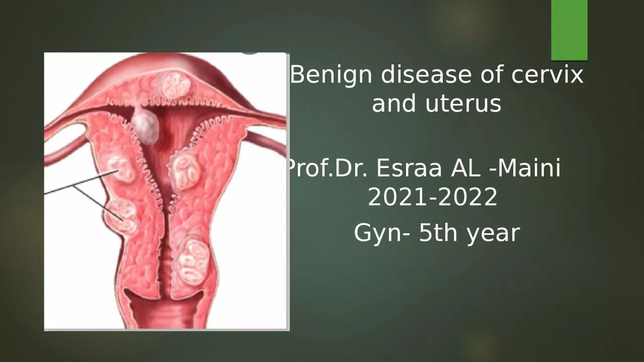
ProfDr Esraa AL Maini 20212022 Gyn 5th year Cervical ectropion Cervical ectropion In women of reproductive age the columnar epithelium replaced squamous epithelium and visible on the ectocervix as a circular red area surrounding the external cervical ID: 932319

fibroids fibroid cervical uterine fibroid fibroids uterine cervical treatment women pregnancy bleeding endometrial size uterus symptoms pain blood intramural

Share:

Link:

Embed:

Download Presentation from below link

Download Presentation The PPT/PDF document "Benign disease of cervix and uterus" is the property of its rightful owner. Permission is granted to download and print the materials on this web site for personal, non-commercial use only, and to display it on your personal computer provided you do not modify the materials and that you retain all copyright notices contained in the materials. By downloading content from our website, you accept the terms of this agreement.


Presentation Transcript

Slide1

Benign disease of cervix and uterus

Prof.Dr. Esraa AL -Maini 2021-2022 Gyn- 5th year

Slide2

Cervical ectropion:

Cervical ectropion In women of reproductive age the columnar epithelium replaced squamous epithelium and visible on the ectocervix as a circular, red area surrounding the external cervical

os

.

This is a normal finding and should not be called ‘cervical erosion’ OR ulcer.

Causes: ‘three Ps’: puberty, pill and pregnancy.

Slide3

Symptoms:

1- Intermenstrual and postcoital bleeding (contact bleeding).2 -Excessive, clear, odorless mucus-type discharge. 3-Associated cervicitis may produce backache, pelvic pain and at times, infertility.Signs: Per speculum—there is a bright red area at external os in the ectocervix. The outer edge is clearly demarcated. The lesion may be smooth or having small papillary folds.

It is neither tender nor bleeds to touch. On rubbing with a gauze piece

Slide4

Treatment :

1-Prior to treatment end -cervical and lower genital tract swabs are taken to exclude chlamydia and other sexually-transmitted infections and normal cervical cytology should be confirmed to exclude cervical premalignancy and malignancy 2-Should be changed from oestrogen-based hormonal contraceptives. 3-Cervical ablation where the visible glandular producing columnar cells are ablated, usually with cryocautery, as an outpatient.

Slide5

Nabothian follicles :Sometimes the columnar glands within the transformation zone become sealed over, forming small, mucus-filled cysts visible on the ectocervix. These are termed ‘

nabothian follicles’ and are of no pathological significance. No treatment is usually required although extremely large ones can be drained using a large-bore needle

Slide6

Cervical polyps: Cervical polyps are benign

tumours arising from the endocervical epithelium and may be seen as smooth, reddish protrusions. They are usually asymptomatic, being identified incidentally during a routine cervical smear, but as with a cervical ectropion they can cause vaginal discharge, IMB and PCB. They are easily removed by avulsion with polyp forceps as an outpatient.

Slide7

Cervical stenosis: refers to pathological narrowing of the endocervical canal

Causes: 1-is usually an iatrogenic phenomenon caused by a surgical event, like treatment of premalignant disease of the cervix using a cone biopsy or loop diathermy , as can endometrial ablation 2-Congenital cervical stenosis3-Chronic infection (chronic cervicitis)4-Stenosis secondary to a tumor/mass polyp5-Post radiation therapy6-Cervical endometriosis

Slide8

Symptoms

1-Hematomatra as menstrual blood accumulates in the endometrial cavity 2-Amenorrhea with severe cyclical dysmenorrhea like pain3-In postmenopausal women, cervical stenosis may give rise to pyometra, in which accumulated secretions become a focus of infection. Treatment 1.Surgical dilatation of the cervix under ultrasound or hysteroscopic guidance.2-Restenosis can occur and sometimes hysterectomy is required to relieve the pain.3-Cervix not completely stenosed but scarred from previous surgery may fail to dilate during

labour (cervical dystocia), necessitating c/s.

Slide9

Benign endometrial lesions

Endometrial polyps : are focal endometrial outgrowths containing a variable amount of glands, stroma and blood vessels ,may be pedunculated or sessile, single or multiple and vary in size (0.5–4 cm). Incidence: 10–20% of women with AUB and 10% of women with subfertility.Risk factors for endometrial polyp development include obesity, late menopause, the use of the partial oestrogen agonist tamoxifen and possibly the use of hormone replacement therapy (HRT).

Slide10

Symptoms;

Asymptomatic Cause abnormal uterine bleeding (AUB) (heavy menstrual bleeding [HMB], IMB and postmenopausal bleeding [PMB]) , unscheduled vaginal bleedingAdversely impact on fertility. Contain hyperplastic foci in 10–25% of symptomatic cases and 1% is frankly malignant. The risk of polyps harboring serious endometrial disease is increased after the menopause and with the use of tamoxifen.

Slide11

Uterine leiomyomata (fibroids )

DefinitionUterine leiomyomata or fibroids are the most common benign tumors of the female genital tract, arising from neoplastic transformation of single smooth muscle cells of the myometrium.

Slide12

The FIGO classification ( according to their location in relation to the uterine wall):

0 pedunculated intracavitary;1 submucosal, <50% intramural;2 submucosal, ≥50% intramural;3 100% intramural, but in contact with the endometrium;

4 intramural;5 subserosal, ≥50% intramural;6 subserosal

, <50% intramural;

7

subserosal

pedunculated;

8 other (e.g. cervical, , uterine ligaments , ovary, parasitic when fibroid attach to nearby structures and derive their blood supply)

Slide13

The incidence of fibroids

Increases with age Afro‐Caribbean women Obese womenReduction in incidence increasing parity prolonged use of the oral contraceptive pill . smoking

Slide14

fibroid degeneration:

Red ,cystic ,hyaline degeneration Red degeneration– hemorrhage and necrosis occurs within the fibroid typically in second trimester of pregnancy 5% of pregnant women with fibroidsS&S: sudden onset of focal pain and tenderness on palpation localized to an area of the uterus over the fibroid ,low-grade fever ,vomiting.Investigations: leukocytosis ,raised ESR, Ultrasound Treatment : is treated conservatively by bed rest ,hydration Analgesia to relieve pain ,Sedatives, Antibiotics: If required most often, acute symptoms subside within a 3-10 days,

but inflammation may stimulate labor. Surgery is rarely necessary during pregnancy.

Slide15

Malignant or sarcomatous degeneration can occur the incidence of malignancy (leiomyosarcoma) is 0.64 per 100 000 women per year (rare)and is extremely uncommon in women below the age of 40 ,The suspicion is greatest in postmenopausal period when there is a rapidly increasing size of the fibroid.

Slide16

Symptoms associated with uterine fibroids

1-Asymptomatic: around 60% are asymptomatic. In the case of small fibroids2-Menstrual upset: menorrhagia and/or dysmenorrhea ,as in case of submucosal fibroid3- Pain is unusual, except in the special circumstance where acute pain as in:1- Acute red degeneration 2- torsion of a pedunculated fibroid

3- Extrusion of submucosal fibroid from the uterus as uterus contract to expels4- Associated endometriosis5- Adhesion to other organ

6- Sarcomatous changes

Slide17

4- pressure symptoms related purely to the size of the fibroid :

1-Abdominal discomfort ,sensation of pelvic pressure or backache ,abdominal distension2-Urinary frequency, difficulty in micturition, incomplete bladder emptying or incontinence3-Bowel problems such as constipation4-Varicosity or edema of lower limb8-Reproductive dysfunction (pregnancy related complication):

Slide18

Effect of the fibroid on the pregnancy:

Subfertility :difficulty in conceiving ,may result from mechanical distortion or occlusion of the Fallopian tubes, and an endometrial cavity grossly distorted by submucous fibroids may prevent implantation of a fertilized ovum. Removal of submucosal fibroids can enhance fertility surgically removing the other types of fibroids is less clear Thus, these fibroids should only be removed if symptomatic, otherwise unexplained, infertility.

Slide19

Early in pregnancy

: miscarriage ,Recurrent pregnancy loss. In late pregnancy, fibroids located in the cervix or lower uterine segment may cause an abnormal lie Malposition and malpresentation of the fetus , Obstructed labour, Cesarean section, rupture of myomectomy scar during pregnancy ,placental abruption ,Red degenerationAfter delivery: postpartum haemorrhage may occur due to inefficient uterine contraction &delayed involution, infection.Myomectomy not done during pregnancy because bleeding may be profuse, resulting in hysterectomy

Slide20

Effects of pregnancy on fibroid:

Fibroids are estrogen-dependent tumors that can enlarge during pregnancy in response to the hyperoestrogenic state Red degeneration They can undergo degenerative change usually in response to outgrowing their blood supply.Torsion of a pedunculated fibroid, may cause gradual or acute symptoms of pain and tenderness usually after delivery Infection during puerperium ,Expulsion ,Necrosis

Slide21

Diagnosis:

General: signs of anemia.Abdominal examination: The uterus is often found to be enlarged and presents as a pelvic mass (often central and mobile) Bimanual examination: enlarged, firm, smooth or irregular, non-tender uterus palpable. tenderness may suggest red degeneration.

Slide22

2-Investigations:

A full blood count : severe anemia indicate significant fibroid Endometrial biopsy may be indicated in patients with abnormal bleeding.Ultrasonography, is very useful as a first‐line diagnostic test . Fibroids are typically well‐defined round or lobulated myometrial lesions. TVUSS: good for detecting and locating submucous fibroids and small intramural fibroids. Transabdominal ultrasound scan (TAUSS): good for detecting larger intramural and subserosal fibroids and excluding hydronephrosis

Color Doppler a fibroid typically has circumferential vascularization Hysteroscopy: good for detecting submucosal fibroids and endometrial polyps;

Slide23

MRI is occasionally used to

Morphology, size and location of uterine fibroids Indicated prior to uterine artery embolization Monitor treatment response Before surgical intervention Is useful for examining large fibroid In obese womenIn cases of suspected malignancy.

Slide24

Features suggested of malignancy:

There are no pathognomonic features 1- A large (≥8cm), heterogeneous myometrial tumour ,Strong and irregular vascularization2-Central necrosis/degenerative cystic changes 3-Absence of calcifications should raise the suspicion of a leiomyosarcoma. 4-Rapid increase in size 6-MRI with contrast enhancement gives an indication of the vascularity and may prove helpful

7-Total lactate dehydrogenase (LDH) are reportedly elevated in leiomyosarcoma.8- Elevated CA125 levels are seen in advanced‐stage leiomyosarcoma only

Slide25

Treatment :

1-Conservative management is appropriate where asymptomatic The patient near menopause ,Small fibroid below 12weeks uterine size 2-Medical treatments medical treatments tending to reduce menstrual bleeding without effect on fibroid size ineffective in the presence of a submucous fibroid or an enlarged uterus that is palpable abdominally (>12 weeks size).Iron supplementation to correct anemia

Slide26

1-Gonadotrophin‐releasing hormone agonists

These drugs lead to the downregulation of pituitary receptors gonadotrophin output reduction and consequent reduction in ovarian steroid production within 2–3 weeks of commencing treatment. These analogues are given as 1‐ or 3‐monthly depot injections or as nasal spray.fibroid volume reduction of 40% .

Slide27

Disadvantages of GnRH analogue

Fibroids regrow when treatment has stopped. Postmenopausal side effects consisting of hot flushing, vaginal dryness and, prologed use significant bone loss. An ‘add‐back’ therapy by administering low‐dose oestrogen replacement therapy

Slide28

GnRH agonists indication

:1-women with severe anemia. 2- Agonists are also useful prior to surgery , over a 3-month period to reduce the bulk and vascularity of the fibroids. Useful benefits of this approach are to enable a supra pubic rather than a midline abdominal incision, lead to more rapid recovery Disadvantages GnRH agonist pretreatment can obscure tissue planes around the fibroid making surgery more difficult , But on other hand decrease blood loss and the likely need for transfusion are reduced.

Slide29

2-Selective progesterone receptor modulators(SPRM ulipristal acetate):

A

re effective in reducing pain, bleeding and fibroid size, and are associated with an improvement in quality of life In addition to being an oral tablet

Unlike GnRH analogues, SPRMs do not lead to

oestrogen

deficiency

The SPRM ulipristal acetate Side effects include headache and breast tenderness.

it is not yet widely accepted into clinical practice.

Slide30

3-Levonorgestrel‐releasing intrauterine system

U

se in the treatment of associated dysfunctional uterine bleeding .

R

elative contraindication, the device is more likely to be expelled during a very heavy bleeding episode and because the presence of a distorted uterine cavity may make insertion of the device more difficult and increase the expulsion risk.

But

if the cavity is regular, a trial with an LNG‐IUS system may be appropriate.

Slide31

3-Surgical treatment:

1-Where a bulky fibroid uterus causes pressure symptoms

2- Where HMB is refractory to medical interventions

3-Complications like torsion

4-Rapid increase in size

5-Ultrasound features suggestive of sarcoma

6-Sub -fertility

7-Asymptomatic fibroid of such size that they are palpable abdominally

Slide32

1-Hysterectomy:

Indications

Patient complete her family , It ensures definitive resolution of symptoms

Major complications occur with hysterectomy are all increased in the presence of uterine fibroids .

Ovaries need not be removed in younger women

,

Women with a strong family history of breast or ovarian cancer should be counseled appropriately.

Slide33

2-Myomectomy :is indicated in the following

1-In women who wish to retain their fertility, Large myomas (especially the submucosal or intramural type)

2.Any symptomatic fibroid (persistent uterine bleeding despite medical therapy, excessive pain or pressure symptoms)

3.Unexplained infertility with distortion of the uterine cavity by fibroid

4.Recurrent pregnancy wastage due to fibroid.

5.When IVF is indicated (especially if the myoma results in the distortion of the uterine cavity) .

This can be carried out as an laparotomy, laparoscopic (

where‘power

morcellation’.

hysteroscopic procedure for Intracavitary fibroids

(FIGO type 0, 1 and some type 2) which decreases blood loss and improves fertility.

Slide34

Complications during hysteroscopic myomectomy :

1-include uterine perforation and the associated potential for visceral damage,

haemorrhage

, infection and fluid overload.

2-Rupture of the uterus in

labour

3-

U

ncontrolled bleeding which could lead to hysterectomy.

Slide35

3-Uterine artery embolization n Radiological

1. near menopause 2. no longer desire fertility3. have a large uterus4. unfit for surgery

Slide36

Uterine artery embolization (UAE)

It involves embolization of both uterine arteries under radiological guidance.

A small incision is made in the groin under local

anaesthesia

and a cannula placed into the femoral artery and guided into the uterine arteries.

Embolization particles are then injected, reducing the blood supply to the uterus, which induces infarction and degeneration of fibroids such that the overall reduction in fibroid volume is around 50%, requiring opiate analgesia.

Slide37

Complications:

Contrast allergy Post‐embolization syndrome( Infection ,Sepsis requiring emergency hysterectomy occurs in <1% Death Radiation exposure , potential ovarian failureNon‐target or mis‐embolization: ovary, bowel or bladder ,Persistent vaginal discharge endometrial atrophy or intrauterine adhesion ,fibroid expulsion Treatment failure: failed cannulation, revascularization or regrowth of fibroids However, one-third of women subsequently require further medical, radiological or surgical intervention within 5 years of UAE

Women wishing to retain their fertility should be counselled carefully as the effects on subsequent reproductive function are uncertain.Pregnancies have been reported , but concerns remain over premature ovarian failure and effects on the endometrium that may lead to abnormal placentation.

Slide38

4-Ablation procedures

Endometrial ablation may be performed with or without myomectomy and is associated with a high rate of amenorrhea.

Provided that the uterine cavity is not too enlarged or distorted

The first reports have shown a significant reduction in fibroid size, together with symptomatic relief in the first 12 months after the procedure

Slide39

5-High‐intensity focused ultrasound

(HIFU)Focused ultrasonic energy is used to induce coagulative necrosis in the fibroid by a thermal effect, cavitation and direct damage to the lesion’s blood vessels.The procedure is MRI‐guided, not associated with an infarction‐like syndrome. the impact of treatment on HMB is less .

Is neither suitable for large fibroids nor large numbers of fibroids, and its impact on the recurrence rate uncertain

Slide40

THANK YOU