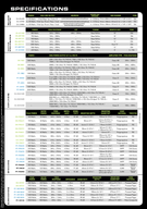
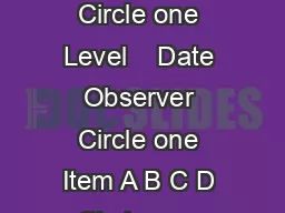
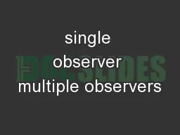
Jieun Kim PhD Florida State University Bridget Algee Hewitt PhD Stanford Univeristy Presenting Author February 20 2018 NIJ RampD Forensic Symposium 1 Introduction The three shapebased computational methods by Slice and ID: 933267
Download Presentation The PPT/PDF document "Analysis of Inter- and Intra-Observer Er..." is the property of its rightful owner. Permission is granted to download and print the materials on this web site for personal, non-commercial use only, and to display it on your personal computer provided you do not modify the materials and that you retain all copyright notices contained in the materials. By downloading content from our website, you accept the terms of this agreement.
Slide1
Analysis of Inter- and Intra-Observer Error Associated with the Use of 3D Laser Scan Data of the Pubic Symphysis
Jieun Kim*, Ph.D., Florida State UniversityBridget Algee-Hewitt, Ph.D., Stanford Univeristy*Presenting AuthorFebruary 20, 2018NIJ R&D Forensic Symposium
1
Slide2Introduction
The three shape-based, computational methods by Slice and Algee-Hewitt (2015) and Stoyanova et al. (2015;2017): Use 3D laser scans of the pubic symphysis Target to minimize subjectivity in age estimation by reducing the effects of observer experience in the age-indicator assessment and methodological bias2
Slide3Introduction
Stoyanova et al. 2015 have shown the improved repeatability of the methods through a single observer error test The computational methods’ performance and reproducibility have not been quantified at the level of multiple observers with different training background and experience levelsParticularly, there is potential for introducing error in the first two steps of data processing: Initial scanning of the pubic symphysisScan editing at different times by different observers. 3
Slide4Introduction
Importance of quantifying observer error associated with these steps An emerging body of research that utilizes computerized, virtual age indicators and high-dimensional image data (e.g. CT/MRI scans): Villa et al. 2013; 2015; Boyd et al. 2015; Navega et al. 2017; Lottering 2013; 2014; Chiba et al. 2014; Curate et al. 2013, etc. 4
Slide5Four Hypotheses
Hypothesis 1. The same observer will edit a set of raw scans inconsistently when editing is done repeatedly overtimeHypothesis 2. Scan editing skills will vary between observers based on their training background and/or levels of experience Hypothesis 3. Edited scans with different margin widths left around the pubic symphyseal face will yield different shape measures and age estimates Hypothesis 4. Edited scans with the ventrally protruding pubic tubercle will yield inaccurate age estimates for the VC method5
Slide6H3: The Effects of Different Margin Widths around the
Symphyseal Face6
6
SB Male Cast Lower Phase V
2mm vs. 4mm vs. 1cm
Region of interest
Slide7H4: The Effects of Editing with/without the Pubic Tubercle
7A pubic symphysis with a tubercle (semi-circled) extending to the face
(SB Female Cast Upper Phase VI)
Showing two different ventral curvatures depending on presence/absence of the tubercle
(SB Male Cast Upper Phase VI)
Slide8Materials
3 replicate scans of the 12 Suchey-Brooks’ male casts taken by a single observer (n=36)8
Slide9Methods
Four observers edited each set of the raw SB scans three timesObservers with various experience levels and training backgroundObserver 1. Biological anthropologist and developer of the scan editing protocolObserver 2. Scientific computing scholar and developer of the methods’ algorithms and software, forAge, (http://morphlab.sc.fsu.edu/software/forAge/index.html)Observer 3. Skeletal biologist with <2 years of experience with scan editingObserver 4. Forensic practitioner new to scan editing 9
Slide10Methods (cont.)
From the edited scans, x,y,z coordinates were retrieved and subjected to a series of the shape analyses3 sets of shape measures Slice-Algee-Hewitt (SAH) scoresBending energy/thin plate splines (BE/TPS) values Ventral curvature (VC) values 5 sets of final age estimates obtained from single variable & multivariate regression analyses
10
Slide11H1 & H2: Intra- & Inter-Observer Error Test
Intraclass correlation coefficients (ICC) Two-way random modelAbsolute agreement on single measuresICC guidelines proposed by Cicchetti 1994
11
ICC value
Interpretation
<0.40
Poor reliability
0.40-0.59
Fair reliability
0.60-0.74
Good reliability
0.75-1.00
Excellent reliability
Slide12H3 & H4: The Effects of Different Editing Conditions on final age estimates
Age estimates vs. Known agesPaired t-test, alpha=0.0512
Slide13Results: Intra-/Inter-observer Error (H1 & H2)
The resulting ICC values were high and mostly fell within the excellent reliability range (0.75-1.0) This demonstrates that the raw scans were edited consistently within and between observers and the derived shape measures and age estimates were highly reliable among observers. 13
Slide1414
Shape measure/Age estimateICC
DF (1,2)
F-Test
Sig. p
95% CI lower
95% CI upper
BE value
0.728
11,22.4
8.42
<0.001*
0.441
0.903
SAH score
0.856
11,22.6
20.5
<0.001*
0.675
0.952
VC value
0.942
11,23.9
48.8
<0.001*
0.859
0.981
BE estimate
0.91
11,23.1
29.7
<0.001*
0.785
0.971
SAH estimate
0.93
11,20
47.5
<0.001*
0.825
0.978
VC estimate
0.942
11,23.8
49
<0.001*
0.859
0.981
VC+BE estimate
0.949
11,24
56.4
<0.001*
0.875
0.984
VC+SAH estimate
0.94511,20.460.3<0.001*0.8610.982
INTRA
-observer error: ICC absolute agreement of within-observer shape measures and age estimates derived from first, second, and third time editing (only showing the ICCs of Observer 1), alpha=0.05
Slide1515
Shape measure/Age estimateICC
DF (1,2)
F-Test
Sig. p
95% CI lower
95% CI upper
BE value
0.865
35,108
26.8
<0.001*
0.790
0.921
SAH score
0.832
35,27.8
29.3
<0.001*
0.696
0.911
VC value
0.756
35,32.8
18
<0.001*
0.594
0.863
BE estimate
0.829
35,60.4
24.1
<0.001*
0.726
0.902
SAH estimate
0.836
35,32.6
28.7
<0.001*
0.711
0.911
VC estimate
0.746
35,28.7
17.8
<0.001*
0.571
0.859
BE+VC estimate
0.811
35,25
26.2
<0.001*
0.656
0.900
SAH+VC estimate
0.85335,18.438.6<0.001*0.700
0.927
INTER-
observer error: ICC absolute agreement of between-observer shape measures and age estimates, alpha=0.05
Slide16BE Value* Comparisons between Observers
16Note the proximity of the BE values generated for each cast by the four observers (ICC= 0.865)
*an average of three trial values was used for each observer
Slide17Comparisons of Final Age Estimates* Generated from the BE values
17*an average of three trial values was used for each observer
All 4 observers yielded consistent age estimates (ICC=
0.829
).
The “X” symbol indicates the documented age for each of the 12 SB casts.
Slide18Results: The Effects of Different Margin Widths on Age Estimates (H3)
The edited scans with 2mm and 4 mm margins did not produce age estimates significantly different from the known ages.However, 1cm margin produced a significantly different mean for all methods (mean diff. b/t estimated & known ages -14-18yrs, p<0.05), except the VC method (mean diff. -9yrs). 18
Age est.
N
Mean age
Mean age est.
Mean
diff.
SE
Upper 95%
Lower 95%
t
DF
Sig.
BE
12
38.833
24.570
14.263
6.023
27.522
1.005
2.368
11
0.037*
SAH
12
38.833
22.684
16.149
5.476
28.203
4.095
2.949
11
0.013*
VC
11
40.454
31.410
9.044
6.986
24.609
-6.520
1.294
10
0.224
VC+BE
11
40.454
22.935
17.519
6.595
32.215
2.823
2.656
10
0.024*VC+SAH1140.45422.48317.9726.06631.4894.4552.962100.014*
Slide19Results: The Effects of the Ventral Tubercle on Age Estimates (H4)
Unlike the expectation, the inclusion of the pubic tubercle for the shape analysis did not yield inaccurate age estimates for the VC methodHowever, it did produce statistically significant mean differences for the SAH-score and TPS/BE methods and the two multivariate regression models (p<0.05). 19
Age est.
N
Mean age
Mean age est.
Mean diff.
SE
Upper 95%
Lower 95%
t
DF
Sig.
BE
12
38.833
21.653
17.181
5.281
28.804
5.557
3.253
11
0.008*
SAH
12
38.833
23.140
15.693
3.804
24.066
7.321
4.125
11
0.002*
VC
11
40.455
36.485
3.969
6.390
18.207
-10.268
0.621
10
0.548
VC+BE
11
40.455
23.052
17.403
5.366
29.358
5.447
3.243
10
0.009*
VC+SAH1140.45524.31016.1453.94124.9257.3644.099100.002*
Slide20Results: The Effects of the Ventral Tubercle on Age Estimates (H4)
A likely reason for this is the fact that the tubercle is located at a different plane from the symphyseal face, and therefore the VC method algorithms are not significantly influenced by the inclusion/exclusion of the feature (mean diff. b/t estimated & known ages -4yrs). However, the methods that assess the complexity of the symphysial face (e.g. SAH-score & TPS/BE methods) may interpret the tubercle as an extra feature to account for, and therefore produce inaccurate (younger, mean diff. b/t estimated & known ages -16-17yrs) age estimates.
As expected, the scans edited without the tubercle yielded age estimates that were not significantly different from the documented ages.
20
Slide21Discussion & Conclusion
The results show high repeatability of the Slice & Algee-Hewitt and Stoyanova et al.’s computational methods regardless of observer’s level of experience or training background.This further supports using a 3D laser scanner and scanned images to aid in resolving the issue of subjectivity and in standardizing data collection and analysis protocols between observers and institutions. Despite the simulated improper editing of the scans with various margin widths remaining, the computational methods were robust enough to self-correct and produce consistent and accurate age estimates. However, 1cm margin seems to be a threshold where the methods start generating incorrect age estimates.Although the inclusion of the pubic tubercle did not necessarily result in inaccurate age estimates for the VC method, we recommend removing the feature as it may “trick” the BE & SAH-score methods and the two multivariate regression models. 21
Slide22Acknowledgements
We thank Dr. Judy M. Suchey for providing the documented chronological ages of the SB male casts. This project is funded by a National Institute of Justice grant (2015-DN- BX-K010) awarded to the senior authors, Slice and Algee-Hewitt.
22
Slide23References
Villa C, Hansen MN, Buckberry J, Cattaneo C, Lynnerup N. Forensic age estimation based on the trabecular bone changes of the pelvic bone using post-mortem CT. Forensic Science International. 2013;233(1):393-402.Villa C, Buckberry J, Cattaneo C,
Lynnerup
N. Reliability of
suchey
‐brooks and buckberry‐chamberlain methods on 3D visualizations from CT and laser scans. American journal of physical anthropology. 2013;151(1):158-63.
Villa C, Buckberry J,
Cattaneo
C, Frohlich B,
Lynnerup
N. Quantitative analysis of the morphological changes of the pubic
symphyseal
face and the auricular surface and implications for age at death estimation. Journal of forensic sciences. 2015;60(3):556-65.
Boyd KL, Villa C,
Lynnerup
N. The use of CT scans in estimating age at death by examining the extent of ectocranial suture closure. Journal of forensic sciences. 2015;60(2):363-9.
Villa C, Buckberry J,
Lynnerup
N. Evaluating osteological ageing from digital data. Journal of Anatomy. 2016.
doi
: 10.1111/joa.12544.
Navega
D, Coelho
JdO
, Cunha E, Curate F. DXAGE: A New Method for Age at Death Estimation Based on Femoral Bone Mineral Density and Artificial Neural Networks. Journal of Forensic Sciences. 2017.
doi
: 10.1111/1556-4029.13582.
Curate F, Albuquerque A, Cunha EM. Age at death estimation using bone densitometry: testing the Fernandez Castillo and Lopez Ruiz method in two documented skeletal samples from Portugal. Forensic Science International. 2013;226(1):296. e1-. e6.
Lottering N, Macgregor DM, Meredith M, Alston CL, Gregory LS. Evaluation of the
suchey
-brooks method of age estimation in an Australian subpopulation using computed tomography of the pubic
symphyseal
surface. Am J Phys
Anthropol
. 2013;150(3):386-99.
doi
: 10.1002/ajpa.22213. PubMed PMID: 23283754.
Lottering N, Reynolds MS, MacGregor DM, Meredith M, Gregory LS. Morphometric modelling of ageing in the human pubic symphysis: Sexual dimorphism in an Australian population. Forensic science international. 2014;236:195. e1-. e11.
Chiba F, Makino Y,
Motomura
A,
Inokuchi
G,
Torimitsu
S, Ishii N, et al. Age estimation by quantitative features of pubic symphysis using multidetector computed tomography. International journal of legal medicine. 2014;128(4):667-73.
Kimmerle
, EH, Prince DA, and Berg GE. Inter‐Observer Variation in Methodologies Involving the Pubic Symphysis, Sternal Ribs, and Teeth. Journal of Forensic Sciences. 2008;53(3): 594-600.
Shirley, NR and Ramirez Montes, PA Age Estimation in Forensic Anthropology: Quantification of Observer Error in Phase Versus Component-Based Methods. Journal of Forensic Sciences. 2015;60(1): 107–111. doi:10.1111/1556-4029.12617.
Slice DE,
Algee
-Hewitt BF. Modeling Bone Surface Morphology: A Fully Quantitative Method for Age-at‐Death Estimation Using the Pubic Symphysis. Journal of Forensic Sciences. 2015;60(4):835-43.
Stoyanova D,
Algee
‐Hewitt BF, Slice DE. An Enhanced Computational Method for Age‐at‐Death Estimation Based on the Pubic Symphysis Using 3D Laser Scans and Thin Plate Splines. American Journal of Physical Anthropology. 2015;158(3):431-40.
Stoyanova D,
Algee
‐Hewitt BF, Kim J, Slice DE. A Fully Computational Framework for Age-at-Death Estimation from the Adult Skeleton: Surface and Outline Analysis of Three-Dimensional Laser Scans of the Pubic Symphysis. Journal of Forensic Sciences.
2017. doi:10.1111/1556-4029.13439.Cicchetti DV. Guidelines, criteria, and rules of thumb for evaluating normed and standardized assessment instruments in psychology. Psychological assessment. 1994;6(4):284.23
Slide24Cristina Figueroa-Soto, MA
University of TennesseeWaukesha County ME OfficeBiological Anthropologycfiguer1@vols.utk.edu24Bridget Algee-Hewitt, PhD
Stanford University
Skeletal Variation, Human Genetics, Computational Biology, Forensics
bridgeta@stanford.edu
Dennis Slice, PhD
Florida State University
Morphometrics, Scientific Computing
dslice@fsu.edu
Detelina
Stoyanova
, PhD
Florida State University
Scientific Computing
detelinastoyanova@gmail.com
Jieun
Kim, PhD
Florida State University
Biological Anthropology jkim17@fsu.edu