/
Fungus Attacks! EOPS 2018, Washington DC Fungus Attacks! EOPS 2018, Washington DC

Fungus Attacks! EOPS 2018, Washington DC - PowerPoint Presentation

Lionheart
Lionheart . @Lionheart
Follow
343 views
Uploaded On 2022-08-04

Fungus Attacks! EOPS 2018, Washington DC - PPT Presentation

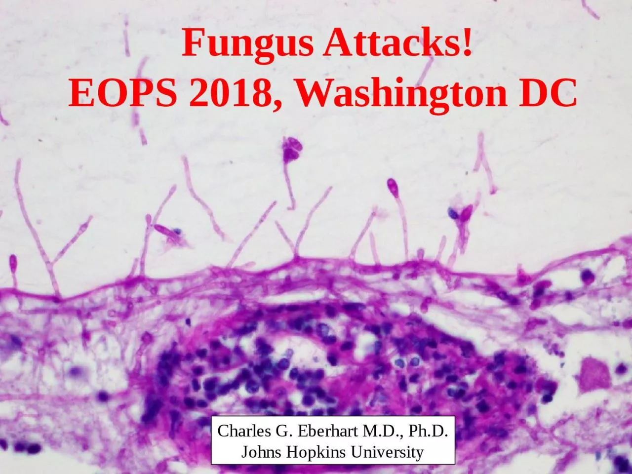
Charles G Eberhart MD PhD Johns Hopkins University Clinical History The patient was a 43 yearold female with a past medical history significant for monocytic acute myeloid leukemia AML with FLT3 ITD mutation ID: 934769

myeloid scedosporium fungal history scedosporium myeloid history fungal brain acute infection 2018 leukemia prolificans erythroid treatment marrow invasive lineage

Share:

Link:

Embed:

Download Presentation from below link

Download Presentation The PPT/PDF document "Fungus Attacks! EOPS 2018, Washington DC" is the property of its rightful owner. Permission is granted to download and print the materials on this web site for personal, non-commercial use only, and to display it on your personal computer provided you do not modify the materials and that you retain all copyright notices contained in the materials. By downloading content from our website, you accept the terms of this agreement.


Presentation Transcript

Slide1

Fungus Attacks!EOPS 2018, Washington DC

Charles G. Eberhart M.D., Ph.D.

Johns

Hopkins University

Slide2

Clinical History

The patient was a 43 year-old female with a past medical history significant for monocytic acute myeloid leukemia (AML) with FLT3 ITD mutation. She was first diagnosed with AML in July 2017, and subsequently had refractory disease despite multiple treatment protocols. An ophthalmic exam performed in February 2018 was normal aside from some vitreous floaters.

Slide3

Slide4

Pathology Report

BONE MARROW (BIOPSY AND ASPIRATE): PERSISTENT ACUTE MYELOID LEUKEMIAMICROSCOPIC DESCRIPTION: The marrow is hypercellular (70-80%). The myeloid:erythroid ratio is markedly increased. The myeloid lineage demonstrates a pronounced left shift with sheets of immature cells andrare maturing granulocytes. The erythroid lineage is markedly hypoplastic with few scattered erythroid lineage cells. Megakaryocytes are decreased in number, and are small with hypolobated and hyperchromatic nuclei.

Slide5

Slide6

Slide7

Clinical History

In early May 2018, she was found to have a right frontal lobe brain mass and CNS involvement of her leukemia.She was treated with multiple courses of intrathecal chemotherapy, antibiotics, and antifungal therapy. Her brain mass progressed, despite treatment, and was concerning for an invasive fungal infection. Blood cultures grew out Scedosporium prolificans (formerly known as Lomentospora prolificans).She was transitioned to comfort care and died in late May 2018.

Slide8

Imaging

T2 Flair

Slide9

Autopsy Findings

Autopsy demonstrated systemic invasive fungal infection, involving the brain, heart, stomach, thyroid, and kidney. The fungal hyphae were septate, with acute angle branching, and on a morphological basis were consistent with the ante mortem culture of ScedosporiumMultiple hemorrhages and infarctions were noted in the cerebral hemispheres, cerebellum, and brainstem secondary to invasive fungal infection. The patient's acute myeloid leukemia was identified in the brain, eye, breast, kidney, and possibly spleen, with persistent disease present in the bone marrow.

Slide10

Thyroid

Slide11

Heart

Slide12

Slide13

Slide14

Slide15

Slide16

Slide17

Slide18

Slide19

Slide20

Scedosporium

Ubiquitous filamentous fungi present in soil, sewage, and polluted watersThe genus Scedosporium contains two medically important species:S. apiospermum (and its sexual state Pseudallescheria boydii)S. prolificansCan colonize soft tissues, damaged lungs or disseminate hematogenously

Slide21

Scedosporium

prolificansScedosporium prolificans was originally named Lomentospora prolificans in 1974. It was transferred to the genus Scedosporium  in 1991.Subcutaneous infections occur following traumatic implantation.Immune impairment is associated with systemic infection. It is resistant to most antifungal drugs and infections are often fatal.

Slide22

Conidia

Conidiophore

Slide23

Slide24

Slide25

Day 8

6 Years

Slide26

Slide27

Slide28

Slide29

Slide30

Of 1,792 culture proven keratitis cases, 10 (0.6%) were

Scedosporium.Trauma history in 8/10 casesTopical natamycin treatment was often successful when started early

Slide31

Scedosporum

apiospermum

Slide32

Thank You