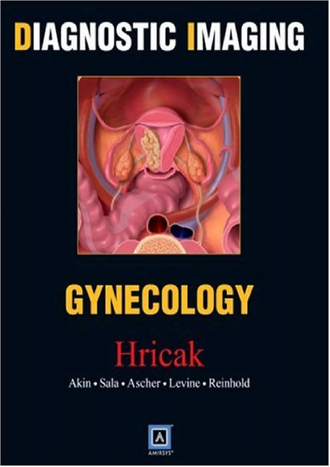
PDF-(BOOK)-Diagnostic Imaging: Gynecology
Author : LoriShaw | Published Date : 2022-09-04
Authored by some of the worlds preeminent authorities in its field this new book represents todays best single source of guidance on gynecological imaging It presents
Presentation Embed Code
Download Presentation
Download Presentation The PPT/PDF document "(BOOK)-Diagnostic Imaging: Gynecology" is the property of its rightful owner. Permission is granted to download and print the materials on this website for personal, non-commercial use only, and to display it on your personal computer provided you do not modify the materials and that you retain all copyright notices contained in the materials. By downloading content from our website, you accept the terms of this agreement.
(BOOK)-Diagnostic Imaging: Gynecology: Transcript
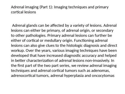
Authored by some of the worlds preeminent authorities in its field this new book represents todays best single source of guidance on gynecological imaging It presents more details for each diagnosis more representative images more case data and more current references than any other reference tool At the same time its userfriendly format lets you access all of this information remarkably quickly Dr Hedvig Hricak has assembled an extraordinary group of world recognized experts in gynecologic imaging They have collated their combined experience to create the most comprehensive review of gynecologic abnormalities ever published It is exquisitely illustrated and contains the most update imaging techniques in US CT and MRICovers the top imaging diagnoses in gynecology including both common and uncommon entities Provides exquisitely reproduced imaging examples for every diagnosisplus concise bulleted summaries of terminology imaging findings key facts differential diagnosis pathology clinical issues a diagnostic checklist and selected referencesIncludes an extensive image gallery for each entity depicting common and variant casesOffers a vivid fullcolor design that makes the material easy to readDisplays a thumbnail visual differential diagnosis for each entity. to Wrongness. John Banja, PhD. Center For Ethics. Emory University. jbanja@emory.edu. Why Be Interested in Diagnostic Error?. Diagnostic errors are the leading cause of medical malpractice suits: 45% of cases. People Management Network. 1. Overview of the session. Introduction to the project . An explanation of the Diagnostic. Conclusions – what I have learnt. Developed . from the Strengthening the Performance Framework Project. New diagnostic tests been studied extensively. Some 90% of immunogenicity. However, even fully therapeutic antibodies has been very successful. Well-known examples of are applied for in ammatory disea We need to shift from functional expertise .... to building . capacity. in organizations.. Project 2: Universal Corporate Diagnostic. We used to look at engagement data to understand culture – that’s playing in the functional expertise zone.. J. Sal Saldivar, M.D.. Gynecology Oncology - Department of OB/GYN. El Paso First Health. 1145 Westmoreland Drive . August 28th, 2014 - 1:00pm. Description of the Project. AAGL approved Fellowship in MIS . 2018. Committee on Acute Care Surgery, Canadian Association of General Surgeons. DIAGNOSTIC IMAGING MODALITIES. 4. Melissa Hanson MD, and. . Jacinthe. . Lampron. MD. Committee . on Acute Care Surgery, Canadian Association of General Surgeons. Professor Errico Zupi Current Position or Job Title Associate Professor of Ob/Gyn Univerity of Tor Vergata Rome Italy Hospital of Affiliation Policlinico Tor Vergata City, Country Rome Official Emai NameEffective Dates ToPractice Area Code 34Version Code 05-2013aInitial privileges initial appointment Renewal of privileges reappointment Expansion of privileges modificationAll new applicants must m Updated 7/9/2021COMPUTED TOMOGRAPHYThisprogram offers radiologic technologists nuclear medicine technologists and radiation therapists a pathway to expand their knowledge in Diagnostic Imaging CT whil FORM - NMC - 2 - PG ( ) - V_2020 Signature of Dean Signature of Assessor 1 STANDARD ASSESSMENT FORM FOR PG COURSES SUBJECT - OBSTETRICS & GYNECOLOGY INSTRUCTIONS FOR DEANS/PRINCIPALS AND ASSESSORS Volume 3 , Issue 3, 2018, PP 7 - 9 ISSN 2456 - 0561 DOI: http://dx.doi.org/10.20431/2456 - 0561.0 3 0 3 00 2 www.arcjournals.org ARC Journal of Gynecology and Obstetrics Page | 7 Case Report: Role Adrenal glands can be affected by a variety of lesions. Adrenal lesions can either be primary, of adrenal origin, or secondary to other pathologies. Primary adrenal lesions can further be either of cortical or medullary origin. Functioning adrenal lesions can also give clues to the histologic diagnosis and direct workup. Over the years, various imaging techniques have been developed that have increased diagnostic accuracy and helped in better characterization of adrenal lesions non-invasively. In the first part of the two part series, we review adrenal imaging techniques and adrenal cortical tumors such as adenomas, adrenocortical tumors, adrenal hyperplasia and oncocytomas. – An Introduction – . Dr . Rudra. . Pratap. . Pandey. Prof. VSR. Conventional radiography. Digital radiography (CR & DR). A. Equipment details. B. Safety issues – Radiation . Advantages . . radiologi. IPSG.1 – . Identifikasi. . pasien. . secara. . benar. IPSG.2 – . Melaporkan. . hasil. . pemeriksaan. yang . kritis. , . serah. . terima. . pasien. ACC.2.2.1 – . Radiologi.
Download Document
Here is the link to download the presentation.
"(BOOK)-Diagnostic Imaging: Gynecology"The content belongs to its owner. You may download and print it for personal use, without modification, and keep all copyright notices. By downloading, you agree to these terms.
Related Documents