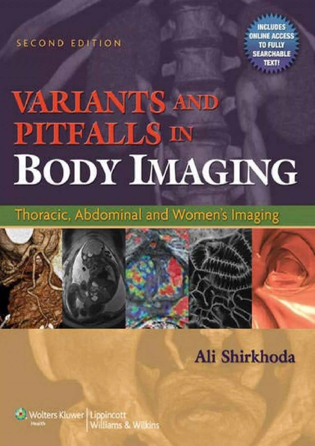
PDF-(BOOK)-Variants and Pitfalls in Body Imaging: Thoracic, Abdominal and Women\'s Imaging

Author : LoriShaw | Published Date : 2022-09-04

Variants and Pitfalls in Body Imaging Second Edition is the key to identifying features on images that can impede accurate diagnosis particularly normal anatomic

Presentation Embed Code

Download Presentation

Download Presentation The PPT/PDF document "(BOOK)-Variants and Pitfalls in Body Ima..." is the property of its rightful owner. Permission is granted to download and print the materials on this website for personal, non-commercial use only, and to display it on your personal computer provided you do not modify the materials and that you retain all copyright notices contained in the materials. By downloading content from our website, you accept the terms of this agreement.

(BOOK)-Variants and Pitfalls in Body Imaging: Thoracic, Abdominal and Women\'s Imaging: Transcript


Variants and Pitfalls in Body Imaging Second Edition is the key to identifying features on images that can impede accurate diagnosis particularly normal anatomic variants and technical artifacts that mimic pathology Covering the abdomen pelvis and thorax and all current imaging modalities this sourcebook explains how to differentiate normal anatomic variants technical artifacts and other diagnostic pitfalls from pathologic conditionsOrganized by site for easy reference the book covers CT MRI ultrasound and nuclear medicine This edition includes advanced technologies such as multidetector CT scanning for cardiovascular imaging CT and MR enterography for enterocolitis virtual colonoscopy CT and MR urography prostate and breast MR imaging and PETCT scanning Wellrespected radiologists walk the reader through specific body areas describing problems solutions and relevant anatomy Users will come away with a clear understanding that will yield ontarget assessments for every patientA companion website will include the fully searchable text and images. Traditionally clinical imaging exploits the contrast due to differences in spin density spinlattice relaxation T and spinspin relaxation T between normal tissues and pathology However other contrast mechanisms such as susceptibility magnetization Cabrera . Guillaume . Lemaitre. Mojdeh. . Rastgoo. CT and MR . Imaging. of . Abdominal . Aortic. . Aneurysm. Presentation. . Outline. Introduction. . to. Abdominal . Aortic. . Aneruysms. Computed. Computed Tomography (CT). CT scanner. Provides high-resolution cross-sectional anatomical images. Table moves through a circular opening in the CT scanner called the gantry, while an x-ray tube emits x-rays as it spins 360 degrees inside the gantry. Chapter 1. Biomedical Engineering. Dr. Mohamed Bingabr. University of Central Oklahoma. INSTRUCTOR. :. Mohamed Bingabr, Ph.D.. CONTACTS:. . Office. : Howell 221D ; . Phone. : 974 5718; . Email. : mbingabr@uco.edu. Elastography. and . Diffusion Acquisitions. : Diffusion-MRE (. dMRE. ). Technology Overview. MR . elastography. (MRE) is an emerging imaging . technology that . is capable of assessing tissue stiffness . Imaging. Evolution . of Medical . Imaging. Types of Medical . Imaging. Medical Radiography. X-Ray Imaging. Nuclear Imaging. Bone Densitometry. Magnetic Resonance Imaging (MRI). Ultrasound. Neuroprosthetics. Define terminology relating to careers in medical imaging. Specify the role of diagnostic medical health care workers including personal characteristics, levels of education, and credentialing requirements. 2018. Committee on Acute Care Surgery, Canadian Association of General Surgeons. DIAGNOSTIC IMAGING MODALITIES. 4. Melissa Hanson MD, and. . Jacinthe. . Lampron. MD. Committee . on Acute Care Surgery, Canadian Association of General Surgeons. Clinical (Diagnostic) Terminology. Radiography. –. Film records (radiographs) of internal structures of the body made by electromagnetic radiation . (X-rays, gamma rays, radio waves. ) passing through the body to act on special film. Computed tomography in oral and maxillofacial radiology. Computed tomography (CT) is today commonly used in imaging of the maxillofacial area. Conventional CT examinations are usually performed in medical X-ray departments. However, a relatively new technique named cone-beam computed tomography (CBCT) or digital volume tomography (DVT) has now also become available for dental purposes. The advantage with this technique is a lower radiation dose compared to conventional CT. Common examples when DVT is used are; for diagnosing the position of impacted canines and suspected root . Febrile Seizures. When to Image Based on . Choosing . Wisely. ®. . and ACR Appropriateness . Criteria. ®. What Is R-SCAN?. 2. C. ollaborative activity . for referring clinicians and radiologists to improve patient . Magnetic Resonance Imaging of Rectal Cancer shwari et al. Journal of Gastrointestinal and Abdominal Radiology ISGAR ol. 2 o. 1/2019 Magnetic Resonance Imaging of Rectal Cancer shwari et al. Journal of BioE. 477/598DP. Module 1. Dipanjan Pan, PhD, FRSC, FAHA, FACC. Department of Bioengineering. Beckman Institute of Advanced Imaging and Science. Department of Materials Science and Engineering. dipanjan@illinois.edu. Resident Board Review. January 23. rd. , 2014. Jo-Annie Letendre, DVM. Outline. Basic principles of CT. Use of CT in ECC. Review of the paper:. Radiology of thoracic . trauma in . the dog and cat. Review of the paper:.

Download Document

Here is the link to download the presentation.
"(BOOK)-Variants and Pitfalls in Body Imaging: Thoracic, Abdominal and Women\'s Imaging"The content belongs to its owner. You may download and print it for personal use, without modification, and keep all copyright notices. By downloading, you agree to these terms.

Related Documents