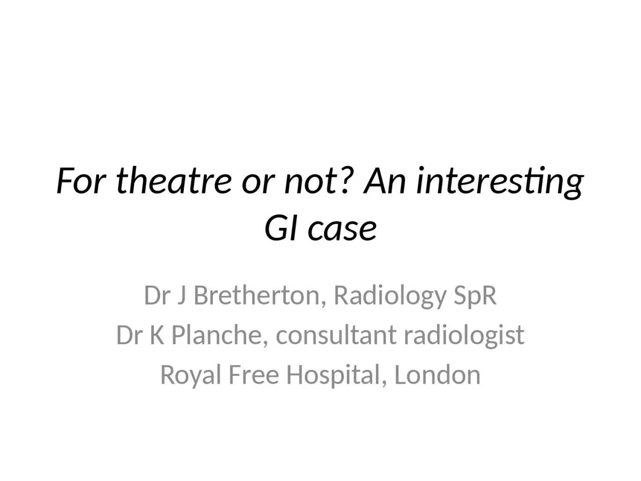
Dr J Bretherton Radiology SpR Dr K Planche consultant radiologist Royal Free Hospital London Clinical Information 54yearold female Presenting complaint abdominal pain and distension ID: 931964
Download Presentation The PPT/PDF document "For theatre or not? An interesting GI ca..." is the property of its rightful owner. Permission is granted to download and print the materials on this web site for personal, non-commercial use only, and to display it on your personal computer provided you do not modify the materials and that you retain all copyright notices contained in the materials. By downloading content from our website, you accept the terms of this agreement.
Slide1
For theatre or not? An interesting GI case
Dr J
Bretherton
, Radiology
SpR
Dr K
Planche
, consultant radiologist
Royal Free Hospital, London
Slide2Clinical Information
54-year-old female
Presenting complaint: abdominal pain and distension
Past medical history:
Vaginal hysterectomy for uterine prolapse
Raynaud’s
Chronic bloating and constipation
Laparotomy at another hospital (details unclear)
Examination findings:
Abdominal distension and generalised abdominal tenderness without guarding
PR examination: stool in rectum.
Stable observations
Slide3Investigations (X-rays)
Slide4Investigations (X-rays)
CXR:
subdiaphragmatic
free gas and dilated bowel loops.
AXR:
Rigler’s
sign
and
dilated bowel loops.
Slide5Investigations (contrast enhanced CT abdomen and pelvis)
Slide6Investigations (contrast enhanced CT abdomen and pelvis)
Large volume free fluid and
locule
of free gas (arrowed).
Slide7Investigations (CT- lung windows)
Slide8Investigations (CT- lung windows)
Dilated small bowel loops, large volume
pneumoperitoenum
and gas containing cystic structures in the bowel wall (arrowed) in keeping with
pneumatosis
No portal venous gas or bowel wall defect was demonstrated.
Slide9Investigations (CT- lung bases)
Slide10Investigations (CT- lung bases)
Patulous, fluid filled oesophagus (asterisk)
Mild dilatation of the lower lobe airways (arrow)
*
Slide11Final diagnosis
= Systemic sclerosis with benign spontaneous
pneumoperitoneum
Slide12How we confirmed the diagnosis
Our institution is a tertiary referral centre for systemic sclerosis and scleroderma, and the referring clinicians and reporting radiologist were familiar with this entity.
The dilated oesophagus
and early basal architectural distortion seen
on the CT
are characteristic features of systemic sclerosis. The clinical team were able to confirm that the patient had cutaneous manifestations of the disease (
calcinosis
and Raynaud’s phenomenon).
Notes from previous hospital admission elsewhere were obtained. The presentation was similar and the patient had a laparotomy for suspected perforation. No site of perforation was found, but the operating surgeon noticed small bowel
pneumatosis, and took an intra-operative biopsy to confirm.Patient improved with conservative management and was referred to Rheumatology.Repeat CT after six months (to investigate weight loss) showed resolution of the pneumoperitoneum but persistent small bowel dilatation and ascites.
Slide13Systemic Sclerosis
Systemic sclerosis is a multisystem autoimmune disease characterised by fibrosis of the skin and internal organs secondary to collagen accumulation, and
vasculopathy
[1].
GI manifestations are common, occurring in approximately 90% of patients, and the spectrum of GI involvement is wide (oesophagus and small bowel most commonly involved) [1].
Atrophy of muscularis propria
replacement by collagen decreased peristalsis stasis and dilatation [1].
Slide14Pneumatosis in Systemic Sclerosis
Aka
pneumatosis
cystoides
intestinalisBenign condition characterised by air filled cysts in bowel wall and mesentery, most commonly occurring in the jejunum and ileum[2].May be associated with portal venous gas.Pathophysiology poorly understood but thought to involve dissection of gas into bowel wall either due to:
- Defect in bowel wall secondary to atrophy/
ischaemia
/bacterial overgrowth [3]
- Gut flora overgrowth and excessive hydrogen production because of bowel stasis[3].May be asymptomatic or associated with abdominal pain and distension[3]. Should not be associated with signs of peritonism/fever/raised inflammatory markers[3].May be exacerbated by immunosuppressive agents.
N.b. these can also mask signs of true perforation.
Slide15Benign spontaneous pneumoperitoneum
in Systemic Sclerosis
Almost always associated with
pneumatosis
.
Thought to occur due to cyst rupture [3].May also occur due to microperforation of jejunal diverticula [3].CT imaging is invaluable- reassuring findings are:
presence of
pneumatosis
absence of other signs such as fat stranding, fluid collection, discrete bowel wall defect
Can persist for years and does not require surgical treatment [3].
Slide16Key learning points
Radiologists and clinicians should be aware of this uncommon condition (benign spontaneous
pneumoperitoneum
in systemic sclerosis) to avoid unnecessary surgery in this high risk patient group.
These patients are also at risk of perforation due to
ischaemia and obstruction therefore clinical correlation is important (signs of peritonism = not benign pneumoperitoneum!). Clinical correlation can be challenging in context of immunosuppressant use.
CT is helpful to look for
pneumatosis
and absence of any signs suggesting other cause of
pneumoperitoneum.
Slide17Key learning points
Remember the classical imaging appearances of
pneumatosis
cystoides
intestinalis
Slide18References
[1] Schreiner et al.
Gastrointestinal Manifestations of Systemic
Sclerosis.
J Scleroderma Relat Disord. 2016; 1(3): 247–256.[2] Hassan & Devgun Pneumatosis Cystoides
Intestinalis
: A Rare Benign Cause of
Pneumoperitoneum. Case Rep Radiol. 2013; 2013: 353245.[3] Vischio et al. Benign Spontaneous Pneumoperitoneum in Systemic
Sclerosis. Journal of Clinical Rheumatology. 16(8):379-381, DEC 2010