/
HISTEROSALPHINGOGRAPHY –COVENTIONAL HISTEROSALPHINGOGRAPHY –COVENTIONAL

HISTEROSALPHINGOGRAPHY –COVENTIONAL - PowerPoint Presentation

RefreshingView
RefreshingView . @RefreshingView
Follow
342 views
Uploaded On 2022-08-04

HISTEROSALPHINGOGRAPHY –COVENTIONAL - PPT Presentation

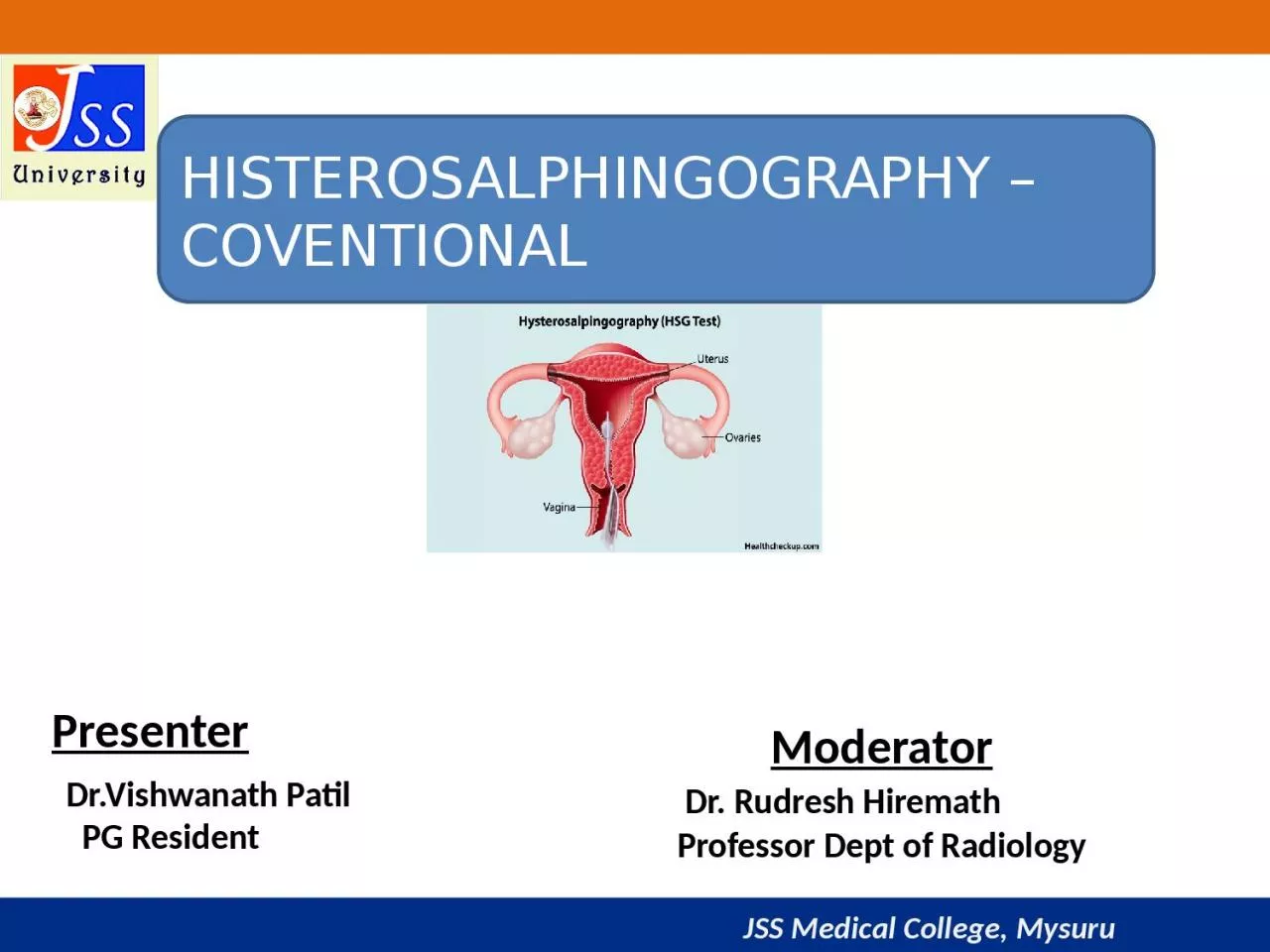
Presenter DrVishwanath Patil PG Resident Moderator Dr Rudresh Hiremath Professor Dept of Radiology Defination Hysterosalpingography is the radiographic evaluation of uterus and fallopian tubes ID: 935571

tubal contrast uterus uterine contrast tubal uterine uterus tube fallopian filling appearance radiograph hsg tubes defects cervical left spot

Share:

Link:

Embed:

Download Presentation from below link

Download Presentation The PPT/PDF document "HISTEROSALPHINGOGRAPHY –COVENTIONAL" is the property of its rightful owner. Permission is granted to download and print the materials on this web site for personal, non-commercial use only, and to display it on your personal computer provided you do not modify the materials and that you retain all copyright notices contained in the materials. By downloading content from our website, you accept the terms of this agreement.


Presentation Transcript

Slide1

HISTEROSALPHINGOGRAPHY –COVENTIONAL

Presenter

Dr.Vishwanath Patil PG Resident

Moderator

Dr. Rudresh Hiremath Professor Dept of Radiology

Slide2

DefinationHysterosalpingography is the radiographic evaluation of uterus and fallopian tubes

under fluoroscopic guidance.

Slide3

INDICATION 

1. Infertility (main role) 2. Recurrent spontaneous abortions

.3. Congenital anomalies of uterus.

4. Postoperative evaluation following (a)tubal ligation (b) reversal of tubal ligation. 5. Suspected case of genital tuberculosis 6. To prove tubal occlusion after insertion of transcervival sterilization micro insert (essure).

HSG also has a potential therapeutic role in increasing the probability of pregnancy ( especially if oil soluble contrast –lipoid is used)

Slide4

CONTRAINDICATIONSuspected pregnancy

Acute pelvic infection Active

vaginal bleeding Recent dilation and curettage Tubal

or uterine surgery within last 6 wks.Contrast sensitivity

Slide5

PATIENT PREPARATION Done in first half of menstrual cycle in proliferative phase between 8th to 12th day

. Patient to avoid unprotected sexual intercourse from the date of her period until investigation is over

.If periods are irregular , do urine B- hcg

.Exclude active pelvic infection .Prophylactic antibiotics not routinely recommended (considered in case of bacterial endocarditis)

Slide6

Accessory & Equipments

Disposable HSG tray is used. SpeculumCotton balls, cup, gauze, drapes. Sponge-holding forceps. 10 ml syringes, lubricating jelly extension tube.Contrast.

Slide7

CONTRAST MEDIA Heuser was the first to report on the use of lipiodol in HSGs.

Lipiodol was gradually replaced by water soluble contrast media for several reasons .

Slide8

CONTRAST MEDIA

LIPID SOLUBLE CONTRAST (lipiodol) Sharp image Minimal painDelayed absorption Risk of lipogranuloma formation in case of tubal

block/hydrosalpynx.Intravasation of contrast and possible risk of oil embolism Need of delayed film Less often used

WATER SOLUBLE CONTRAST (iohexol-omnipaque,meglumine diatrizoate-urograffinAmpullary rugae clearly visualised Gets absorbed within hours, does not leave residueGranuloma formation rare Pain persists after procedure Prompt demonstration of tubal patency, delayed film not needed. Widely used and preferred

Slide9

PROCEDUREInformed consent is taken .

Patient is asked to empty bladder immediately before procedure .Scot film may be taken. Patient is placed in lithotomy position.The perineum is cleaned with antiseptic solution (Betadine)and draped with sterile towel.

The cervix is localized and cleansed with povidine-iodine solution. A speculum is inserted into the vagina. Cervix is cannulated with any of available cannulas which is made air free before administration of

contrast.

Slide10

PROCEDURETenaculm

is used to hold anterior lip of cervix .Speculum is removed & Patient is placed in slight trendelenburg position and contrast is slowly given 3 ml contrast to fill uterine cavity and another 3 ml to fill tube. ( up to 10 ml)

Slide11

PROCEDURE4 spot films are taken .

1.Early filling -any filling defect 2. uterus fully distended- shape of the uterus. 3. Evaluate the fallopian tubes. 4. free intraperitoneal spillage of contrast material.Additional oblique views may be taken for optimal visualization of pelvic pathology and tortuous fallopian tubes( to see retroverted or anteverted

).After end of the procedure , antibiotic course is given and patient is informed about vaginal spotting for 1-2 days.

Slide12

COMPLICATION Pain (because of dilatation of uterus , spillage into peritoneum).

Infection (pelvic). Bleeding. Vascular or lymphatic Intravasation .Vasovagal episode. Allergic reaction (to iodinated contrast media

).Uterine perforation.

Slide13

NORMAL HSGThe uterine cavity is shown during HSG as a triangular contrast-filled

structure.The uterine fundus on top, which can be flattened, concave or slightly convex . Free spillage of the contrast to the peritoneum noted

Slide14

NORMAL HSG

Slide15

NON PATHOLOGIC FINDINGS

Air bubble- round, often multiple, welldefined mobile filling defect ,usually displaced to fallopian tubes if additional contrasts given.

Slide16

UTERINE FOLDS

Uterine folds. HSG spot radiograph demonstrates uterine folds (arrows) as linear filling

defects that parallel the longitudinal axis of the uterus.

Slide17

Previous caesarean section scar 

Previous caesarean section scar: linear appearance (as in this case) or can occasionally manifest as a wedge-shaped outpouching or diverticulum 

Slide18

PROMINENT CERVICAL GLANDS

Prominent cervical glands-tubular structure with their origin in both cervical walls.

Slide19

DETECTABLE PATHOLOGY

UTERINE1. Uterine anomaly 2. Fibroid (

submucosal) 3. Adenomyosis 4.Endometrial polyp 5.Intrauterineadhesions/synaechiae .6.Endometrial TB 7

. Cervical incompetenceTUBAL1. tubal block 2. Tubal spasm 3. Tubal polyp 4.Hydrosalpinx 5.Salpingitis isthmic nodosum (SIN). 6. Peritubal adhesions. 7. TB salpingitis .

Slide20

UTERINE ANOMALIES

Slide21

UTERINE ANOMALIES

Slide22

Unicornuate uterusSpot radiograph demonstrates

a single uterine horn with an irregular medial contour. HSG cannot be used to exclude the presence of a noncommunicating rudimentary horn .Single right uterine horn with single right fallopian

tube.

Slide23

UTERUS DIDELPHYS

2 Uterine cavities, 2 cervical canals, 2 vagina. (nonfusion of the two Müllerian ducts.)Vaginal obstruction may manifest shortly after menarche, lead to complications, and require intervention.

Slide24

 BICORNUATE UNICOLLIS

Widely splayed uterine horns with intercornual angle >100.2 uterine cavities, 1 cervical canal Incomplete fusion of the cephalad extent of the uterovaginal horns with resorption of the uterovaginal septum.Often asymptomatic .Surgery usually not indicated

Slide25

BICORNUATE BICOLLI

Two cervical canals; central myometrium extends to external cervical os

Slide26

Slide27

Septate UterusHistory of midtrimester pregnancy loss .

Surgical resection may be considered if recurrent fetal loss occurs

Slide28

SEPTATE UTERUSSlight separation forming acute angle.

Slide29

Bicornuate and Septate Uteri

Bicornuate Fundus indented – Cavities widely separated( > 100 degree) – Partial fusion of mullerian ducts.

Definite diagnosis by MRI Intervening cleft > 1 cm & intercornual distance > 5cm in bicornuate uterus. Septate

Normal external surface – Cavities are close together – Defect in canalization or resorption of midline septum between mullerian ducts. Angle of less than 75° between.

Slide30

Classification criteria for USG

Bicornuate Septate

When the apex of the fundal contour is more than 5 mm (arrow) above a line drawn between the tubal ostia, the uterus is septate.

When the apex of the fundal contour is below or less than 5 mm above a line drawn between the tubal ostia, the uterus is bicornuate .

Slide31

Arcuate Uterus

Near reabsorption of the uterovaginal septum and is characterized at imaging by a mild indentation of the external fundal contour.HSG: Saddle-shaped indentation at the uterine fundus is seen.

Slide32

DES UterusDES-related anomaly of the uterus involves a hypoplastic or T-shaped uterus.

Slide33

Abnormalities of Uterine ContourAdenomyosis is a condition in which endometrium extends into the myometrium.

At HSG, adenomyosis appears as small diverticula extending into the myometrium that is irregular outline with multiple diverticulum.

Slide34

 FIBROID UTERUS

Leiomyomas manifest as well-defined filling defects at HSG and can have a variety of appearances depending on their size and their location within the uterus.

Slide35

Luminal Filling Defects

SynechiaeSpot radiograph shows a central oval irregular filling defect within the uterus, a finding that represents a synechia.Multiple synechiae associated with infertility is known as Asherman syndrome.

Multiple filling defects are observed in the uterine cavity with irregular edges.

Slide36

Virtual Hysterosalpingography (VHSG)

Multiplanar reconstructions show irregular elevated lesions with soft tissue density which extend from the uterine walls.Sagittal maximum intensity projection image that shows an anteverted uterus, which presents multiple filling defects compatible with synechiae. 

Virtual endoscopy image which illustrates endoluminal lesions.(c,d). 3D volume rendering images which exhibit irregularities on the wall corresponding to synechiae.

Slide37

Luminal Filling Defects

Endometrial polypThey usually manifest as well-definedfilling defects and are best seen during the early filling stage.

Small polyp on the right lateral wall of the uterine silhouette

Slide38

Fallopian Tubes10–12 cm in length.Salpingitisisthmicanodosum (

SIN).Cornual spasm.Tubal occlusion.Per tubal adhesionsHydrosalpinx.Irreversible tubal occlusion with a micro insert.Tubal

polyps.

Slide39

Salpingitis isthmica

nodosum (SIN)Spot radiograph demonstrate SIN as small outpouchings or diverticulum from the isthmic portion of the fallopian tubes. Unknown cause.

A/W 1.infertility 2.PID 3.Ectopic pregnancySINcan be either unilateral or bilateral.

Slide40

Cornual spasm

Early filling stage of the uterus, the right fallopian tube does not opacify

beyond the cornual portion.After the instillation of additional

contrast material, the right fallopian tube opacified to the ampullary portion.

Slide41

Tubal occlusionSpot radiograph demonstrates abrupt cutoff of the left fallopian tube.

Spot radiograph demonstrates cutoff of contrast material in the isthmic portions of both fallopian tubes, with bulbous dilatation.

Slide42

Hydrosalpinx(a)

Steep right oblique spot radiograph shows dilatation of the ampullary portion of the right fallopian tube (arrow).(b) Spot radiograph shows dilatation of the ampullary portion of the left fallopian tube, a finding that is consistent with a hydrosalpinx.

Slide43

Peritubal adhesions

Spot radiograph demonstrates a round collection of contrast material adjacent to the left fallopian tube, a finding that suggests per tubal adhesions. Note the free contrast material spillage on the right side.

Slide44

Irreversible tubal occlusion with a microinsert

(a) Scout radiograph obtained prior to the instillation of contrast material shows a micro insert. (b) Radiograph obtained after instillation shows no contrast material filling of the fallopian tube beyond the micro insert

Slide45

Tubal polyp.Small smooth filling defect (arrow) in the proximal left

fallopian tube, a finding that typically represents a tubal polyp.Without concomitant dilatation or tubal occlusion.Rare.

Asymptomatic

Slide46

HSG finding in women with TB

Genital tuberculosis (TB) is an important cause of health problem and infertility.It remains the initial diagnostic procedure in the evaluation of tubal, uterine cavity, and peritoneal factors leading to infertility.1.Multiple small diverticular like appearance surrounding the ampulla produced by caseous ulceration gives the tubal outline a

Rosette-like appearance.

Slide47

TB Salphagitis isthemica

nodosaPenetration of contrast medium between the mucosal folds produces small diverticular-like outpouchings with a bizarre pattern.

 Cotton-wool plug appearanceDistribution of contrast medium in a reticular pattern.

Slide48

BEADED TUBE Multiple constrictions along the fallopian tube giving rise to a " beaded" appearance

 .GOLF CLUB TUBE

Sacculation of both tubes in distal portion with an associated hydrosalpinx giving a Golf club-like appearance.

Slide49

PIPE STEM APPEARANCEAbsence of normal tortuosity and a curved or straight pipe like appearance show

fibrotic stage of tuberculous salpingitis. FLORAL APPEARANCE 

Twisted hydrosalpinx resembles a floral appearance of left side tube.

Slide50

LEOPARD SKIN APPEARANCEMultiple rounded filling defects following intraluminal granuloma formations

within the hydrosalpinx, resembling a " leopard skin" appearance.

Slide51

COBBLE STONE APPEARANCE

Intraluminal scarring of the tube gives rises a cobblestone like appearance which is an effective radiographic sign of intraluminal adhesionsCORK SCREW APPREANCE Vertically fixed tubes secondary to dense

peritubal adhesions. Dense connective tissue causes the lack of tubal mobility. The hyperconvulated right tube and manifests a " cork screw" like appearance

Slide52

PERITUBAL HALOThickening of the tubal walls due to peritubal

adhesions (arrows) represents a cloudy sign on hysterosalpingograms. TOBACCO POUCH APPREANCE 

Terminal hydrosalpinx with the conical narrowing is seen in the right tube.Eversion of the fimbria secondary to adhesions, with a patent orifice produces the tobacco pouch appearance in the left terminal. 

Slide53

Pseudo-unicornuate uterus.

Unilateral scarring of the cavity makes an asymmetric intrauterine obliteration, resembling a unicornuate uterus. the irregular contour and vertical orientation of long axis.True unicornuate uterus. the smooth contour, more horizontal orientation of long axis and normal ipsilateral fallopian

tube.

Slide54

TRIFOLIATE SHAPED UTERUS Synechiae formation at the uterine borders and partial obliteration in the fundus produce a

trifoliate like appearance. Both tubes are obstructed in the isthmic portion.

Slide55

ConclusionHSG remains the front-line imaging modality in the investigation

of infertility.Has a low sensitivity for the diagnosis of pelvic adhesions, which is why it cannot replace laparoscopy.

Slide56

ReferencesPathology of the Uterine Cavity: Clinical key.

Hysterosalpingographic findings in women with genital tuberculosis; Donya Farrokh, Parvaneh Layegh, Monavvar

Afzalaghaee, Mohaddeseh Mohammadi, Yalda Fallah RastegarIran J

Reprod Med. 2015 May; 13(5): 297–304.Simpson Jr WL, Beitia LG, Mester J. Hysterosalpingography: a reemerging study. Radiographics. 2006 Mar;26(2):419-31.Imaging of Müllerian Duct Anomalies Spencer C. Behr, Jesse L. Courtier, Aliya Qayyum Online:Oct 4 2012https://doi.org/10.1148/rg.326125515

Slide57

Slide58

?

Slide59

AnswerThe cornua, isthmic and proximal 2/3rd of ampullary part of right fallopian tube are normal in calibre and show normal contrast opacification n. Rest of the distal 1/3

rd of ampullary and infundibular parts of the right fallopian tube is dilated.

Slide60

?

Slide61

AnswerNON VISUALIZATION OF THE LEFT FALLOPIAN TUBE IN ITS ENTIRE LENGTH BEYOND THE CORNUA - S/O LEFT CORNUAL BLOCK.

Slide62

?

Slide63

AnswerThere is intravasation of contrast into the myometrial-parametrial vessels extending into paracaval

veins occurring immediately – S/O Level 3 intravasation. 

Related Contents


Next Show more