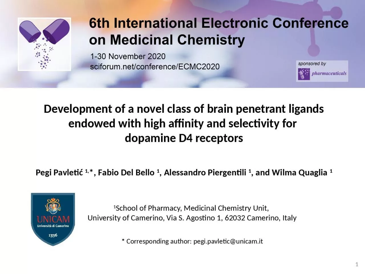
dopamine D4 receptors Pegi Pavletić 1 F abio Del Bello 1 Alessandro Piergentili 1 and Wilma Quaglia 1 1 1 School of Pharmacy Medicinal Chemistry Unit University of ID: 935291
Download Presentation The PPT/PDF document "Development of a novel class of brain pe..." is the property of its rightful owner. Permission is granted to download and print the materials on this web site for personal, non-commercial use only, and to display it on your personal computer provided you do not modify the materials and that you retain all copyright notices contained in the materials. By downloading content from our website, you accept the terms of this agreement.
Slide1
Development of a novel class of brain penetrant ligands endowed with high affinity and selectivity for dopamine D4 receptorsPegi Pavletić 1,*, Fabio Del Bello 1, Alessandro Piergentili 1, and Wilma Quaglia 1
1
1
School of Pharmacy, Medicinal Chemistry Unit,
University of Camerino, Via S. Agostino 1, 62032 Camerino, Italy
*
Corresponding author:
pegi.pavletic@unicam.it
Slide2Graphical AbstractDevelopment of a novel class of brain penetrant ligands endowed with high affinity and selectivity for dopamine D4 receptors2
77-LH-28-1
Slide3AbstractDopamine is a catecholamine neurotransmitter involved in a variety of physiological functions, through interaction with five different G-protein-coupled receptors (D1-D5). Among dopamine receptors, the D4R subtype has recently emerged as a potential target for the treatment of eating disorders, drug addiction and cancer. Classical D4R ligands are characterized by a common pharmacophore, consisting of a lipophilic moiety linked by a spacer to a piperidine or piperazine basic function and an aromatic terminal. It has been demonstrated that the known M1 muscarinic bitopic agonist 77-LH-28-1 also behaved as a potent D4R ligand and showed an unexpected D4 selectivity over D2 and D3 subtypes. The structure of 77-LH-28-1 differs from other known selective D4R ligands (characterized by the presence of a substituted aromatic group at position 4 of the piperidine ring
) by having a butyl
aliphatic chain. Since
77-LH-28-1 is the first example of a D4R selective ligand with a unique structural feature, an extensive structure-activity relationship study has been undertaken to evaluate the importance of the butyl aliphatic chain for the interaction with D4R. From a preliminary study, potent, selective and brain penetrant D4R antagonists were identified. The results prompt
ed us to further investigation of 77-LH-28-1 structure and D4R selectivity.Keywords: D4R, dopamine, 77-LH-28-1
3
Slide4Introduction
Catecholamine neurotransmitter involved in physiological functions in the central and peripheral
nervous systemsInteracts with five different G-protein-coupled receptors (GPCRs), namely D1-D5 receptors (D1R-D5R)
DOPAMINE
DOPAMINE
RECEPTORS
D
ivided
into D1-like and D2-like subfamilies on the basis of sequence similarity and signal transduction properties
(
Figure 1
)
a) D1-like
subfamily
:
-
D1R and D5R subtypes
b)
D2-like
subfamily: -D2R, D3R, and D4R subtypes
Figure 1
.
Depiction
of
D1-
like
and
D2-
like
dopamine receptor
subfamilies
Slide5IntroductionD4R subtype is encoded by DRD4 geneLocated in the frontal cortex, amygdala, hippocampus
, globus pallidus, substantia nigra pars compacta and thalamus; and at the periphery in retina, kidney, adrenal glands, sympathetic ganglia, blood vessels, heart and gastrointestinal tract
Potential target for the treatment of widespread diseases: addiction
, eating disorders, Parkinson’s disease and cancer Given the limitations of current drugs, innovative treatments that improve efficacy and safety are an urgent need! Compounds belonging to different chemotypes, including aryl-linked piperazines, amide-linked piperazines, piperidines, morpholines and imidazolines, have been reported as selective D4R ligands (
Figure 2)
D4
R
Figure 2
.
Depiction of different chemotypes of selective D4
R
ligands
Slide6IntroductionKnown as M1 muscarinic bitopic agonistWe demonstrated that it also behaves as a potent and selective D4R ligand
77-LH-28-1
structure fits all the features of the pharmacophoric model proposed for the classical selective D4R ligands and consisting
of a lipophilic moiety linked by a spacer to a basic function and an aromatic terminal (Figure 3A), except for the presence of a butyl chain instead of the aromatic terminal
(Figure 3B)77-LH-28-1 → model for synthesis of novel D4R molecules
77-LH-28-1
Figure 3A)
G
eneral
pharmacophoric model deduced from
the
structures
of
classic dopamine D4R drug
s.
Figure
3
B) Chemical structure of 77-LH-28-1, fitting all the features of the pharmacophore except for the aromatic terminal, which is replaced by a butyl chain.Del Bello F. et al. J Med Chem. 2018, 61:
3712
Compound
p
K
i
D2R
D3R
D4R
D4/D2
D4/D3
77-LH-28-1
6.17±0.166.21±0.139.01±0.04691631
77-LH-28-1
No aromatic terminal!
Slide7IntroductionThe quinolinone lipophilic portion has been replaced by other moieties, based on known
D4-selective ligands (Figure 4A)P
ropyl linker has been replaced by chains of different lengths and nature
to assess the role of the distance between the basic function and the quinolinone moiety (Figure 4B)
Piperidine has been replaced by a piperazine nucleus, which proved to be suitable for high affinity D4R ligands (Figure 4C)77-LH-28-1 butyl chain has been replaced by different alkyl, arylalkyl and aryl groups (Figure 4D)
AIM OF THE STUDY
Figure 4.
Modifications to the structure of 77-LH-28-1.
A)
M
odifications of the lipophilic area;
B)
M
odifications
of
the linker;
C)
M
odifications
of
the basic function
;
D)
M
odifications
of
the n-butyl terminal.
Synthesis and biological evaluation of new 77-LH-28-1 analogues,
to better understand the structural features required for the selective interaction with D
4
R.
The following modifications have been performed:
Slide8Results and discussion8
FIRST SERIES OF COMPOUNDS
Slide9Results and discussionFrom the study of the first series of compounds it emerges that the aliphatic chain of 77-LH-28-1 can be successfully replaced by an aryl or arylalkyl chain without affecting high D4R affinity and selectivity over D2R and D3R subtypes (compounds 9 and 12) (Figure 5) Compound 9 behaves as a potent D4R antagonist and, unlike 77-LH-28-1, as a weak partial antagonist at M₁ muscarinic receptor. It shows very high D4R affinity and selectivity over the other D2-like subtypes
and over M₁-M₅ muscarinic receptors.
Piperazine ring causes a sharp decrease in affinity, except for the N-phenyl derivative 12, which shows high affinity for D4R and selectivity over D2R and D3R
, M₁-M₅ subtypes and other selected off-targets, namely α1a
, α1b, α1d-, β1- and β
2
-adrenoceptors, σ
1
receptor, dopamine and serotonin transporters (DAT and SERT)
9
Figure 5
. Modifications to structure of 77-LH-28-1 that resulted in synthesis of compounds
9
and
12
MOST INTERESTING RESULTS FROM THE STUDY OF THE FIRST SERIES OF COMPOUNDS
Slide10Results and discussionInterestingly, in in vivo pharmacokinetic studies, a relevant brain penetration characterizes compound 12 (Figure 6) From functional studies, 12 shows a biased behavior, potently and partially activating Gi protein and inhibiting β-arrestin2 recruitment (Table 1)
Future studies with 12 might reveal mechanistically related behaviors and the interplay between G-protein- and β-arrestin-mediated signaling in D4R-related physiological effects
10
Figure
6
. Plot of mean concentration with standard
devations
of
12
in plasma and brain after subcutaneous administration (3 mg/kg).
Table 1
. Potency Values (Expressed as pEC
50
or pIC
50
) and Efficacy Values of
12
and Dopamine for D4R Expressed in HEK293T Cells.
MOST INTERESTING RESULTS FROM THE STUDY OF THE FIRST SERIES OF COMPOUNDS
Slide11Results and discussion11
Due to its interesting biological profile,
12
has been selected as a model for the synthesis of analogues, to define extensive SARs for this class of potent and selective D4R ligands
(
Figure 7
)
Before
engaging
in
the
synthesis
of
a
new
series
of
derivatives
, a
preliminar
in
silico
analysis
was
performed
on
the
selected
compounds
P1-P7, to collect useful infomation on the structural requirements for an optimal interaction with D4R P1-P7Figure 7. Compound 12 (left) as a model for the synthesis of ligands P1-P7 (right)
Slide12Results and discussionName
StructureM.W. / g/mol
P1349,43
P2
319,44P3320,43
P4
337,48
12
Table
2
. Structure and characteristics of D4R ligands P1- P4
Slide13Results and discussionName
StructureM.W. / g/mol
P5335,49
P6
320,43P7321,42
13
Table
3
. Structure and characteristics of D4R ligands P5- P7
Slide14Results and discussion143D
models of the compounds
P1- P7 were created using
Avogadro program, and were stabilized using
MMFF94s force field Structure of the human D4R in complex with the antipsychotic drug Nemonapride (PDB ID: 5WIU) was used for docking analysis (program: AutoDock Vina)5WIU D4R crystal structure
is
a monomer
in
which
mutations
in
a
form
of
cytochrome b562
(
UniProtKB - P0ABE7) originating from Escherichia Coli were introduced at the positions 238, 333 and 337For the purpose of analysis of
compounds
P1-P7,
all
water
and
phospholipid
molecules
are
removed
from
the
surroundings of the D4RWe tried to determine how successfully can the newly sythesized compounds bind to the D4R in a simulation, in order to take the next steps in the synthesis of D4R antagonistsIN SILICO ANALYSIS
Slide15Results and discussion15
IN SILICO
ANALYSIS
To
better understand the binding of P1-P7, we have analyzed the
properties
of
the
D4R
orthosteric
binding
pocket
(OBP)
and
the
extended binding pocket (EBP)Figure 8. depicts the hydrophobicity surface of both the OBP and the EBP. EBP is highly
hydrophobic
,
due
to
close
presence
of
VAL 87, PHE 91
and
MET 112
surrounding
the
pocket. Slightly hydrophilic and neutral surfaces can be found on the transition to the OBP.OBP is predominantly neutral, with the exception of three polar regions (ARG 186, ASP 115, HIS 414) dividing it from the EBP (Figure 8, arrows) and lateral hydrophobic surface caused by VAL 116Figure 8. Orthosteric binding pocket (OBP) and extended binding pocket (EBP) colored by hydrophobicity: brown surfaces are non-polar (hydrophobic), and blue surfaces are polar (hydrophilic). Arrows show polar areas between OBP and EBPOBPEBP
Slide16Results and discussion16
IN SILICO
ANALYSIS
Coulombic surface coloring indicates that the OBP is slightly negatively charged, except the surface of positive charge on the crossing to EBP due to ARG 186 in the chain ending and negative charge caused by ASP 115 on the internal crossing to EBP (Figure 9A)EBP is slightly negatively charged throughout the entire surface (
Figure 9B)Figure 9. Coulombic surface coloring
of
the
orthosteric
binding
pocket
is
shown
in
the picture A) and of the extended binding pocket in the picture B). Red surface
presents
negative
charge
while
the
blue
surface
represents
positive charge. Arrow indicates the same structure shown in both pictures, for space orientation.A)B)
Slide17Results and discussion17
IDENTIFIED DESIRABLE LIGAND PROPERTIES
High
D4R selectivityMolecular
weight below 500 DaBlood Brain Barrier penetrationAbility of
forming
stable
H-
bonds
within
OBP
Penetration into the EBP and forming hydrophobic interactions
-
desirable, not necessary!
Slide18Results and discussion18
Table 4. Docking results for compounds P1-P7
Name
Nr
.
of
conformations
in
extended
pocket
Nr
. of conformations establishing
H-bonds
Highest
number
of
H-bonds per conformationScore valueRMSD l.b. Aminoacid that forms H-bond
P1
9
3
1
-9,1
2,142
ASP 115
P2
9
1
1
-8,5
2,241
HIS 414
P3
921-9,01,429ASP 115P4972-9,80,0TYR 438,ASP 115P5911-9,91,091ASP 115P6973-9,90,00ASP 115P7912-8,36,373ARG 186
Slide19Results and discussion19P1, P3, P5, P6 bounded to ASP 115 in the chain A of D4R (
Figure 10A)P2 bound to HIS 414 in the chain A of D4R (Figure 10B
)P4 bound to ASP 115 and TYR 438 in the chain A of D4R (Figure 10C)P7 bound to ARG 186 in chain A of D4R (Figure 10D
)P3, P4, P5, P6 and P7 produce stable bonds, while P1, P2 show unstable H-bonds present at slightly different distances than expected for H-bonds (2,6-3,1 Å)
Hydrogen
bonds
in
the
orthosteric
pocket
A)
B)
A)
C)
D)
Figure 10 A)
H-bond between P1 ligand and ASP 115 of the D4R;
B)
H-bond between P2 ligand and HIS 414 of the DRD4;
C)
H-bonds between the P4 ligand and TYR 438 and ASP 115 of the DRD4; picture
D)
2 H-bond between ligand P7 and ARG 186 of the DRD4.
Slide20Results and discussion
20
HYDROGEN BONDS IN THE ORTHOSTERIC POCKET
A)
B)
A)
C)
Figure 11A)
H-bond between hidrogen bound to piperazine nitrogen of the P3 ligand and ASP 115 of the D4R;
B)
H-bond between hidrogen bound to piperazine nitrogen of the P5 ligand and ASP 115 of the DRD4;
C)
H-bond between the hidrogen bound to the benzoimidazole of the P6 ligand and ASP 115 of the DRD4
Most
compounds
for
which
multiple
conformations
were
calculated
during
docking
show
secondary
binding
possibility
to HIS 414,
like
in the case of P2Most stable H-bonds are formed between the negatively charged ASP 115 and nitrogen bound hydrogens in either piperazine (P1, P3, P5) or lipophilic area of the molecule (P4, P6), examples of which are shown in Figure 11
Slide21Conclusions21
Considering the renewed interest garnered by D4R, recently emerged as a potential therapeutic target for diseases such as cancer, drug addiction, as well as Parkinson’s disease, in which D4R antagonists can attenuate L-DOPA-induced dyskinesias, the selective D4R compounds
9
and
12
might help to better clarify the role played by this subtype in the above disorders and are good candidates for further evaluation in in vivo animal models for D4R-mediated pathologies
12
shows a biased behavior, potently and partially activating Gi protein and inhibiting β-arrestin2 recruitment
-
due
to
this
specificity
,
12
makes
an
interesting
model for future drug design
and
synthesis
!
Slide22Conclusions22
In
silico
analysis
of
the
D4R
provided
insight
into
the
potential
target
structure of the novel
drugs
:
lipophilic
area
of
the
molecule
must
bind
within the
orthosteric pocket, while the basic function probably serves to orient or additionally bind the molecule to the polar aminoacids of the DRD4 receptorsHydrophobic interactions within the EBP might additionally help in ligand binding to the receptor, which is in line with the previously reported data for aromatic groups (Wang S et al., Science 2017).P7 exhibited 2 H-bonds between benzoxazole nitrogen and the ARG186 in the protein chain ending, which is very flexible, so this result can be disregarded taking into consideration that AutoDock Vina cannot take into consideration the flexibility of proteins.Molecules of water and their impact on ligand- binding were not assessed in this research and could provide additional insight into binding of the ligand to D4RP7P7
Slide23Acknowledgments23
Dr. Stefano Fontana
Dr. Valerio Mammoli
This work was supported by grants from the University of
Camerino
(
Fondo
di Ateneo per la
Ricerca
2019)
Dr. Rosanna
Matucci
Dr. Amy H. Newman
Dr. Alessandro Bonifazi
Dr.
Hideaki
Yano