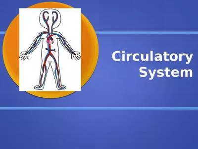
/
Circulatory System I s composed of two separate components: Circulatory System I s composed of two separate components:

Circulatory System I s composed of two separate components: - PowerPoint Presentation

SimplySweet
SimplySweet . @SimplySweet
Follow
342 views
Uploaded On 2022-08-02

Circulatory System I s composed of two separate components: - PPT Presentation

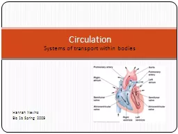
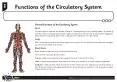
Cardio vascular system Lymphatic vascular system Cardio vascular system is concerned with the transport of blood and lymph through the body The main function of the blood are to transport 02 nutrients and hormones to the tissues and to collect the ID: 932344

tunica elastic blood arteries elastic tunica arteries blood lamina vessels system cells media internal large muscle fibers endothelial layer

Share:

Link:

Embed:

Download Presentation from below link

Download Presentation The PPT/PDF document "Circulatory System I s composed of two s..." is the property of its rightful owner. Permission is granted to download and print the materials on this web site for personal, non-commercial use only, and to display it on your personal computer provided you do not modify the materials and that you retain all copyright notices contained in the materials. By downloading content from our website, you accept the terms of this agreement.


Presentation Transcript

Slide1

Circulatory System

Slide2

I

s composed of two separate components:

_Cardio vascular system

_Lymphatic vascular system

Cardio vascular system is concerned with the transport of blood and lymph through the body.

The main function of the blood are to transport 02, nutrients and hormones to the tissues and to collect the waste products( carbon dioxide and waste metabolites) for removal from the body via excretory system.

Slide3

The cardio vascular system consist of the:

_Heart (muscular pump).

_Pulmonary circulation (system of blood vessels to and from the lungs).

_Systemic circulation (system of blood vessels bringing blood to and from all the other organs of the body).

Blood Vessels were classified in to:1-Arteries2-Capillaries3-Veins

Slide4

.

Are efferent vessels that transport blood away from the heart to the capillary beds.

There are two major arteries pulmonary and aorta.

_The arterial wall is composed of three main layers or tunics:

1-Tunica Intima(internal tunic) consisting of: _Endothelium-single lining layer of endothelial cell (simple squamous epithelium) lining the lumen of blood vessels rest on a basal lamina. _Sub endothelial layer lies immediately beneath the endothelial cells, it is composed of loose connective tissue and a few scattered smooth muscle cells.

_Arteries

Slide5

_Internal elastic lamina beneath the sub endothelial layer ,is composed of elastin which is fenestrated sheet that permits the diffusion of substances in to the deeper region of the arterial wall to nourish the cells there.

2-Tunica media(middle tunic):

Is the thickest layer of the vessels wall, consisting of circular smooth muscle or(spiral), concentric elastic lamina (have external elastic lamina) formed by the smooth muscle cells circularly arranged, which more delicate than the internal elastic lamina.

3-Tunica adventitia (external tunic): Composed of connective tissue (fibroblast, collagen fibers and elastic fibers) surrounding the vessel.

Slide6

Vein Artery

Slide7

Small muscular artery

Slide8

The arterial blood vessels are classified according to their diameter in to :

1-Large (elastic) arteries

2-Medium(muscular) arteries

3-Arteriols Classification of Arteries

Slide9

-Are arterial vessels with a diameter below 0.1-0.5mm .

-have relatively narrow lumen.

-endothelial cells are smaller than in larger arteries, the endothelium still rests on a internal elastic lamina.

-the internal elastic lamina is absence of small arterioles . -the tunica media consist of 1-3 concentric layers of sooth muscle cells. -it is difficult to identify an external elastic lamina. -the tunica adventitia, which consist mainly of c.t fibers, this layer blends with the connective tissue surrounding the vessel.

1-Arteriol

Slide10

Arteriol

Slide11

2-Medium(Muscular) arteries or( distributing):

_The tunica intima have sub endothelial layer that is thicker than that of arterioles, alayer of loose c.t beneath the endothelium allows the tunica intima to move independently from other layers as the elastic arteries distend with the increase in systolic blood pressure.

_Internal elastic lamina is prominent.

_T media contain layers of smooth muscle cell, these cell are intermix with various number of elastic lamellae, external elastic lamina is present only in large muscular arteries

.

Slide12

3-Large elastic arteries (Conducting):

_There are large arteries closest to the heart (aorta, renal artery).

_Tunica intima is thicker than in other arteries.

_An internal elastic laminae.,although present. Tunica media consist of elastic fibers and elastic laminae ,between them present smooth muscle cells and reticular fibers.

_Tunica adventitia appears thinner than the tunica media and contains collagen fibers.

Slide13

The walls of these large arteries are so thick that their peripheral parts can not derive enough 02 and nutrient from the blood of the vessel that they from large vessels are therefore accompanied by smaller blood vessel which supply the tunica adventitia and outer part of tunica media of the vessel wall ,these vessels are called vasa vasorum ,that nourished the deeper cells of the tunica media and adventitia.