/
Isolation and identification of Staphylococci Isolation and identification of Staphylococci

Isolation and identification of Staphylococci - PowerPoint Presentation

SultrySiren
SultrySiren . @SultrySiren
Follow
342 views
Uploaded On 2022-08-03

Isolation and identification of Staphylococci - PPT Presentation

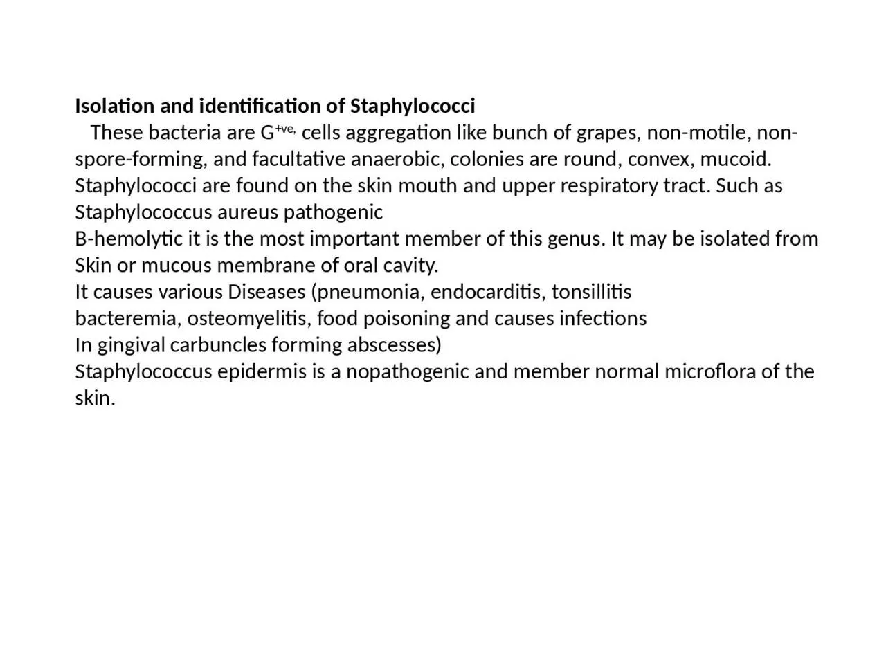
These bacteria are G ve cells aggregation like bunch of grapes nonmotile nonsporeforming and facultative anaerobic colonies are round convex mucoid Staphylococci are found on the skin mouth and upper respiratory tract Such as Staphylococcus ID: 933164

catalase test staphylococci agar test catalase agar staphylococci dnase coagulase colonies bacteria aureus pathogenic figure plate mannitol staph broth

Share:

Link:

Embed:

Download Presentation from below link

Download Presentation The PPT/PDF document "Isolation and identification of Staphylo..." is the property of its rightful owner. Permission is granted to download and print the materials on this web site for personal, non-commercial use only, and to display it on your personal computer provided you do not modify the materials and that you retain all copyright notices contained in the materials. By downloading content from our website, you accept the terms of this agreement.


Presentation Transcript

Slide1

Isolation and identification of Staphylococci

These bacteria are

G

+ve

,

cells aggregation like bunch of grapes, non-motile, non-spore-forming, and facultative anaerobic, colonies are round, convex,

mucoid

. Staphylococci are found on the skin mouth and upper respiratory tract. Such as Staphylococcus

aureus

pathogenic

Β-hemolytic it is the most important member of this genus. It may be isolated from Skin or mucous membrane of oral cavity.

It causes various Diseases (pneumonia, endocarditis, tonsillitis

bacteremia, osteomyelitis, food poisoning and causes infections

In gingival carbuncles forming abscesses)

Staphylococcus epidermis is a

nopathogenic

and member normal

microflora

of the skin.

Slide2

Procedures:

Swab from the throat around the

tonsillar

area see (fig. 32) or rotate

amoisted

with (saline) swab around entire perimeter of both nares.

Roll the swab near the edge of a blood agar plate, and

mannitol

salt agar.

Streak with sterile loop, label the plates.

Repeat the same steps for Gingivae and

tonque

label the plate.

Incubate the plates at 35°c for 24-48 hr.

Observe the growth

Slide3

blood agar

Staph.

aureus

colonies are large , smooth β-hemolytic (clear zone around the colonies).Colonies are small, white, opaque, and non-hemolytic. Staph. epidermis colonies are smooth white , opaque , non-hemolytic Staph. saprophyticus  

Slide4

On

mannitol salt agar

Figure -

(23)

Slide5

Staph.

aureus colonies and culture medium turn to yellow due to fermentation of

mannitol

Nonpathogenic staphylococci do not use mannitol and produce no color change. (Fig. 31 b),Prepare slides from different colonies for gram stain, stain and examine under microscope, Record the observations and results in a report.

8-for identification the following biochemical tests must be done; Coagulase test, D-nase test and catalase test.

Catalase

: +

Coagulase:

S.aureus

+

S .

epidermidis

_

DNase

+

_

Slide6

Slide7

Catalase Test

: Some bacteria contain

flavoproteins

that reduce O2 resulting in the production of hydrogen peroxide (H2O2) or superoxide (O2). These are toxic for obligate aerobes and facultative anaerobes .Many bacteria produce enzymes to protect themself against superoxide (O2), these enzymes are catalase, peroxidase or superoxide dismutase, which catalyze the destruction of H

2O2 or O2 as follows

:

Catalase production can be noted by mixing H

2

O

2

with the tested bacteria. Bubbles of O

2

represent a positive catalase test, and the absence of bubbles represents a negative catalase test. Most strict anaerobes lack both enzymes and cannot tolerate O

2

2O

2

+ 2H

+

O

2

+ H

2

O

2

dismutase

catalase or

2H

2

O

2

2 H

2

O + O

2

peroxidase

Slide8

Coagulase Activity Test

: This test is used to distinguish between pathogenic and non-pathogenic staphylococci . Coagulase is enzyme produced by pathogenic Staphylococci , that clot blood plasma . Citrate is usually added to act as anticoagulant and prevent false positive results.

Procedure

Add 0.5 ml of citrated rabbit plasma to two small test tubes, label the tubes with name of bacteria .Add 0.5 ml broth culture of S. aureus to one tube and 0.5 ml broth culture of S. epidermis to the other tube.

Incubate the broth cultures at 37ºc for 1 to 4 hours in water bath.Examine the broth cultures for the presence (coagulase positive) or absence (coagulase negative) of clouding and clots.

Record the results. (fig.50).

Slide9

Figure

(24

) a-Catalase test on slant ,b- Catalase test on slide.


Figure

(25)-

Coagulase test

Slide10

DNAse

Activity Test

: Most pathogenic strains of staphylococci produce a nuclease enzyme called

DNase degrades host DNA and increases the pathogenicity of staphylococci.ProcedureDivide a DNase agar plate in half with a wax pencil and label each part with the name of bacteria .On one part of the agar plate , put heavily spot-inoculate of

S.aureus and the other part inoculated by the same way with S

.

epidermidis

.

Incubate at 37ºc for 18 to 24 hours.

Flood the

DNase test agar with 1 N

HCl

. A zone of clearing around the colony indicates a positive

DNase

test.

Record the result.(fig. 26).

.


Slide11

Figure

(26)

DNase

Test (a and b)