/
Basic Embryology for pharmacist Basic Embryology for pharmacist

Basic Embryology for pharmacist - PowerPoint Presentation

WiseWolf
WiseWolf . @WiseWolf
Follow
342 views
Uploaded On 2022-08-02

Basic Embryology for pharmacist - PPT Presentation

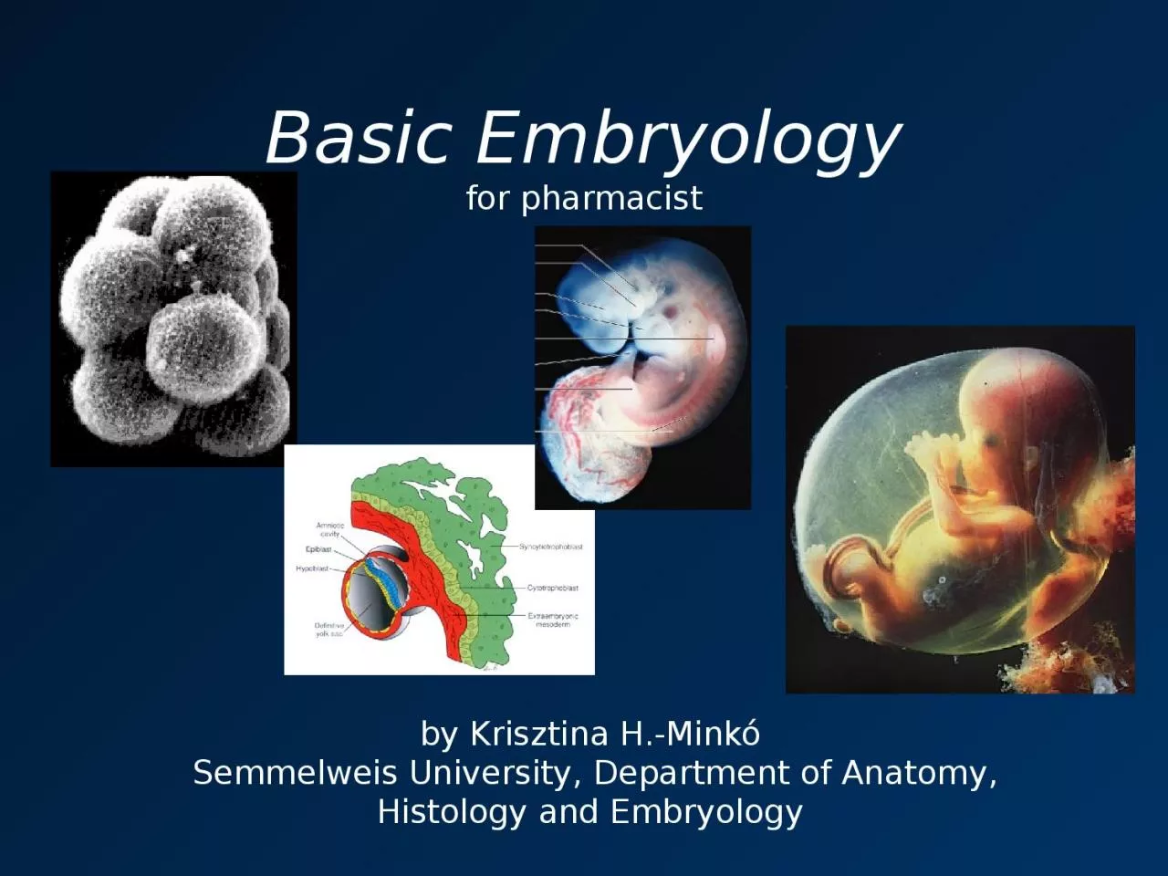
by Krisztina HMinkó Semmelweis University Department of Anatomy Histology and Embryology Question of time anatomy vs gynecology Zero time Fertilization anatomy clinically usually impossible to determine uncertain ID: 932761

fertilization cells weeks days cells fertilization days weeks layer day cell zona cleavage ovulation genetic produced system pellucida number

Share:

Link:

Embed:

Download Presentation from below link

Download Presentation The PPT/PDF document "Basic Embryology for pharmacist" is the property of its rightful owner. Permission is granted to download and print the materials on this web site for personal, non-commercial use only, and to display it on your personal computer provided you do not modify the materials and that you retain all copyright notices contained in the materials. By downloading content from our website, you accept the terms of this agreement.


Presentation Transcript

Slide1

Basic Embryologyfor pharmacist

by

Krisztina

H.-Minkó Semmelweis University, Department of Anatomy, Histology and Embryology

Slide2

Question of time (anatomy vs. gynecology)

Zero time?

Fertilization (anatomy) – clinically usually impossible to determine, uncertain

Last menstruation (gynecology) – certain, but just an estimate, not actual age (ovulation usually 14 days after mestr., fertilization within 2 days)

Length of pregnancy: 280 days (40 weeks) after the last menstruation, 266 days (38 weeks) after conception

Trimesters:

1st: 0-12 weeks

2nd: 12-25 weeks

3rd: 25-40

Slide3

Slide4

Developmental events in the first 3 weeks after fertilizationFertilization

CleavageGastrulationNeurulationPlacenta formation(Spermio- and oogenesis)

Slide5

Mitosis vs. meiosis

Number of chromosomes in somatic cells: 22 pair XX (females) or 22 p. XY (males).

During mitosis equal, diploid daughter cells are produced.

During meiosis the number of chromosomes is halved (22X or 22Y – haploid cells). This is achieved by DNA duplication followed by two cell divisions.Normal chromosome number is restored at fertilization, where two haploid cells fuse.Increases variability!

Slide6

Crossing over

Identical regions of homologous pairs of chromosomes exchanged. Further increases variability to infinitely high values.

Slide7

Oogenesis

Starts in the embryonic period, arrested

a

t the first prophase till puberty. The first meiotic division completed upon ovulation, the second division at fertilization. In humans, usually only one oocyte is ovulated in each menstruational period, between puberty and menopausa, when further maturation cases.Four cells are produced, but only one is functional, the rest (polar bodies) are defective. The oocyte gives the cell organelles the cytoplasm, mitochondria and yolk to the embryo.

Some oocytes may rest in the vulnerable period of arrested meiosis for 40-50 years, resulting in increased ratio of genetic defects with maternal age.

Slide8

Spermiogenesis

Starts at puberty, continuously active, high number of sperms are produced, ideally no age limit exists.

Four functional, mobile cells are produced.

22X or 22Y: determines the genetic sex of the embryo.

Slide9

Spermiohistogenesis

Production of mobile cells from spermatids.

Chromosome condensation to decrease the amount of transportable materials, tail formation (motor), acrosome formation (digestive enzymes to penetrate zona pellucida), mitochondria (energy).

Only the genetic material is used!Strong selection: defective cells will not usually reach the site of fertilization

Slide10

ovulation

fertilisation

Slide11

1st week

Ovulation, fertilization, cleavage, transport to uterus, implantation

Slide12

Slide13

Fertilization 1

Spermia must penetrate the corona radiata

and the zona pellucida by active movement and by the help of acrosomal enzymes. When the first sperm reaches the cell membrane, they fuse, and the genetic material (male pronucleus) enters the cell. This is the activating signal for the oocyte to finish the second meiotic division.

Slide14

Fertilization 2

Two pronuclei unite

Cleavage starts

Slide15

Slide16

Cleavage

2 cells 4 cells 16 cells: morula cavity: blastula

Inner cell mass = embryoblast

Slide17

Timing of Cleavage

Embryo reaches the lumen of uterus by the

4

th day after fertilization – at a blastula stage. By this time zona pellucida has disintegrated, the trophobalst layer invades the endometrium, implantation begins.

The embryo may implant anywhere once the zona pellucida has disappeared. If there is a problem with transportation, it will implant to the uterine tube or other abdominal organs. This state is referred to as extra uterine (ectopic) pregnancy.

Slide18

Extrauterine (ectopic) pregnancy

Slide19

Slide20

Implantation (d6-d9)

Trophobalst layer invades the endometrium

App. at the 6

th

day after fertilizationUterine mucosa must be in receptive stage

(secretory phase)

Slide21

Slide22

Slide23

Slide24

Slide25

Slide26

Slide27

Slide28

Slide29

2nd week

Bilaminar germ disc

Slide30

Gastrulation 1(7-14 days a.f.)

Implantation proceeds, trophoblast differentiates.

Embryoblast cells differentiate, bilaminar germ disc appears, with epiblast and hypoblast layers

.

Slide31

3rd week

Trilaminar germ discNeurulation begins

Slide32

Gastrulation 2(trilaminar germ disc)

14

th

-16th days after fert.Epiblast cells proliferate, move in a central direction, form an invagination (primitive streak and node). Cells migrate to the hypoblast and to the intermediate layer to form the endoderm and mesoderm respectively.

Animation of gastrulation:

https://www.youtube.com/watch?v=sUuX-4fEF3A

Slide33

Ectoderm

Neural fold

17th day: early differentiation of mesoderm

Notochord will induce the development of the nervous system from ectoderm.

Slide34

Late differentiation of mesoderm, neurulation (day 17-21)

Slide35

D 21

Developing aorta, and heart tube is visible, heart pulsation is detectable in ultrasound imaging.

Slide36

D 21

Dermatome: connective tissue derivates (dermis) of dorsal skin

Intermediate mes

oderm: urinary system (male genital tract)Somatopleura: parietal layer of serous membranes, lateral and ventral body wall (except for epithelia of skin)Splanchnopleura: visceral layer of serous membranes CT and smooth muscles of intestine

Sclerotome: vertebrae

Myotom

e

: axial muscles

Slide37

Neurulation (Day 17-29)(4.w)

Neural tube: central nervous system

Neural crest: peripheral nervous system, etc…

Slide38

Neurulation

Animation of neurulation:

https://www.youtube.com/watch?v=iHmBIJs77ZQ&t=91s

Slide39

Folding

(3.-6.w)

Slide40

Slide41

Thank You for attention!

Slide42

Literature

T.W. Sadler: Langmann’s Medical Embryology, 7th edition, 1995, Baltimore, Maryland, USA - pictures

Papp Z: Szülészet és nőgyógyászat tankönyve, Semmelweis Kiadó, 1999, Budapest

Previous authors of the lecture: Ákos Lukáts, Nándor Nagy