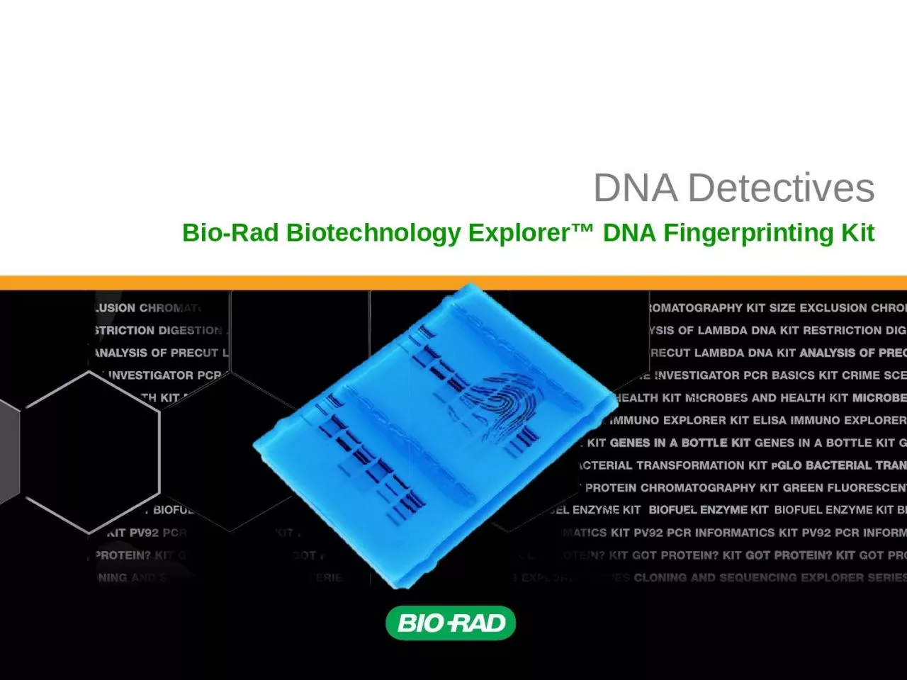
Explorer DNA Fingerprinting Kit Crime Scene Have fun setting up your own crime scene Be as elaborate or as simple as you wish Dye Electrophoresis Could you eliminate any suspects using dye electrophoresis ID: 932504
Download Presentation The PPT/PDF document "DNA Detectives Bio-Rad Biotechnology" is the property of its rightful owner. Permission is granted to download and print the materials on this web site for personal, non-commercial use only, and to display it on your personal computer provided you do not modify the materials and that you retain all copyright notices contained in the materials. By downloading content from our website, you accept the terms of this agreement.
Slide1
DNA Detectives
Bio-Rad Biotechnology
Explorer™ DNA Fingerprinting Kit
Slide2Crime Scene
Have fun setting up your own crime scene. Be as elaborate or as simple as you wish.
Slide3Dye Electrophoresis
Could you eliminate any suspects using dye electrophoresis?
What other methods might be more conclusive?
Slide4Innocence Project
302 DNA exonerations in the U.S. since 1989 (48 in TX)
Exonerees served an average of 13.6 years in prison
Flawed eyewitness testimony to blame for many cases
Slide5Innocence Project - Resources
Innocence Project:
www.innocenceproject.org
Innocence Project of Texas: www.ipoftexas.org
Houston Chronicle profiles:
www.chron.com/exonerees
Slide6Crime scene
Human relatedness
Paternity
Animal relatedness
Anthropology studies
Disease-causing organisms
Food identification
Human remains
Monitoring transplants
DNA Fingerprinting – Real World Applications
Slide7DNA Fingerprinting Lab – Day 1
Slide8DNA Fingerprinting Lab – Day 2
Slide9DNA Fingerprinting Lab – Day 3
Slide10How to use a micropipet
Play video demonstration or demonstrate live
http://www.bio-rad.com/webroot/web/html/lse/support/tutorial_micropipet_wndw.html
Slide11Let’s Get Started!
Place your crime scene (CS) and suspect DNA (S1-5) in your foam rack. Write your initials on your tubes.
Pipet
10
ul
of enzyme (ENZ) into each of your tubes.
Use a separate tip for each sample!
ENZ
10 ul
Slide12Let’s Get Started!
Cap the tubes, flick the bottom of each one to mix, and then bring contents to bottom by tapping on the table.
Place your tubes (in the foam rack) in a 37 degree water bath.
Slide13DNA Structure
Slide14DNA Schematic
Slide15Student DNA Model
Slide16Evolved
by bacteria to protect against viral DNA infection
Endonucleases
= cleave within DNA strands
Over
3,000 known enzymes
Restriction Enzymes
Slide17Restriction Buffer provides optimal conditions
NaCI
provides the correct ionic strength
Tris
-HCI provides the proper pH
Mg
2+
is an enzyme co-factor
DNA Digestion Reaction
Slide18Each
enzyme digests (cuts) DNA at a specific sequence =
restriction site
Enzymes
recognize 4- or 6- base pair, palindromic sequences
(
eg
GAATTC)
Palindrome
Restriction site
Fragment 1
Fragment 2
Enzyme Site Recognition
Slide19Enzyme cuts
5 Prime Overhang
Slide20E
co
RI
–
Eschericha coli
– 5 prime overhang
P
st
l
–
Providencia stuartii
– 3 prime overhang
Common Restriction Enzymes
Slide21Classroom Obstacle Course
Slide22Restriction Fragment Length PolymorphismRFLP
Allele 1
Allele 2
GAATTC
GTTAAC
GAATTC
GTTAAC
CTGCAG
GAGCTC
CGGCAG
GCGCTC
PstI
EcoRI
1
2
3
3
Fragment 1+2
Different
Base Pairs
No restriction site
+
M
A-1
A-2
Electrophoresis of restriction fragments
M
: Marker
A-1
: Allele 1 Fragments
A-2
: Allele 2 Fragments
Slide23How to load an agarose gel
Play video demonstration or demonstrate live
http://www.bio-rad.com/webroot/web/html/lse/support/tutorial-agarose-gel-electrophoresis-wndw.html
Slide24Gel Electrophoresis
Collect your DNA samples from the water bath.
Add
4
ul
of
Uview
loading
dye (LD) into each of your tubes.
Use a separate tip for each sample!
Cap the tubes and mix by flicking with your finger.
Slide25Gel Electrophoresis
Place an
agarose
gel in the gel box. Make sure the wells are near the black (-) electrode.
Using a separate tip for each sample, load your gel:
Lane 1: M, DNA size marker, 10
μ
l
Lane 2: CS, green, 20
μl
Lane 3: S1, blue, 20
μl
Lane 4: S2, orange, 20
μ
l
Lane 5: S3, violet, 20 μl
Lane
6: S4, red, 20
μl
Lane 7: S5, yellow, 20
μl
Slide26Gel Electrophoresis
Place the lid on the gel box, and plug the electrodes into the power supply. Electrophoresis at 200V for 20 minutes.
Slide27How important is each step in the lab protocol?
What part of the protocol can I manipulate to see a change in the results?
Possible variables:
enzyme concentration
substrate concentration
incubation temp or time
enzyme or DNA UV exposure
methylated plasmid
agarose
concentration
buffer concentration
running time.
How do I insure the changes I make is what actually affects the outcome (importance of controls).
Write the protocol. After approval – do it!
Student Inquiry – Question to Consider
Slide28What can I learn about these plasmids?Can I use these plasmids to successfully transform bacteria?
Can I ligate these plasmids together and successfully transform bacteria?Can I do a restriction digest on pGLO plasmid?
Can I determine the plasmid map using different enzymes?
Student Inquiry – Advanced Question
Slide29What materials and equipment do I have on hand, and what will I need to order?Extra agarose, DNA, different / more restriction enzymes? Water bath (different temps)
Other supplies depending on student questions (mini prep, thermal cyclers, etc)Consider buying extras in bulk or as refills – many have 1 year + shelf life.
What additional prep work will I need? Order suppliesPour gels
How much time do I want to allow?
Limited time? Have students read lab and come up with inquiry questions and protocol before they start. Collaborative approach.
Will you need multiple lab periods?
Will everyone need the same amount of time?
Student Inquiry – Teacher Considerations
Slide30BamHI:
EcoRI:
HindIII:
EcoRI+Hind III:
1 linear fragment; 7367bp
2 fragments; 863bp / 6504bp
3 fragments; 721bp/2027bp/3469bp
5 fragments; 721bp/863bp/947bp/1659bp/2027bp
BamHI
7367bp
EcoRI
863bp
6504bp
Hind III
721bp
2027bp
3469bp
EcoRI+ HindIII
2027bp
1659bp
947bp
863bp
721bp
Plasmid Map and Restriction Sites
Slide31Electrical current carries negatively-charged DNA through gel towards positive (red) electrode
Power Supply
Buffer
Dyes
Agarose gel
Slide32Agarose gel separates DNA fragments according to size
Electrical current carries (-) charged DNA through gel to (+) electrode.
Small fragments move faster than large fragments
Power Supply
Agarose
Electrophoresis
Buffer
DNA &
Loading Dye
Agarose
gel
Slide33Analysis of Stained Gel
Determine restriction fragment sizes
Create standard curve using DNA markerMeasure distance traveled by restriction fragments
Determine size of DNA fragments
Identify the related samples
Slide34Size (bp) Distance (mm)
23,000 11.0
9,400 13.0
6,500 15.0
4,400 18.0
2,300 23.0
2,000 24.0
Fingerprinting Standard Curve: Semi-log
Molecular Weight Determination
Slide35Why incubate at 37°C?
Body temperature is optimal for these and most other enzymes
What happens if the temperature is too hot or cool?
Too hot
= enzyme may be denatured (killed)
Too cool
= enzyme activity lowered, requiring longer digestion time
DNA Digestion Temperature