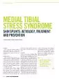
/
150 Written by Maarten Moen The NetherlandsABSTRACTOne of the most 150 Written by Maarten Moen The NetherlandsABSTRACTOne of the most

150 Written by Maarten Moen The NetherlandsABSTRACTOne of the most - PDF document

abigail
abigail . @abigail
Follow
342 views
Uploaded On 2022-08-19

150 Written by Maarten Moen The NetherlandsABSTRACTOne of the most - PPT Presentation

L pain may even be provoked by activities of daily living S TMENT A 33 Hypothesis I bone overloadWith the bone overload hypothesis it is assumed that MTSS is caused by overload of the tibial cortex ID: 938022

threshold bone oedema pain bone threshold pain oedema marrow studies athletes damage showed periosteal mtss loading overload mri erent

Share:

Link:

Embed:

Download Presentation from below link

Download Pdf The PPT/PDF document "150 Written by Maarten Moen The Netherla..." is the property of its rightful owner. Permission is granted to download and print the materials on this web site for personal, non-commercial use only, and to display it on your personal computer provided you do not modify the materials and that you retain all copyright notices contained in the materials. By downloading content from our website, you accept the terms of this agreement.


Presentation Transcript

L – Written by Maarten Moen, The NetherlandsABSTRACTOne of the most common causes of exercise-induced leg pain is medial tibial stress syndrome. This review focuses on its aetiology, patient evaluation, treatment and prevention. INTRODUCTIONExercise-induced lower leg pain is a frequent complaint in athletes and medial tibial stress syndrome (MTSS) or shin splints is one of the most common of its causes. Incidences vary from % in dierent sports. This condition is most frequently seen in runners and athletes involved in jumping, for example basketball players and rhythmic gymnasts.In the past, many dierent authors used dierent names to describe MTSS: shin soreness, tibial stress syndrome, pain may even be provoked by activities of daily living S, TMENT A 33 Hypothesis I: bone overloadWith the bone overload hypothesis it is assumed that MTSS is caused by overload of the tibial cortex. Several studies have shown that MRI can depict bone marrow and periosteal oe

dema as a sign of bone remodelling. CT scan studies showed osteopenia on the posteromedial side of the tibia in symptomatic legs. Magnusson et al performed dual X-ray absorptiomerty (DEXA) scans in athletes, which showed decreased bone density in symptomatic legs compared to their controlsIn addition to the observations described above, it has been shown that even in asymptomatic legs of athletes, bone marrow oedema and periosteal oedema can be present. Bergman et al performed a study in which an MRI scan was performed asymptomatic runners who ran to miles each week. The MRI scans showed % (ve unilateral ndings and four bilateral ndings). The ndings varied from mild to moderate periosteal oedema to periosteal oedema and bone marrow oedema on T images. None of the runners developed complaints -month follow-up period. Moen et al and the study by Batt et al also showed bone marrow and periosteal oedema in asymptomatic legs of athletes with MTSSThese MRI 

ndings raise the question of whether bone marrow oedema represents normal remodelling of the bone after loading. It is possible that bone marrow oedema in athletes represents a normal sign of bone remodelling, which may become symptomatic once the loading exceeds a certain threshold.Loading can aect remodelling of the bone in a variety of ways. Loading applied by muscle forces and axial loading transferred through the joint can deform the bone tissue and create strains which inuence cellular processes in the cortex. This concept was placed in a theoretical framework by Frost who called it the ‘Mechanostat Theory’ (Figure . When the load is minimal and below a genetically derived threshold, the bone becomes weaker (disuse threshold: MESy) by resorbing trabecular or endocortical bone. When the load is higher than the disuse threshold and below a second threshold (maintenance threshold: MESm) the bone maintains its strength. When the load is between the maintena

nce threshold and a threshold above which cortical micro-damage occurs (damage threshold: MESp), the bone strengthens. Loads above this last threshold lead to microscopic fatigue damage (micro-damage), which accumulates when the loads and subsequent micro-damage are too great to recover from. Bone pain mechanismWhen the bone is overloaded and micro-damage accumulates, pain may occur. The pain mechanism of overloaded bone has only recently been established. Previously, the prevailing opinion was that bone pain was derived from the densely innervated periosteum. However, more recent studies showed that sensory nerve bres also innervate the mineralised bone and bone marrow. Several studies suggested that these nerve bres are stimulated by mechanical distortion. Other studies suggested that sensory nerve bres could be activated by protons, which are released by osteoclasts when resorbing bone. That is because osteoclasts form a highly acidic compartment between themse

lves and mineralised bone. The acidic environment is sensed by aerent nerves that innervate the mineralised bone and induces a pain signal. Furthermore, microscopic cracks could also provide direct sensory input to intra-cortical nerves. When the crack disrupts the nerve, signalling is interrupted which can potentially lead to (referred) pain (personal communication:Weinans Hypothesis II: tractionThe alternative theory on the aetiology of MTSS is that the syndrome is caused by traction. In the past, it was generally accepted that the posteriortibial muscles caused traction on the periosteum, leading to periostitis. However, signicant evidence for periostitis was lacking, as shown in studies which performed histological . Furthermore, the location of the posteriortibial muscles (proximal and lateral) does not resemble the location of complaints in patients with MTSS MESyDWMOWPOWAWMESmMESpFx W=disuse window, W=mild overload window, W=pathologic overload window. MESy=disu