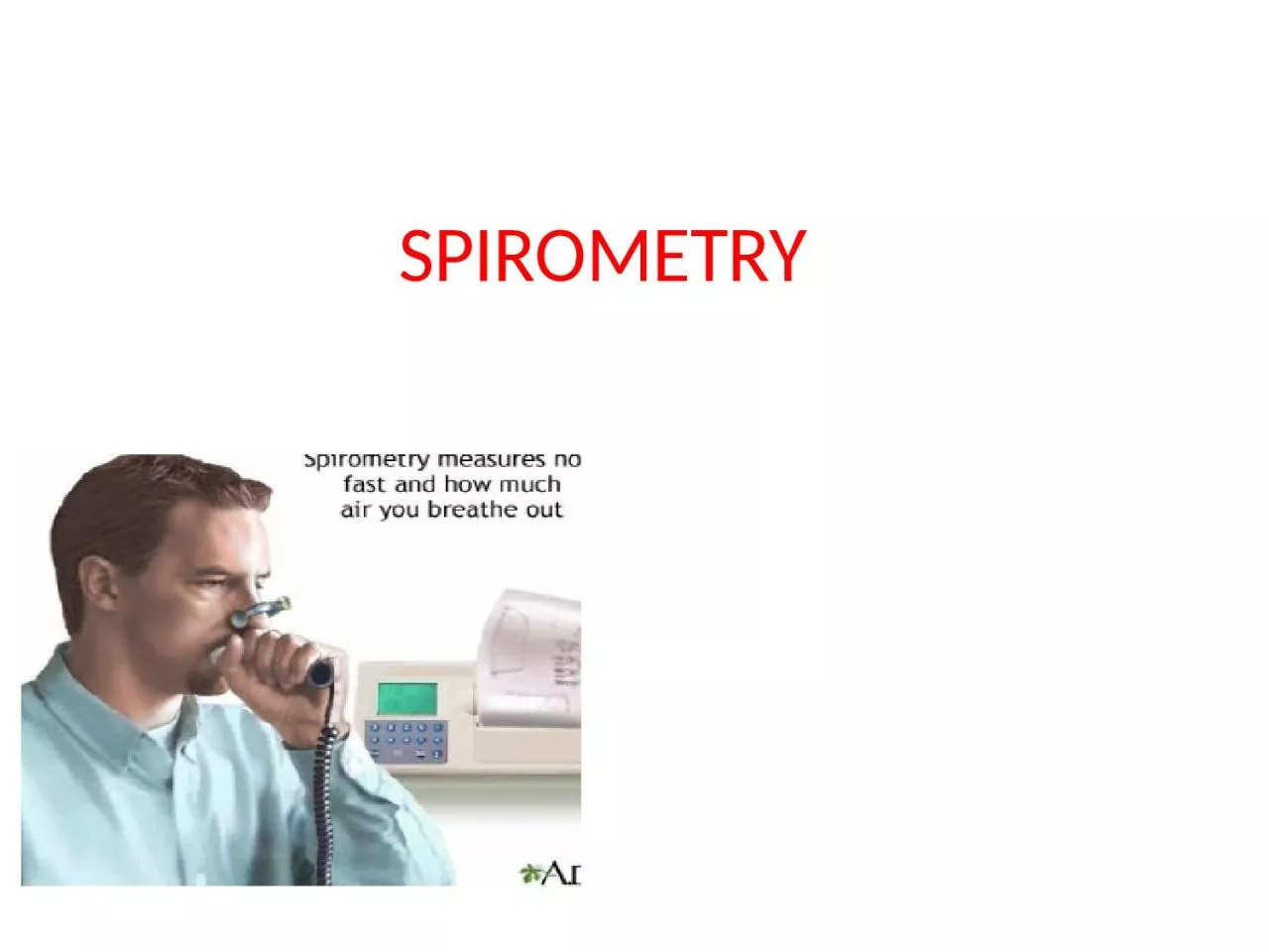
/
SPIROMETRY Spirometry  is the measurement of the flow and volume of air entering and leaving SPIROMETRY Spirometry  is the measurement of the flow and volume of air entering and leaving

SPIROMETRY Spirometry is the measurement of the flow and volume of air entering and leaving - PowerPoint Presentation

adah
adah . @adah
Follow
0 views
Uploaded On 2024-03-15

SPIROMETRY Spirometry is the measurement of the flow and volume of air entering and leaving - PPT Presentation

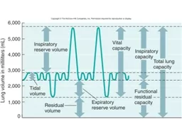
Static lung volumes and capacities Tidal Volume TV The volume of air inhaled amp exhaled at each breath during normal quiet breathing Inspiratory Reserve Volume IRV The volume of air that can be forcefully inspired following a normal quiet inspiration ID: 1048392

air volume lungs lung volume air lung lungs fev1 fvc amount normal disease pulmonary capacity amp expiration predicted forced

Share:

Link:

Embed:

Download Presentation from below link

Download Presentation The PPT/PDF document "SPIROMETRY Spirometry is the measuremen..." is the property of its rightful owner. Permission is granted to download and print the materials on this web site for personal, non-commercial use only, and to display it on your personal computer provided you do not modify the materials and that you retain all copyright notices contained in the materials. By downloading content from our website, you accept the terms of this agreement.


Presentation Transcript

1. SPIROMETRY

2. Spirometry is the measurement of the flow and volume of air entering and leaving the lungs

3. Static lung volumes and capacitiesTidal Volume TV The volume of air inhaled & exhaled at each breath during normal quiet breathingInspiratory Reserve Volume IRV The volume of air that can be forcefully inspired following a normal quiet inspirationExpiratory Reserve Volume ERVThe volume of air that can be forcefully expired after a normal or resting expiration

4. Vital Capacity VC The maximum amount of air that can be exhaled after the fullest inspiration possible (TV + ERV + IRV)Inspiratory Capacity ICThe maximum amount of air that can be inhaled after a normal exhalation (TV + IRV)

5. Residual Volume RV: The volume of air remaining in the lungs after a forceful expiration.Total Lung Capacity TLC :The total volume of the lungs (VC + RV)Functional Residual capacity FRC:The amount of air remaining in the lungs after a normalquiet expiration (ERV + RV)

6.

7.

8. Lung volumes & capacities depend on:• Age• Body size (height & weight)• Gender• Pulmonary health• Altitude• Irritants

9. Dynamic lung volumes:• Valuable in following the progress of a patient withchronic lung disease.• Assessing the results of a treatment• Epidemiological surveys to assess industrialhazards or to document the incidence of disease inthe community• NOT a key factor in making a definitive diagnosis

10. Forced Vital Capacity FVC:The total volume expired by a forced maximal expiraion from a position of maximal inhalation Forced Expiratory Volume in 1 sec FEV1.0:The volume of air expired in the first second ofmaximal forced expiration from a position of fullInspiration

11. Calculating FVC

12. Calculating FEV1.0

13. Maximal Ventilatory Volume (MVV): maximal amount of air that a person can breathe in or out in a short period of time, typically 10, 12, or 15 secs.Other volumes & capacities

14.

15. Spirometry• the measurement of the flow and volume of air entering and leaving the lungs• test of pulmonary function (PFT)• Indicator of health status or disease• Exercise fitnes

16. Spirometer• Apparatus used to measure static & dynamic lungvolumes/capacities using a closed system.• Registers the amount and rate of air moved intoor out of the lungs

17. 2 main types:1. Volume: records the amount of air exhaled orinhaled within a certain time*2. Flow: measures how fast the air flows in or out asthe volume of air inhaled or exhaled increases

18. Respiratory Diseases• Bronchitis• Asthma• COPD• Emphysema• Influenza

19. Obstructive Airways DiseaseFor obstruction, review the FEV1/FVC ratio first. If this is less than Lower Limit of predicted, think obstruction,and then observe the FEV1 to determine degree ofobstruction.When FEV1/FVC is below the Lower Limit of Normal (LLN)…….Mild : FEV1 above LLN (>70% predicted)Moderate : FEV1 40-60% predictedSevere : FEV1 < 40% predictedPlease check current guidelines for asthma and COPD as thesemay change, depending on source.

20. Obstructive (Bronchial Asthma)• Abnormally large TLC, but expiration endsprematurely.• Early airway closure brought about by theincreased smooth muscle tone of the bronchii(asthma) or loss of radial traction from surrounding parenchyma (emphysema).• Other causes include edema of bronchial walls orsecretions in the airway.

21.

22.

23. Restrictive Lung Disease: For restriction, review the FEV1/FVC ratio first. If this is normal, think restriction, and then observeFVC to determine degree of restriction. When FEV1/FVC is normal Mild : FVC below LLN (>60% predicted) Moderate : FVC 50-60% predicted Severe : FVC < 50% predicted

24. Restrictive Lung Disease Affects the lung tissue The inability of the lungs to expand. The lungs are small because of Fibrosis or scarring Full inflation is not possible (disease of lung lining, chest wall or abdomen.Inspiratory respiratory muscles are weak

25.

26.

27. Compliance• the ease with which the chest wall and lungs expand, degree of stiffness.• Direct effect on the amount of pressure required to increase of decrease lung volume• Low compliance = ‘stiffer’ lungs ∴more effort required to inflate alveoli• Pulmonary fibrosis → restrictive lung disease

28. Resistance• Degree of ease in which air can pass through the Airways• Determined by the number, length and diameter of the airways.• High resistance → individual may not be able to exhale fully ∴ some air trapped in lungs

29. Restrictive (Pulmonary Fibrosis):• Loss of pulmonary and chest compliance, orweakness of the respiratory muscles

30.