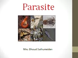
Mrs Ohoud Salhumaidan Introduction Paraitology Parasitism chapter 21 Parasite is an organism baring food and shelter temporarily or permanent and living in or on another organism ID: 611371
Download Presentation The PPT/PDF document "Parasite" is the property of its rightful owner. Permission is granted to download and print the materials on this web site for personal, non-commercial use only, and to display it on your personal computer provided you do not modify the materials and that you retain all copyright notices contained in the materials. By downloading content from our website, you accept the terms of this agreement.
Slide1
Parasite
Mrs. Ohoud S.alhumaidanSlide2
Introduction
Paraitology : Parasitism : (chapter 21)
Parasite :
is an organism baring food and shelter temporarily or permanent and living in or on another organism
.
Kind of parasite ( according to habitat )
Ectoparasites
:
(chapter
21
)
Endoparasites
:
(chapter
21)Slide3
Endoparasite
EctoparasiteSlide4
Parasites can
be:
1-Facultative
parasite
:
parasites able to live both free living and parasite living e.g.
Strongyloides
species.2-Obligate parasite: parasite living permanently in a host and cannot live without a host e.g. Trichomonos species.3-Coprozoic (spurious) parasites:foreign organisms which have been swallowed merely pass along alimentary canal of man (without establishment) to be recovered in faeces. (without affect)Slide5
HOST
Host :organism harboring the parasite species may be affected or not
.
Classification of
Hosts
1-
Definitive
host or final host
: ( sexual development) Eg: man. (see page 358 chapter 21) 2-Intermediate host: ( asexual development)
Eg
:
Taenia>>>>>>> adult------ man
Larva
–---
cattle
(see page 358 chapter 21
)
3
-Reservoir host (carrier):
the carrier host is well adapted to the parasite and tolerates the
infection but serve as source of the infection to other
organisms
4-Vector
:
an
arthropod which carries the parasite from one host to another.Slide6
Classification of parasites
General classification: animal parasites are classified according to international code taxonomy – Each parasite belong to a:
Kingdom
Phylum
Class
Order
Family
Genus Species Some have further divisions to: Sub – order, super family, sub – speciesin classification, scientific parasitic name is of 2 parts: Genus name and species name. Ex: Plasmodium Falciperum Genetic name (one word): plasmodium Species name (two words):
plasmodium
falciperum
.
Genus:
means group of close related species.
Species:
means population with the same genetic characters.Slide7
Mode of parasitic infections
Congenital from mother to fetus.Sexually transmission
Ingestion
of contaminated food and
water
or undercooked meat in which
the infective stage has developed.Penetration of the skin due to contact with infected soil or water stream.Inhalation of dust carrying the infective stage of parasite.Vectors: through the bite or faeces of infected vector or by swallowing the vector.Slide8
Classification of parasites
Parasites
Protozoa
Helminthes
Intestinal
Blood and tissue
Urogenital
Cestodes
Trematodes
Nematodes
arthropodsSlide9
ProtozoaSlide10
Protozoa
See page 72 chapter 5
Protozoa life cycle consist of two stage :
Trophozoite
Vegetative
stage. Can be motile.
Cyst
Infective stage, dormant, immotile.Slide11
Protozoa are classified
(according to their method of locomotion)
See page 73 , chapter 5 , table 5-3)
Flagellates
(e.g.
Giardia
lamblia
).Amoeboids (e.g. Entamoeba histolytica).Sporozoans (e.g. Plasmodium).Ciliates (e.g. Balantidium coli).Slide12
Protozoa infection
Intestinal
Blood and tissue
Entamoeba
histolytica
Giardia
lamblia
Cryptosporidium
Malaria
Toxoplasma
Trypanosoma
Leishmaina
Urogenital tract
Trichononas
VaginalisSlide13
Entamoeba histolytica
Name of Disease:
Amoebiasis (Amebic dysentery)
Parasite:
It possess both
trophozoite
and cyst forms.
It moves by Pseudopodia (false feet)
Dysentery Stool with blood and mucus. Slide14
Entamoeba histolytica
Geographical distribution: Word wide, but more common in tropical and subtropical countries and in countries with poor sanitation
Habitat
:
T
he lumen of the large intestine
.
Reservoir
: Mainly: humans Rarely: dogs, pigs, monkeysSlide15
Amoebiasis
Mode of transmission :See page 362 , chapter 21
, table
21-
3)
Slide16
Life cycle:
Cyst:
infective stage
Inters mouth through contaminated food, drink, fly, or through using human stool as fertilizer
To L.I. lumen and change into
trophozoite
(pathogenic stage)
Produce lytic enzymes (capable of doing
lysis
and produce ulcer)
Flask shape ulcer
Can do erosion through B.V. to liver and other organsSlide17
Cause ulceration of colon,
Can further spread to other organ and cause abscesses (mainly liver)
Trophozoites
form cysts that passes in feces.
Cyst Ingested
Cyst passes to small bowel
Cyst wall disintegrates and release
trophozoites
.
Trophozoites
colonize colon. Slide18
Morphology: Entamoeba
histolyticaCyst:
Infective
stage
Trophozoite
Pathogenic stag
NucleiSlide19
Clinical picture:
Dysentery:
blood+mucous
diarrhea (as a result of
flask shape
ulcer
wall invasion)
Sever abdominal painTenesmus: sense of incomplete evacuation (the patient at this point should be seeking medical advice)Complication:intestinal: peritonitis, appendicitis, HemorrhageExtra intestinal: Most commonly: liver hepatitis (sever right abdominal pain) Fever
amoebic liver abscess (sever pathology in the
liver because the inflammation spots came together)
shoulder pain and Toxemic manifestations
Also in lung, skin, and brainSlide20
Plasmodium sp. (Malaria)
Name of Disease:
Malaria
systemic infection with malaise, fever, chills, sweating, headache, and nausea.
The frequency
wi
th
which the cycle of chills, fever and sweating is repeated is referred to as periodicity and depends on the particular species of plasmodium. Parasite: four species are known to infect humanPlasmodium falciparum (the most deadly and dangerous ,)Plasmodium vivax. (the most common species, ) Plasmodium ovale. Plasmodium malaria.
RBC
Plasmodium sp. Slide21
Plasmodium sp. (Malaria)
Approximately 300 million people worldwide are affected by malaria and between 1 and 1.5 million people die from it every year
Geographical distribution:
Previously
extremely widespread, the malaria is now mainly confined to Africa, Asia and Latin America
The problems of controlling malaria in these countries are aggravated by inadequate health structures and poor socioeconomic conditions. The situation has become even more complex over the last few years with the increase in resistance to the drugs normally used to combat the parasite that causes the disease. Slide22
Plasmodium sp.
Mode of transmission :See page 366 , chapter 21 , table 21
-4)Slide23
Malaria life cycleSlide24
Plasmodium sp. (Malaria)
Pathology and clinical significance:When
merozoits
invade the blood cells, using hemoglobin as a nutrient, eventually, the infected red cells rupture, releasing
merozoits
that can invade other erythrocytes. If a large numbers of red cells rupture at roughly the same time,
a paroxysm
(sudden onset) of fever can result from the massive release of toxic substance.
Plasmodium falciparum is the most dangerous species. P. malriae, P. vivax, and P. ovale cause milder form of the disease, probably because they invade either young or old red cells, but not both. This is in contrast to P. falciparum, which invades cells of all ages.Plasmodium falciparum is characterized by persistent high fever and orthostatic hypertension. Infection can lead to capillary obstruction and death if treatment is not introduced.Slide25
Toxoplama
gondiiName of the disease:
Toxoplasmosis.
Geographical distribution:
World wide. Approximately 50% of human
Population of USA has been infected.
Reproduction
:
Sexually reproduction (Definitive host) In Cats, where Oocysts are released in feces of cat. Asexual reproduction (intermediate host) In worm blooded animals (cats, mice, humans, and birds).Slide26
Mod of Transmission
1.eating row, undercooked meat of sheep and cow containing viable
trophozoits
(
bradyzoits
)
2. swallowing
food and water contaminated with infected cat
feces3.Congenital transmission, through placenta (fatal) especially when infection occurs during pregnancy4. person to person: ex. By blood transfusion or organ transmissionSlide27
Toxoplama gondii
Mod of Transmission :See page 361 , chapter 21 , table 21-2)Slide28
Clinical symptoms
Infection of normal human hosts are common and usually asymptomatic.
Infection in
immunocompromised
individuals is very sever and they my suffer relapse of the infection.
Congenital
infections can also be sever,
it can result in still births, brain lesions, and they are a major cause of blindness in newborns. Slide29
Helminthes ( worms)Slide30
Helminthes ( worms)
Eukaryotic and multicellular parasites
ranging
from barely visible roundworms (0.3 mm) to huge tapeworms 25 meters
long
.
h
elminthes grouped them into three categories: Nematodes (roundworms), Trematodes (flukes)Cestodes (tape- worms)Slide31
Helminthes
live inside their host. They are worm-like organisms that live and feed off living hosts, receiving nourishment and protection while disrupting their hosts' nutrient absorption, causing weakness and disease
Sources for human infection are contaminated food, soil, and water or infected animals,
routes
of infection are by oral intake or penetration of unbroken skin Slide32Slide33
Nematodes (Roundworms)
elongate, cylindrical shape.Nonsegmented
and tapered at both ends .
Sexes
are separate.
the vast majority are free- living soil and freshwater worms, but around 200 are parasitic, including 50 species that affect humans.
Nematodes divided into:
intestinal nematodes (
e,g, Ascaris lumbricoidesTissue nematodesSlide34
Ascaris lumbricoides
(Roundworm) Ascaris
lumbricoides
is the largest nematode (roundworm)
parasitizing
the human intestine
1/3 the world population is infected with this wormGeographical distribution: world wide, common among people with low standard of living and among childrenMorphology: Adult: in small intestine adult egg: infective stageSlide35
Life cycle:
2 phases: lung
and
intestinal
Egg ingested, hatches in
duodenum
; larvae penetrate intestine wall, enter blood vessels and
embolize
through liver to lungs. They then migrate into airspaces, up trachea and are swallowed, taking up permanent adult residence in the small intestine; ~ 2 months from egg to mature adult Each female produce 200,000 eggs per dayAdult worms can live 1 to 2 years. Slide36Slide37
Clinical Symptoms:
Ascaris lumbricoides
Lung phase
It
causes hemorrhage, inflammation, bacterial infection.
Symptoms:
Bloody
sputum, cough , wheezing, and fever.
Intestinal phaseSymptoms: Related to number of worms; Small numbers: asymptomatic. Large numbers: Passing of worm in stool, vomit, nausea, malabsorption and abdominal pain.Complications: If untreated, can cause intestinal obstruction (blockage) and malnutrition.
Deaths form
ascariasis
range from 8000 to 100,000 annually worldwide. Slide38
Cestodes (tapeworms)
Long, ribbon-like, segmented worms.Primarily intestinal parasites.
Can reach 15 m in length.
human
species
are :
Taenia
saginata ( the beef tapeworm )T. solium, ( the swine tape- worm )Diphyllobothrium latum ( the fish tapeworm )Name of the disease: Teniasis. Slide39
Taenia
saginata or Beef Tape-Worms
Habitat:
is the small
intestine (the ileum )
Transmission:
acquired in humans through the ingestion of raw or poorly cooked meat of infected cows. These cows have been infected via the ingestion of human feces containing the eggs of the parasite Slide40
Taenia
saginata Morphology:
Adult
is divided into three parts,
1- a head
: round and small. It has four suction disks
2- neck: A small, slender neck, about an inch long 3- number of segments. Adult tapeworms and can grow up to 25 meters in the lumen of the intestine, but are usually closer to 5 meters in length Egg present in feces Slide41
Taenia
saginata: life cycleSlide42
Taenia saginata
Habitat: - adult in small intestine of man
only
(
devenitive
host )
- Egg in feces
- larva stage in muscle of thigh, shoulder, neck and heart of
cattle only (intermediate host)Clinical symptoms:High infection: diarrhea and consitipationVomitingLoss of appetiteAnemiaSlide43
Trematodes (fluke)
Small (about 1 cm) flat , leaf-like worms.Infest various organs of the human host (e.g. intestinal veins, urinary bladder, liver, or lung)
All parasitic trematodes use freshwater snails as an
intermediate host. Slide44
Schistosoma spp.
Name of the disease:Schistosomiasis (Bilharzia)– it is a disease of the venous system.
Transmission:
By direct skin penetration, when people come in contact with contaminated water.
Schistosoma is
NOT
acquired by ingestion of contaminated food, it directly penetrates the skin of swimmers in contaminated rivers and lakes. Currently more than 200 millionPeople are infected.Slide45
Bilharzia (Schistosomiasis)
Disease of the
venous system
, acquired by people when they come in contact with contaminated water
Adult
Schistosomes
take up residence in various abdominal veins, depending on the species; they are, therefore called
(Blood Flukes)Very common among childrenTransmission: Direct skin penetrationFresh water becomes contaminated by Schistosoma eggs when infected people urinate or defecate in the water. The eggs hatch and the parasites grow and develop inside snails.types of Schistosomiasis: intestinal Schistosomiasis Urinary tract SchistosomiasisSlide46
Schistosoma spp. Life CycleSlide47
Bilharzia (Schistosomiasis
)Clinical symptoms:Most people have no symptoms when they are first infected.
After
few days,
rash
or itchy
skin due to hypersensitivity reaction to the parasite.
within
1-2 months, other symptoms may develop including: fever, couph , urticaria, splenomegaly, diarrhea and abdominal pain. Chronic Schistosomiasis:Without treatment, schistosomiasis can persist for years. Symptoms: - Intestinal: GI bleeding, diarrhea, pain, and enlarged liver. - Urinary Tract: Heamaturia (blood in urine) and dysuria (painful urination).