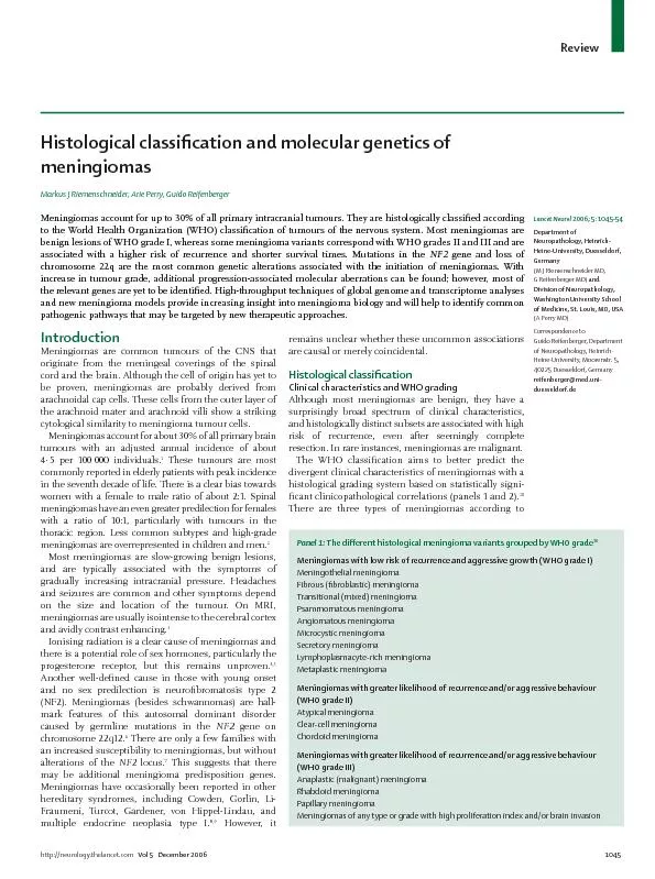
/
http://neurology.thelancet.comVol 5   December 2006 Review http://neurology.thelancet.comVol 5   December 2006 Review

http://neurology.thelancet.comVol 5 December 2006 Review - PDF document

alida-meadow
alida-meadow . @alida-meadow
Follow
458 views
Uploaded On 2016-06-12

http://neurology.thelancet.comVol 5 December 2006 Review - PPT Presentation

cation and molecular genetics of Markus J Riemenschneider Arie Perry Guido Reifenberger Meningiomas account for up to 30 of all primary intracranial tumours They are histologically classi ID: 358880

cation and molecular genetics

Share:

Link:

Embed:

Download Presentation from below link

Download Pdf The PPT/PDF document "http://neurology.thelancet.comVol 5 De..." is the property of its rightful owner. Permission is granted to download and print the materials on this web site for personal, non-commercial use only, and to display it on your personal computer provided you do not modify the materials and that you retain all copyright notices contained in the materials. By downloading content from our website, you accept the terms of this agreement.


Presentation Transcript

http://neurology.thelancet.comVol 5 December 2006 Review cation and molecular genetics of Markus J Riemenschneider, Arie Perry, Guido Reifenberger Meningiomas account for up to 30% of all primary intracranial tumours. They are histologically classi“ ed according to the World Health Organization (WHO) classi“ cation of tumours of the nervous system. Most meningiomas are Meningiomas are common tumours of the CNS that cation Lancet Neurol Panel : erent histological meningioma variants grouped by WHO grade http://neurology.thelancet.comVol 5 December 2006Review malig nancy grades: benign (WHO grade I), atypical (WHO grade II), and anaplastic (malignant; WHO grade III) meningiomas. Benign meningioma: WHO grade IAbout 80% of all meningiomas are slow-growing tumours of WHO grade I. Any histological variant is compatible with WHO grade I, except for the chordoid, clear-cell, papillary, and rhabdoid meningiomas, which are consistently associated with more aggressive clinical features. The histological variants most commonly diagnosed in pathology specimens are meningothelial, brous, and transitional meningioma (“ gure 1, panel 1). Meningothelial meningiomas are histologically composed of characteristic uniform tumour cells that form lobules surrounded by thin collagenous septae. Within the lobules, ned cell borders that resemble a syncytium. Characteristic nuclear changes include clear spaces (that seem empty of karyoplasm) and rounded eosinophilic cytoplasmic protrusions, referred to as pseudoinclusions. Fibrous meningiomas are mainly bro-blasts and form intersecting fascicles embedded in a collagen-rich and reticulin-rich matrix. Transitional (mixed) meningiomas combine features of both subtypes and usually present with extensive whorl formation, wherein tumour cells wrap around each other forming concentric layers. The latter have a tendency to hyalinise and calcify to form the characteristic concentric cations known as psammoma (sand-likeŽ, based on their gritty, gross appearance) bodies. Tumours that are mostly composed of the latter are referred to as psammomatous meningiomas. In addition to these common meningioma subtypes, several other benign gure 1, panel 1). bromatosis type 2 protein is the only rmly implicated in the development of benign meningiomas. However, the gene mutations varies in the di erent broblastic and trans- mutations in up to 80% gure 1).compartments, such as orbit, soft tissue, and skin. cult to resect the tumour, they are not considered as atypical or malignant. By contrast, brain invasion is associated Although more common Atypical meningioma and other WHO grade II variantsAtypical meningiomas constitute 15…20% of meningi-omas. Following gross total resection, benign men- Histology of the di erent benign meningioma variants (WHO grade I) Variants of WHO grade I: meningothelial (A), “ brous (B), transitional (C), psammomatous (D), angiomatous (E), microcystic (F), secretory (G), lymphoplasmacyte-rich (H), and metaplastic (I). (A…D, F, H, I: hematoxylin-eosin; E: immunostaining for endothelial cells with anti-CD34 antibody; G: periodic acid Schi stain). Note syncytial growth of meningothelial cells (A), fascicular growth of “ broblast-like spindle cells (B), formation of multiple meningeal whorls (C), presence of numerous calci“ ed psammoma bodies (D), numerous densely packed blood vessels (E), prominent microcystic degeneration (F), production of periodic acid Schi positive pseudopsammoma bodies (G), extensive ammatory in“ ltrates (H), and xanthomatous changes of tumour cells (I). Panel : WHO criteria for meningioma grading Benign meningioma (WHO grade I) Histological variant other than clear-cell, chordoid, papillary, or rhabdoid Lacks criteria of atypical and anaplastic meningiomaAtypical meningioma (WHO grade II) (any of three criteria) Mitotic index  four mitoses/ten high-power “ elds (HPF) At least three of “ ve parameters: Increased cellularity High nuclear/cytoplasmatic ratio (small cells) Prominent nucleoli Uninterrupted patternless or sheet-like growth Foci of spontaneous necrosis (ie, not induced by embolisation or radiation) Brain invasionAnaplastic (malignant) meningioma (WHO grade III) (either of two criteria) Mitotic index 20 mitoses/10 HPF Anaplasia (sarcoma, carcinoma, or melanoma-like histology) http://neurology.thelancet.comVol 5 December 2006 Review Thus, the diagnosis of atypical elds (“ gure 2). Nevertheless, in the absence of increased mitotic activity, other With the WHO 2000 de“ nitions, presence of three of the ve following criteria may lead to the diagnosis of atypical meningioma: increased cellularity, high nuclear ed in the Clear-cell and chordoid variants of meningioma are Thus, these meningiomas nition (panel 1, gure 2). Clear-cell meningioma is composed of sheets of polygonal cells with clear, glycogen-rich cytoplasm , and dense perivascular gure 2). Chordoid gure 2). Clear-cell meningiomas are tentorial. Although genetic features associated with clear- der(1)t(1;3)(p12…13;q11) has been proposed c cytogenetic marker for the chordoid variant.However, this “ nding is still to be validated and the genes Anaplastic (malignant) meningioma and other WHO Anaplastic meningiomas account for 1…3% of all These tumours have clinical charac-teristics similar to other malignant neoplasms, which can ltrate neighbouring tissues and form metastatic deposits. Anaplastic meningiomas are associated with recurrence rates of up to 50…80% after surgical resection Histologically, anaplastic meningiomas have features elds being diagnostic gure 3). Some anaplastic meningiomas are di cult to necrosis. However, therapeutic embolisation (iatrogenic) Certain meningioma variants are consistently associated with malignant behaviour and therefore invariably gure 3). Papillary meningiomas, which usually occur in children, show invasion of the brain and other local structures in 75% of patients, recurrence in 55%, and metastasis in 20%. ned by a discohesive growth, which results in a perivascular pseudopapillary pattern and even pseudorosette-like gure 3). Another highly aggressive meningioma variant is the rhabdoid meningioma, which contains rhabdoid cells with abundant eosinophilic cytoplasm, eccentrically placed nuclei, and paranuclear inclusions that ultrastructurally laments gure 3). Both papillary and rhabdoid features may be viewed as progression-associated changes, because they rst time at recurrence and increase Immunohistochemistry may be of particular diagnostic value in cases of anaplastic meningioma, although it should be emphasised that even immunohistochemical evidence of meningothelial di erentiation can be lacking in some patients. Moreover, there is also a role for immuno-histochemistry in meningioma by Histology of WHO grade II meningiomas Atypical meningioma with increased mitotic activity (A), clear-cell meningioma with clear, glycogen-rich cytoplasm (B), and chordoid meningioma showing chordoma-like growth of tumour cells in a myxoid matrix (C). All are stained with hematoxylin-eosin.Tumour cells with an eosinophilic, vacuolated cytoplasm embedded in a basophilic, mucin-rich matrix. Physaliferous cells are typical for The unequal exchange of material between two chromosomes resulting in loss or gain of genetic Histology of WHO grade III meningiomas Anaplastic meningioma with cellular anaplasia and numerous mitotic “ gures (A), rhabdoid meningioma containing large rounded tumour cells with eccentric nuclei (B), and papillary meningioma that shows a pseudopapillary growth pattern (C ); A and C are stained with hematoxylin-eosin and B is toluidine blue, http://neurology.thelancet.comVol 5 December 2006Review membrane antigen, which yields at least patchy positivity in most Depending on the di erential diagnostic context, vimentin staining can also be helpful, as all meningiomas strongly express this intermediate lament. Unfortunately, epithelial membrane antigen or c and therefore additional markers erentiation are sorely needed.Another important role for immunohistochemistry in meningioma diagnostics lies in the assessment of the proliferative index, which in clinical pathology is usually measured with the antibody MIB-1, the clone that n-embedded tissue. Raised MIB-1 labelling indices are associated with increased risk of recurrence. Although c counting techniques and cut-o levels are ned and bear a considerable degree of interlaboratory variability, MIB-1 labelling indices above 5% suggest a greater likelihood of recurrence and can be especially helpful as an adjunct to grading in Expression of the pro-gesterone receptor is inversely associated with meningioma grade and therefore may also be useful in borderline cases. However, the role of this receptor in routine meningioma diagnostics is not well c antibody gures and helps to focus the pathologists attention on the most mitotically active erentiation of mitotic “ gures from apoptotic Molecular genetics of meningiomas Chromosome 22q, the Monosomy of chromosome 22 is the most common rst cytogenetic alterations described in solid tumours.About half of meningiomas have genetic losses that ed as the major gene in this region, with ed in virtually all NF2-associated The results of one study of sporadic alterations The gene product belongs to the 4.1 family of It shows strong similarity Merlin is also known as Merlin contains an aminoterminal protein 4.1 cell- Merlin is localised to the cell membrane at regions that regulate cell…cell contact and motility. Several merlin-interacting proteins have been ed. One class includes cell-surface proteins that Lastly, molecules have been ed that may be important for regulation of ion regulatory factor, and endocytosis, such as the hepatocyte growth factor-regulated, tyrosine kinase substrate. ects merlin-mediated inhibition of cell proliferation and cell motility. The cytoplasmic tail of CD44 binds to This In addition, the association between merlin and the actin cytoskeleton is important for the have mutations with loss of merlin expression, other protein 4.1 family members are also down regulated although one group found it pre- By contrast to loss of These “ ndings suggest that similarly found loss of heterozygosity in a cohort of tumours from Recent data suggest that protein 4.1B acts as a tumour suppressor by activating Rac1-dependent c-Jun-NH Of note, (tumour suppressor in lung cancer-1), a gene originally identi“ ed as deleted http://neurology.thelancet.comVol 5 December 2006 Review Moreover, another family member, Putative alternative tumour suppressor genes on is the most frequently altered gene and mutations in meningioma. Furthermore, reports of gene mutations were As a chromosome 22q may exist. Several candidate genes gene is a member of the human -adaptin gene family, transport of proteins in the trans-Golgi network. In a However, a Other possible candidates are gene, and gene is located on Protein function, however, seems to be that of a transcription factor rather than a tumour suppressor. As gene on 22q12.3 is one of and mutational analysis is still needed. Lastly, the gene on 22q11.23 was analysed There is strong evidence that most gene alterations discussed above are early events in meningioma tumorigenesis. For instance, in atypical and anaplastic gene mutations occur in about the same number of cases as in benign WHO grade I is involved in meningioma initiation rather than progression. However, the genetic alterations associated with atypical and anaplastic meningioma are complex, with cations gure 4). Losses on chromosomes 1p, 6q, 10, 14q, and 18q and gains (DNA copy number increases) on 1q, 9q, 12q, 15q, 17q, and 20q are common in atypical meningiomas. Anaplastic meningiomas share these chromosomal aberrations, but show more frequent losses on 6q, 10, and 14q, additional losses on 9p, and cations on 17q23.abnormality in meningiomas. Two main target regions Recent “ ne-mapping The region on 1p36 spans Several candidate genes on 1p have been screened, However, none of these genes showed any methylation of multiple promoter-associated The gene was detected in 13 of 30 cases with 1p Several putative target genes located on other examples clearly associated with speci“ c genes; namely, /S-phase checkpoint through negative e ects on the Arachniodal (meningeal) cell Atypical meningioma 4.1B, 4.1R, TSLC1 loss of expressionNotch, WNT, IGF pathway activationPR loss of expression telomerase/hTERT activation deletion)ampli“cation on 17q23 ( Genetic alterations associated with meningioma initiation and Meningiomas probably arise from arachnoidal cap cells or meningothelial progenitor cells. The most common genetic events that lead to meningioma initiation comprise losses on chromosome 22q, of other members of the protein 4.1 superfamily. In addition, a plethora of molecular changes and genetic pathways that conveys the progression from benign to atypical and anaplastic meningioma have been identi“ ed. Loss of a circumscribed genomic region from within a A region of DNA rich in CpG dinucleotide sites, often located in the 5-end of genes and of CpG sites may reduce the is thus critical to the regulation http://neurology.thelancet.comVol 5 December 2006Review cyclin-dependent kinases Cdk4 and Cdk6, p14 gure 5).Homozygous deletions or mutations of are found in most anaplastic meningiomas, checkpoint is essential for malignancy. About 70% of (encoding p18 nding in high-grade meningiomas, the gene on 10q23 has been studied extensively. However, mutations Alterations of the gene are By contrast, cations are common in anaplastic Two independent studies addressed the Gains and ampli“ cations could be detected in a few anaplastic meningiomas. However, Nowadays, advances in high-throughput expression ling techniques allow simultaneous screening of thousands of genes within a single tumour. In other cation may correlate better with cation. High- cation. Of note, cellular proliferation) accurately distinguished higher- Also, Moreover, expression pro“ ling has ed new candidate genes involved in meningioma progression. For example, the gene localised to Furthermore, loss of NDRG2 expression was signi“ cantly Telomerase activation is implicated as a further mechanism in promotion of tumour progression. Telomerase is an enzymatic complex, which is cally involved in maintenance of telomeres, the very ends of chromosomes. Activation of the reverse transcription subunit hTERT (reverse telomerase transcriptase) has been extensively studied in Although activation of hTERT was only found in up to 37% of benign meningiomas, it was cantly increased in WHO grade II and III tumours (up to 95%), which correlates with higher risk of recurrence and malignancy. Moreover, hTERT may serve as a potential predictor of recurrence because its cantly correlated with a shorter Pathway-associated alterationsin meningioma tumorigenesis is still limited. High-grade gure 5). The pRB pathway has a central role in the to S-phase transition. Under mitogenic Ds bind either to Cdk4 or Cdk6, thereby phosphorylating In anaplastic meningiomas, inhibition of the Cdk4/cyclinD complex is Most often used to describe a piece of DNA that has been synthesised using ampli“ cation techniques such as PCR. In the context of tumour-associated genetic alterations the term refers to a DNA segment with increased copy number that contains one or more ampli“ ed and overexpressed genes. p53 pathway pRB pathway The interrelationships of pRB and p53 cell cycle regulation pathwaysARF, homozygous deletions and mutations, indicating that inactivation of the G1/S-phase cell-cycle checkpoint is essential for malignancy. ) is only rarely mutated in cases of atypical or anaplastic inhibit cell-cycle progression at the G1/S-phase checkpoint through ects on the cyclin-dependent kinases Cdk4 and Cdk6, ARF functions as a negative regulator of the Mdm2 oncoprotein, thereby inhibiting p53 degradation. Molecules found to be involved in meningioma progression are shaded in grey. More detailed information is provided in the text.For information on http://neurology.thelancet.comVol 5 December 2006 Review The pRB pathway is connected to the p53 Physiologically, type of emergency brakeŽ on the cell cycle in case of aberrant pRB pathway activation. However, in anaplastic Recently, gene expression pro“ ling data indicated that the Notch signalling pathway might also be dysregulated in meningiomas. HES1 (hairy/enhancer of split), the Notch2 and Notch1 receptors, and the Jagged1 ligand transcripts and proteins were expressed in meningiomas of all grades, whereas TLE2 (transducin-like enhancer of split) and TLE3 were only expressed in higher-grade Another study implicated several members of the WNT (wingless) and insulin-like growth factor (IGF) signalling cascades, associated with losses on chromosomes 10 and 14 in higher grade Both IGF-II and IGFBP2 are expressed II associated with invasiveness and malignant gure 4).Numerous studies have shown increased expression of kinases. For instance, the epidermal growth factor Moreover, meningioma cells are capable of growth factor receptor, such as TGF- and EGF, Also, the platelet- and aberrantly stimulated by an autocrine receptor. This problem Meningioma cell lines chromosome 22q loss. Moreover, when transplanted knockout mice have provided insight gene during development, they in the formation of meningiomas. Recently, a promising conditional knockout mouse was reported, using Cre cally inactivate the These conditional ects of gene inactivation. genic pathways and may contribute to a better Summary and outlook cation of meningiomas is still primarily this review, however, there has been tremendous progress Several chromosomal regions and new candidate genes ed; these must be further validated as diagnostically and prognostically relevant markers, cation. The use of array-formed between two epithelial characterised by dense plaques of protein into which laments in the References for this review were identi“ ed by searches of PubMed from 1958 until July 2006. Main search terms were meningiomaŽ, meningioma pathologyŽ, meningioma geneticsŽ, NF2Ž, merlinŽ, and meningioma progressionŽ. The new related link functionŽ in PubMed was also used to detect relevant literature crosslinks. Articles were also ed through searches of the authors own “ les and the cation of Tumours of the Nervous System. Only literature published in English was reviewed. http://neurology.thelancet.comVol 5 December 2006Review MJR wrote the Review, composed the “ gures and tables, and did the literature search. AP edited the Review and contributed to the “ gures and literature selection. GR coordinated authors activities, edited the Review, and contributed to the literature search and composition of gures and tables. icts of interestWe have no con” icts of interest.References1 CBTRUS (2005). Statistical report: primary brain tumors in the United States, 1998-2002. Central Brain Tumor Registry of the 2 Jaaskelainen J, Haltia M, Servo A. Atypical and anaplastic meningiomas: radiology, surgery, radiotherapy, and outcome. Surg Neurol3 Sheporaitis LA, Osborn AG, Smirniotopoulos JG, Clunie DA, Howieson J, DAgostino AN. Intracranial meningioma. 29…37.4 Harrison MJ, Wolfe DE, Lau TS, Mitnick RJ, Sachdev VP. Radiation-induced meningiomas: experience at the Mount Sinai Hospital and 5 Bickersta ER, Small JM, Guest IA. The relapsing course of certain 6 Baser ME, DG RE, Gutmann DH. Neuro“ bromatosis 2. 7 Louis DN, Ramesh V, Gusella JF. Neuropathology and molecular bromatosis 2 and related tumors. Brain Pathol8 Louis DN, von Deimling A. Hereditary tumor syndromes of the Brain Pathol9 Asgharian B, Chen YJ, Patronas NJ, et al. Meningiomas may be a Clin Cancer Res10 Kleihues P, Cavenee WK, International Agency for Research on Cancer. Pathology and genetics of tumours of the nervous system. Lyon: IARC Press, 2000.11 Wellenreuther R, Kraus JA, Lenartz D, et al. Analysis of the bromatosis 2 gene reveals molecular variants of Am J Pathol12 Hartmann C, Sieberns J, Gehlhaar C, Simon M, Paulus W, von Brain Pathol13 Perry A, Scheithauer BW, Sta ord SL, Lohse CM, Wollan PC. MalignancyŽ in meningiomas: a clinicopathologic study of 116 Cancer14 Weber RG, Bostrom J, Wolter M, et al. Analysis of genomic Proc Natl Acad Sci 15 Perry A, Sta ord SL, Scheithauer BW, Suman VJ, Lohse CM. Meningioma grading: an analysis of histologic parameters. Am J Surg Pathol16 Zorludemir S, Scheithauer BW, Hirose T, Van Houten C, Miller G, Meyer FB. Clear cell meningioma. A clinicopathologic study of a Am J Surg Pathol17 Couce ME, Aker FV, Scheithauer BW. Chordoid meningioma: a Am J Surg Pathol18 Steilen-Gimbel H, Niedermayer I, Feiden W, et al. Unbalanced erentiation. Genes Chromosomes Cancer19 Paulus W, Meixensberger J, Hofmann E, Roggendorf W. E ect of J Clin Pathol876…77.20 Ludwin SK, Rubinstein LJ, Russell DS. Papillary meningioma: a Cancer21 Pasquier B, Gasnier F, Pasquier D, Keddari E, Morens A, Couderc P. Papillary meningioma: clinicopathologic study of seven cases and Cancer22 Kepes JJ, Moral LA, Wilkinson SB, Abdullah A, Llena JF. Rhabdoid indication of increased proliferative activity: report of four cases. Am J Surg Pathol23 Perry A, Scheithauer BW, Sta ord SL, Abell-Ale PC, Meyer FB. RhabdoidŽ meningioma: an aggressive variant. Am J Surg Pathol24 Schnitt SJ, Vogel H. Meningiomas. Diagnostic value of Am J Surg Pathol25 Artlich A, Schmidt D. Immunohistochemical pro“ le of meningiomas Hum Pathol26 Nakasu S, Li DH, Okabe H, Nakajima M, Matsuda M. Signi“ cance Am J Surg Pathol27 Perry A, Sta ord SL, Scheithauer BW, Suman VJ, Lohse CM. The cance of MIB-1, p53, and DNA ” ow cytometry in Cancer28 Carroll RS, Glowacka D, Dashner K, Black PM. Progesterone Cancer Res29 Hsu DW, E“ rd JT, Hedley-Whyte ET. Progesterone and estrogen 30 Perry A, Cai DX, Scheithauer BW, et al. Merlin, DAL-1, and 31 Ribalta T, McCutcheon IE, Aldape KD, Bruner JM, Fuller GN. The c antibody anti-phosphohistone-H3 (PHH3) Am J Surg Pathol32 Zang KD. Meningioma: a cytogenetic model of a complex benign human tumor, including data on 394 karyotyped cases. Cytogenet Cell Genet33 Seizinger BR, de la Monte S, Atkins L, Gusella JF, Martuza RL. Molecular genetic approach to human meningioma: loss of genes Proc Natl Acad Sci USA34 Dumanski JP, Carlbom E, Collins VP, Nordenskjold M. Deletion Proc Natl Acad Sci USA35 Harada T, Irving RM, Xuereb JH, et al. Molecular genetic bromatosis type 2 tumor suppressor 36 Ruttledge MH, Xie YG, Han FY, et al. Deletions on chromosome 22 Genes Chromosomes Cancer37 Leone PE, Bello MJ, de Campos JM, et al. NF2 gene mutations and 38 Merel P, Hoang-Xuan K, Sanson M, et al. Predominant occurrence Genes Chromosomes Cancer39 Lomas J, Bello MJ, Arjona D, et al. Genetic and epigenetic alteration Genes Chromosomes Cancer40 Gusella JF, Ramesh V, MacCollin M, Jacoby LB. Merlin: the bromatosis 2 tumor suppressor. Biochim Biophys Acta41 Trofatter JA, MacCollin MM, Rutter JL, et al. A novel moesin-, ezrin- bromatosis 2 tumor suppressor. Cell42 Rouleau GA, Merel P, Lutchman M, et al. Alteration in a new gene bromatosis type 2. 43 Shimizu T, Seto A, Maita N, Hamada K, Tsukita S, Hakoshima T. bromatosis type 2: crystal structure of 44 Kang BS, Cooper DR, Devedjiev Y, Derewenda U, Derewenda ZS. The bromatosis type Acta Crystallogr D Biol Crystallogr45 Obremski VJ, Hall AM, Fernandez-Valle C. Merlin, the bromatosis type 2 gene product, and beta1 integrin associate erentiating Schwann cells. http://neurology.thelancet.comVol 5 December 2006 Review 46 Morrison H, Sherman LS, Legg J, et al. The NF2 tumor suppressor Genes Dev47 Scoles DR, Huynh DP, Morcos PA, et al. Neuro“ bromatosis 2 Nat Genet48 James MF, Manchanda N, Gonzalez-Agosti C, Hartwig JH, Ramesh V. The neuro“ bromatosis 2 protein product merlin selectively binds laments through a 49 Jannatipour M, Dion P, Khan S, et al. Schwannomin isoform-1 50 Xu HM, Gutmann DH. Merlin di erentially associates with the 51 Murthy A, Gonzalez-Agosti C, Cordero E, et al. NHE-RF, a regulatory cofactor for Na(+)-H+ exchange, is a common interactor for merlin 52 Gutmann DH, Haipek CA, Burke SP, Sun CX, Scoles DR, Pulst SM. The NF2 interactor, hepatocyte growth factor-regulated tyrosine kinase substrate (HRS), associates with merlin in the openŽ conformation and suppresses cell growth and motility. Hum Mol Genet53 Sherman LS, Gutmann DH. Merlin: hanging tumor suppression on the Rac. Trends Cell Biol54 Kissil JL, Johnson KC, Eckman MS, Jacks T. Merlin phosphorylation ects of phosphorylation on merlin 55 Shaw RJ, Paez JG, Curto M, et al. The Nf2 tumor suppressor, merlin, functions in Rac-dependent signaling. Dev Cell56 Xiao GH, Beeser A, Cherno J, Testa JR. p21-activated kinase links Rac/Cdc42 signaling to merlin. 57 Fernandez-Valle C, Tang Y, Ricard J, et al. Paxillin binds ect on cell morphology. Nat Genet58 Gutmann DH, Donahoe J, Perry A, et al. Loss of DAL-1, a protein 4.1-related tumor suppressor, is an important early event in the Hum Mol Genet59 Nunes F, Shen Y, Niida Y, et al. Inactivation patterns of NF2 and Cancer Genet Cytogenet60 Martinez-Glez V, Bello MJ, Franco-Hernandez C, et al. Mutational analysis of the DAL-1/4.1B tumour-suppressor gene locus in Int J Mol Med61 Heinrich B, Hartmann C, Stemmer-Rachamimov AO, Louis DN, MacCollin M. Multiple meningiomas: Investigating the molecular Int J Cancer62 Gerber MA, Bahr SM, Gutmann DH. Protein 4.1B/di erentially suppressor in meningioma cells by activating Rac1-dependent c-Jun-Cancer Res63 Surace EI, Lusis E, Murakami Y, Scheithauer BW, Perry A, Gutmann DH. Loss of tumor suppressor in lung cancer-1 (TSLC1) 1015…27.64 Robb VA, Li W, Gascard P, Perry A, Mohandas N, Gutmann DH. cation of a third Protein 4.1 tumor suppressor, Protein 2003; 65 Lomas J, Bello MJ, Alonso ME, et al. Loss of chromosome 22 and Hum Pathol66 Peyrard M, Fransson I, Xie YG, et al. Characterization of a new member of the human beta-adaptin gene family from chromosome Hum Mol Genet67 Peyrard M, Pan HQ, Kedra D, et al. Structure of the promoter and genomic organization of the human beta-adaptin gene (BAM22) Genomics112…17.68 Lekanne Deprez RH, Riegman PH, Groen NA, et al. Cloning and 69 Peyrard M, Seroussi E, Sandberg-Nordqvist AC, et al. The human LARGE gene from 22q12.3-q13.1 is a new, distinct member of the glycosyltransferase gene family. Proc Natl Acad Sci USA70 Schmitz U, Mueller W, Weber M, Sevenet N, Delattre O, von Br J Cancer71 Buschges R, Ichimura K, Weber RG, Reifenberger G, Collins VP. cation on the long arm of chromosome 17 in Brain Pathol72 Lamszus K, Kluwe L, Matschke J, Meissner H, Laas R, Westphal M. Allelic losses at 1p, 9q, 10q, 14q, and 22q in the erentiated Cancer Genet Cytogenet73 Ozaki S, Nishizaki T, Ito H, Sasaki K. Comparative genomic hybridization analysis of genetic alterations associated with 74 Bello MJ, de Campos JM, Vaquero J, Kusak ME, Sarasa JL, Rey JA. High-resolution analysis of chromosome arm 1p alterations in Cancer Genet Cytogenet75 Sulman EP, White PS, Brodeur GM. Genomic annotation of the 76 Murakami M, Hashimoto N, Takahashi Y, Hosokawa Y, Inazawa J, Mineura K. A consistent region of deletion on 1p36 in cation and relation to malignant progression. Cancer Genet Cytogenet77 Lomas J, Bello MJ, Arjona D, et al. Analysis of p73 gene in Cancer Genet Cytogenet78 Bostrom J, Meyer-Puttlitz B, Wolter M, et al. Alterations of the Am J Pathol79 Mendiola M, Bello MJ, Alonso J, et al. Search for mutations of the Mol Carcinog80 Niedermayer I, Feiden W, Henn W, Steilen-Gimbel H, Steudel WI, Zang KD. Loss of alkaline phosphatase activity in meningiomas: a rapid histochemical technique indicating progression-associated deletion of a putative tumor suppressor gene on the distal part of 1997; 81 Bello MJ, Aminoso C, Lopez-Marin I, et al. DNA methylation of multiple promoter-associated CpG islands in meningiomas: Acta Neuropathol 82 Lomas J, Aminoso C, Gonzalez-Gomez P, et al. Methylation status Cancer Genet Cytogenet83 Ruas M, Peters G. The p16INK4a/CDKN2A tumor suppressor and Biochim Biophys ActaF115…77.84 Prives C, Hall PA. The p53 pathway. J Pathol85 Perry A, Banerjee R, Lohse CM, Kleinschmidt-DeMasters BK, Scheithauer BW. A role for chromosome 9p21 deletions in the Brain Pathol86 Peters N, Wellenreuther R, Rollbrocker B, et al. Analysis of the Neuropathol Appl Neurobiol87 Wang JL, Zhang ZJ, Hartman M, et al. Detection of TP53 gene immunohistochemistry, polymerase chain reaction/single-strand n-embedded samples. Int J Cancer88 Cai DX, James CD, Scheithauer BW, Couch FJ, Perry A. PS6K cation characterizes a small subset of anaplastic Am J Clin Pathol89 Nutt CL, Mani DR, Betensky RA, et al. Gene expression-based cation of malignant gliomas correlates better with survival cation. Cancer Res1602…07.90 Wrobel G, Roerig P, Kokocinski F, et al. Microarray-based gene ling of benign, atypical and anaplastic meningiomas es novel genes associated with meningioma progression. J Cancer http://neurology.thelancet.comVol 5 December 2006Review 91 Watson MA, Gutmann DH, Peterson K, et al. Molecular ling using high-density oligonucleotide microarrays. Am J Pathol92 Lusis EA, Watson MA, Chicoine MR, et al. Integrative genomic es NDRG2 as a candidate tumor suppressor gene Cancer Res93 Kalala JP, Maes L, Vandenbroecke C, de Ridder L. The hTERT 273…77.94 Leuraud P, Dezamis E, Aguirre-Cruz L, et al. Prognostic value of 95 Simon M, Park TW, Koster G, et al. Alterations of INK4a(p16-96 Maes L, Lippens E, Kalala JP, de Ridder L. The hTERT-protein and Cell Prolif97 Nevins JR. The Rb/E2F pathway and cancer. Hum Mol Genet98 Bates S, Phillips AC, Clark PA, et al. p14ARF links the tumour 99 Cuevas IC, Slocum AL, Jun P, et al. Meningioma transcript pro“ les reveal deregulated Notch signaling pathway. Cancer Res100 Nordqvist AC, Peyrard M, Pettersson H, et al. A high ratio of insulin-like growth factor II/insulin-like growth factor binding Cancer Res101 Nordqvist AC, Mathiesen T. Expression of IGF-II, IGFBP-2, -5, erent brain invasiveness. 102 Johnson MD, Horiba M, Winnier AR, Arteaga CL. The epidermal Hum Pathol103 Jones NR, Rossi ML, Gregoriou M, Hughes JT. Epidermal growth Cancer104 Weisman AS, Raguet SS, Kelly PA. Characterization of the Cancer Res105 Carroll RS, Black PM, Zhang J, et al. Expression and activation of 106 Hsu DW, E“ rd JT, Hedley-Whyte ET. MIB-1 (Ki-67) index and transforming growth factor-alpha (TGF alpha) immunoreactivity are cant prognostic predictors for meningiomas. Neuropathol Appl Neurobiol107 Linggood RM, Hsu DW, E“ rd JT, Pardo FS. TGF alpha expression 108 Shamah SM, Alberta JA, Giannobile WV, et al. Detection of Cancer Res4141…47.109 Todo T, Adams EF, Fahlbusch R, Dingermann T, Werner H. Autocrine growth stimulation of human meningioma cells by platelet-derived growth factor. 110 Adams EF, Todo T, Schrell UM, Thierauf P, White MC, Fahlbusch R. Autocrine control of human meningioma proliferation: secretion of platelet-derived growth-factor-like molecules. Int J Cancer111 Puduvalli VK, Li JT, Chen L, McCutcheon IE. Induction of apoptosis Cancer Res112 Lee WH. Characterization of a newly established malignant 113 Baia GS, Slocum AL, Hyer JD, et al. A genetic strategy to overcome 114 Puttmann S, Senner V, Braune S, et al. Establishment of a benign meningioma cell line by hTERT-mediated immortalization. 115 Kalamarides M, Niwa-Kawakita M, Leblois H, et al. Nf2 gene Genes Dev116 Gutmann DH, Giovannini M. Mouse models of neuro“ bromatosis