/
Journal of Clinical and Diagnostic Research. 2011 August, Vol-5(4): 78 Journal of Clinical and Diagnostic Research. 2011 August, Vol-5(4): 78

Journal of Clinical and Diagnostic Research. 2011 August, Vol-5(4): 78 - PDF document

alida-meadow
alida-meadow . @alida-meadow
Follow
415 views
Uploaded On 2016-08-14

Journal of Clinical and Diagnostic Research. 2011 August, Vol-5(4): 78 - PPT Presentation

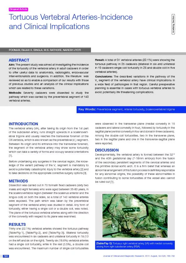
780 780 Tortuous Vertebral ArteriesIncidence Key WorPrevertebral segment Arterial tortuosity Scalenovertebral trigoneThe present study was aimed at investigating the incidence of the tortuosity of ID: 446506

780 780 Tortuous Vertebral Arteries-Incidence Key WorPrevertebral

Share:

Link:

Embed:

Download Presentation from below link

Download Pdf The PPT/PDF document "Journal of Clinical and Diagnostic Resea..." is the property of its rightful owner. Permission is granted to download and print the materials on this web site for personal, non-commercial use only, and to display it on your personal computer provided you do not modify the materials and that you retain all copyright notices contained in the materials. By downloading content from our website, you accept the terms of this agreement.


Presentation Transcript

Journal of Clinical and Diagnostic Research. 2011 August, Vol-5(4): 780-782 780 780 Tortuous Vertebral Arteries-Incidence Key WorPrevertebral segment, Arterial tortuosity, Scalenovertebral trigoneThe present study was aimed at investigating the incidence of the tortuosity of the vertebral artery in adult cadaversin order POONAM, RAJAN K. SINGLA, M.S. RATHORE, NARESH JYOTI The vertebral artery (VA), after taking its origin from the 1st part [Table/Fig-1]: Tortuous right vertebral artery (VA) with medial convexity arising from right subclavian artery (RSA) were observed in the transverse plane (medial convexity in 15 cadavers and lateral convexity in four), followed by tortuosity in the sagittal plane (ventral convexity in four and dorsal in three cadavers). Among the double coil tortuosities, two in the transverse plane, two in the sagittal plane and one in the transverse-sagittal plane were reported. O Journal of Clinical and Diagnostic Research. 2011 August, Vol-5(4): 780-782 www.jcdr.netPoonam et al., Tortuous vertebral arteries 781 vertebro-basilar insufciency. These asymptomatic loops may be corrected by a bypass procedure [1]. The intervertebral for a men is often widened and its appearance may be confused with the congenital absence of the pedicle or the presence of a neurobroma, which is the most common cause of this lesion [6].The reported incidences of tortuous vertebral arteries by earlier authors are shown in [Table/Fig-1]. Earlier, Tschabitscher et al [7] and Matula et al [1] encountered tortuous vertebral arteries in 47.2% and 39.1% of their dissections respectively [Table\Fig-4]. The incidence which was noted by them is much more than the 22.1% incidence which was encountered with this anomaly in the present study. A possible explanation which has been given for such a high incidence in their study is the larger age group (59–82 years) of their study material, which might have been responsible for the atherosclerotic changes in the carotid and the subclavian arteries and also the extension into their branches as well resulting in the lengthening and dilatation of the same (8) (9). But, other authors have suggested that the deection of the VA may result from cervical osteophytosis [10] and yet others have suggested suggested Most of the tortuosities (48.3%) which were encountered in our study were found in the transverse plane, with the direction of their convexities towards the medial side. The details of the tortuosities with their planes and the direction of their convexities which were encountered in our studies have been described in [Table/Fig 5]. Curylo et al suggests that during the anterior decompression surgery of the cervical spine, the medially located vertebral artery carries the risk of VA injury [8].The operative indications for surgery in the cervical region include spondylosis, a herniated inter-vertebral disc, tumour, infection and trauma. Although the lateral position of the VA does not hinder the anterior approach to the cervical vertebra, the presence of an abnormally tortuous VA, by using anatomic landmarks to guide The tortuosity in the VA was not recognized until 1958, when Hadler described four cases in 21 subjects who were aged between 45 and 91 years and were examined post mortem [4].Clinically, the tortuosity of the V segment does not have a haemodynamically signicant consequence. However, the loops can cause radiculopathy via nerve root compression [5]. Cervical spinal fractures have been reported to be secondary to the bony erosion from a V loop which is in contact. Also, a vertebral loop which is caused by the displacement from a mass lesion in the scalenovertebral trigone may be compressed and it may lead to Tschabitscher et al Present Study[Table\Fig-4]: Type of TortuousityDirection of Percentage TransverseVentralTransverseProximal medialProximal dorsalTransverse Proximal medial[Table\Fig-5]: [Table/Fig-2]: Left vertebral artery (VA) in common (CTthyroid artery (ITA) arising from left subclavian artery (SCA) showing tortuousity in sagittal plane with proximal dorsal and distal ventral [Table/Fig-3]: Tortuous right vertebral artery (VA) in transverse sagittal plane with proximal medial and distal dorsal convexity taking origin from right subclavian artery (RSA) entering into C7 transverse Journal of Clinical and Diagnostic Research. 2011 August, Vol-5(4): 780-782 Poonam et al., Tortuous vertebral arterieswww.jcdr.net 782 decompression may not prevent iatrogenic injury to the VA. The failure to recognize this anomaly during the preoperative planning planning The laceration of the VA is the most challenging surgical dilemma which is faced during anterior cervical spinal surgery [12]. Such an injury may cause catastrophic consequences, especially in those patients who present with an inappropriate blood decit by the contralateral artery (e.g. by atherosclerotic occlusion) [13], which may cause brain stem infarction [14] and central respiratory espiratory In a nutshell, apart from understanding the clinical implications, the data which is derived from the gross anatomical dissections of cadavers can be a useful guideline to the surgeons for careful pre-operative planning in cases with an unusual course of VA and can help them in avoiding potentially life threatening complications. eatening complications.    Matula C, Trattnig S, Tschabitscher M et al. The course of the prevertebral segment of the vertebral artery: anatomy and clinical clinical    Heary RF, Albert TJ, Ludwig SC et al. Surgical anatomy of the vertebral vertebral    Yamazaki M , Koshi T , Mannoji C et al. Traumatic C6–7 subluxation with the anomalous course of the vertebral arteries treated with pedicle screw/rod xationCase report. Dr. Dr. Dr. M.S. RathoreDr. Naresh JyotiPARTICULARS OF CONTRIBUTORS:Corresponding Author.Associate Professor (Anatomy), Department of Anatomy, Govt Medical College, Amritsar, Punjab, India.Professor (Anatomy), Department of Anatomy, Jhalawar Medical College, Jhalawar, Rajasthan, India.Assistant Professor (Pharmacology), Department of Pharmacology, Jhalawar Medical College, Jhalawar, Dr Poonam, Assistant Professor (Anatomy), Department of Anatomy, Jhalawar Medical College, Jhalawar, Rajasthan, India-326001DECLARATION ON COMPETING INTERESTS:No competing Interests.ests.   Hadler M. Tortuosity and deection of the vertebral artery. .    Kricun R, Levitt LP, Winn HR. Tortuous vertebral artery shown by MR and CT. .    Waldron T and Antoine D. Tortuosity or Aneurysm? The Palaeopathology of Some Abnormalities of the Vertebral Artery. Int. J. Osteo- Published online in Wiley InterScience (www.interscience.wiley.com). DOI: 10.1002/oa.586 2002; 12: 79–88.com). DOI: 10.1002/oa.586 2002; 12: 79–88   Tschabitscher M, Fuss FK, Matula C et al. Course of arteria vertebralis in its segment V1 from the origin to its entry into the foramen processus ocessus    Curylo LJ, Mason HC, Bohlman HH et al. Tortuous course of the vertebral artery and anterior cervical decompression: a cadaveric and clinical case study. .    Ranganatha SV, Manjunath KV. The course of the V1 vertebral segment artery. .    Oga M, Yuge I, Terada K. Tortuosity of the vertebral artery in patients with cervical spondylotic myelopathy. .    Babin E, Haller M. Correlation between the bony radiological signs and the dolichoarterial loops of the cervical vertebral artery. .    Lu J, Ebraheim NA. The vertebral artery: Surgical anatomy. .    Kajimoto BHJ, Addeo RLD, Campos GC et al. Anatomical study of the vertebral artery path in the human lower cervical spine. Acta ortop ortop    Cho KH, Shin YS, Yoon SH et al. Poor surgical technique in cervical plating, leading to vertebral artery injury and brain stem infarction – case report. eport.    Lanczik O, Szabo K, Lecei O et al. Central respiratory dysfunction