/
Polydactyly of the Foot Polydactyly of the Foot

Polydactyly of the Foot - PDF document

alis
alis . @alis
Follow
342 views
Uploaded On 2022-09-05

Polydactyly of the Foot - PPT Presentation

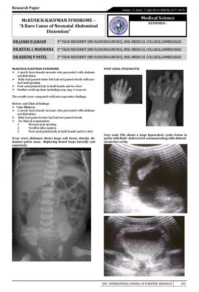
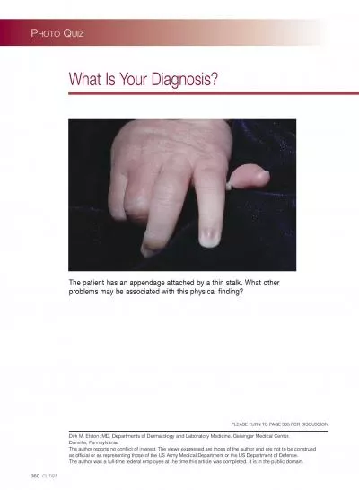
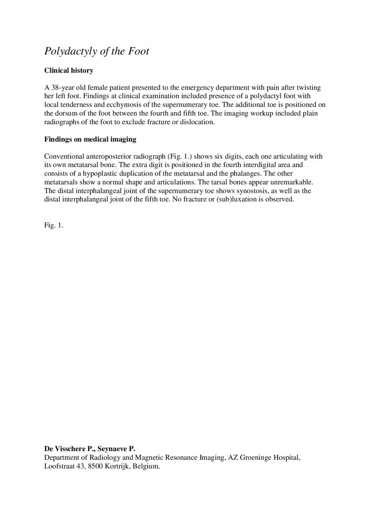
Clinical history A 38 year old female patient presented to the emergency department with pain after twisting her left foot Findings at clinical examination included presence of a polydactyl foot w ID: 949662

ray foot metatarsal polydactyly foot ray polydactyly metatarsal toe duplication cases type lateral digits imaging supernumerary distal department patients

Share:

Link:

Embed:

Download Presentation from below link

Download Pdf The PPT/PDF document "Polydactyly of the Foot" is the property of its rightful owner. Permission is granted to download and print the materials on this web site for personal, non-commercial use only, and to display it on your personal computer provided you do not modify the materials and that you retain all copyright notices contained in the materials. By downloading content from our website, you accept the terms of this agreement.


Presentation Transcript

Polydactyly of the Foot Clinical history A 38 - year old female patient presented to the emergency department with pain after twisting her left foot. Findings at clinical examination included presence of a polydactyl foot with local tenderness and ecchymos is of the supernumerary toe. The additional toe is positioned on the dorsum of the foot between the fourth and fifth toe. The imaging workup included plain radiographs of the foot to exclude fracture or dislocation. Findings on medical imaging Conventio nal anteroposterior radiograph (Fig. 1.) shows six digits, each one articulating with its own metatarsal bone. The extra digit is positioned in the fourth interdigital area and consists of a hypoplastic duplication of the metatarsal and the phalanges. The other metatarsals show a normal shape and articulations. The tarsal bones appear unremarkable. The distal interphalangeal joint of the supernumerary toe shows synostosis, as well as the distal interphalangeal joint of the fifth toe. No fracture or (sub)lux ation is observed. Fig. 1. De Visschere P., Seynaeve P. Department of Radiology and Magnetic Resonance Imaging, AZ Groeninge Hospital, Loofstraat 43, 8500 Kortrijk, Belgium. The radiographic findings in this patient are compatib le with a metatarsal type postaxial polydactyly of the foot with fifth ray duplication . Comment Polydactyly of the foot is a condition in which there are extra digits. It is generally classified into three major groups based on topographic criteria: medi al ray (preaxial), central ray and lateral ray (postaxial) polydactyly. The lateral ray polydactyly is divided into fifth ray duplication and sixth ray duplication, depending on medial or lateral position of the supernumerary toe relative to the little toe . Each ray can be further divided according to the level of duplication, i.e. metatarsal type, proximal - , middle - and distal - phalangeal type. The metatarsal type often shows a hypoplastic metatarsal and proximal phalanx. The complexity of the deformity ra nges from a simple soft - tissue problem to a completely developed accessory ray. It is not uncommon for an extradigit to be fused in some way to the neighbouring digits (webbing

between the digits or osseous fusions), a condition called polysyndactyly. Pol ydactyly of the foot is one of the most common congenital foot anomalies. Although the frequency varies widely among populations, a slightly higher incidence is seen in Africa and Asia. Polydactyly of the foot occurs in approximately 2 - 3 newborns out of 10 00 live births. The condition is considered to be isolated and idiopathic in most cases, but in approximately 15% associated anomalies can be found. A positive family history is found in 10 to 30% of the cases. The deformity is bilateral in 25 to 50%. Adul t cases are more rare because most individuals are treated during childhood. Watanabe et al. reported in 1992 a series of 330 feet in 265 patients. Lateral ray polydactyly occurred in 86% of the cases, far more frequent than medial ray (8%) and central r ay (6%) polydactyly. 173 patients in their series had fifth ray duplication (i.e. 76% of the lateral ray polydactylies). Only 10 of these patients had the complete metatarsal duplication type. (i.e. 5.7% of fifth ray duplications and 3.8% of all polydactyl ies). Treatment of polydactyly may be indicated for shoe problems, pain or cosmetic reasons. Surgery can be performed at any age but is generally done before walking age, when the infant is between 9 and 12 months of age. In most cases the surgical proce dure causes no difficulties and prognosis is excellent without complications. Key word Polydactyly References 1. Galois L., Mainard D., Delagoutte J.P.: Polydactyly of the foot. Literature review and case presentations. Acta Orthopaedica Belgica , 2002, 68: 376 - 380 2. Hart E.S., Grottkau B.E., Rebello G.N., Albright M.B.: The Newborn Foot. Diagnosis and Management of Common Conditions. Orthopaedic Nursing, 2005, 24(5): 313 - 321 3. Theodorou S.J., Theodorou D.J., Farooki S.: An Unusual Case of Polydactyly of the Foot . Am J Roentgenol ., 2003, 181(6): 1721 - 1722 4. Watanabe H., Fujita S., Oka I.: Polydactyly of the Foot: An Analysis of 265 Cases and a Morphological Classification. Plast Reconstr Surg , 1992, 89(5): 856 - 877 De Visschere P., Seynaeve P. Department of Radiolog y and Magnetic Resonance Imaging, AZ Groeninge Hospital, Loofstraat 43, 8500 Kortrijk, Belgium