/
Oral  Maxillofacial Pathology  Journal  OMPJ Oral  Maxillofacial Pathology  Journal  OMPJ

Oral Maxillofacial Pathology Journal OMPJ - PDF document

alyssa
alyssa . @alyssa
Follow
348 views
Uploaded On 2022-10-13

Oral Maxillofacial Pathology Journal OMPJ - PPT Presentation

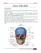
127 Vol 2 No 1 J an Jun 201 1 ISSN 0976 1225 ADENOID CYSTIC CARCINOMA OF HARD PALATE A CASE REPORT Aarthi Mahajan 1 Meena Kulkarni 2 Mitesh Parekh 3 Mehrunisha Khan 4 Arpan Shah 4 Mah ID: 959439

adenoid cystic oral carcinoma cystic adenoid carcinoma oral salivary tumor cells palate pathology tumors gland prognosis left swelling malignant

Share:

Link:

Embed:

Download Presentation from below link

Download Pdf The PPT/PDF document "Oral Maxillofacial Pathology Journal ..." is the property of its rightful owner. Permission is granted to download and print the materials on this web site for personal, non-commercial use only, and to display it on your personal computer provided you do not modify the materials and that you retain all copyright notices contained in the materials. By downloading content from our website, you accept the terms of this agreement.


Presentation Transcript

127 Oral & Maxillofacial Pathology Journal [ OMPJ ] Vol 2 No 1 J an - Jun 201 1 ISSN 0976 - 1225 ADENOID CYSTIC CARCINOMA OF HARD PALATE : A CASE REPORT Aarthi Mahajan 1 , Meena Kulkarni 2 , Mitesh Parekh 3 , Mehrunisha Khan 4 , Arpan Shah 4 , Mahesh Gabhane 4 1 Reader, 2 Professor, 3 Lecturer, 4 PG student, Dept of Oral Pathology, M.G.V’s K.B.H. Dental College & Hospital, Nashik, India Correspond ing Author: Aarthi Mahajan , email : rt_mahen@yahoo.com A bstract Brief Background : This article describes a case of adenoid cystic carcinoma in the left posterolateral portion of the hard palate, in a 41 - year - old woman. Materials & Methods : Clinical examination aided by CT scan findings to assess the size and the extent of the lesion, and to check the evidence of antral involvement. Final diagnosis based on histopathological findings. D iscussion : Discuss ion centered on adenoid cystic carcinoma, its synonyms, histomorphology, grading, biological behavior , treatment & prognosis. Summary and Conclusion : Adenoid cystic carcin oma is a c ommon salivary gland malignancy, frequently showing recurrence, neural invas ion and not uncommonly showing locoregional metastasis. The tumor is known for its varieties of histomorphologies. Its histopathological variety helps its grading and predicts prognosis. KEY WORDS : Adenoid cystic carcinoma, intra - oral , metastasis, progno sis. I ntroduction Adenoid cystic carcinoma is one of the common and best recognized salivary gland malignancies. This tumor was first described by Billroth as ‘cylindroma’ in 1859. 1 The term ‘adenoid cystic carcinoma’ was introduced by Ewing (Foote and Frazell) in 1954. It is a relentless tumor that is prone to local recurrence and eventual distant metastases. 2 It is clinically deceptive by virtue of its small size and slow growth, which belies its extensive subclinical invasion and marked ability for ea rly metastasis, the factors which make the prognosis of the neoplasm questionable when present. 3 Among all adenoid cystic carcinomas, 50% occur in intraoral sites, with definite predilection for the posterolateral portion of the hard palate. 2 Here, we repo rt a case of Adenoid cystic carcinoma in the left posterolateral portion of the hard palate, in a 41 - year - old woman. Case Report A 41 years old female patient visited the department of Oral Medicine and Radiology, M.G.V’s K.B.H Dental c ollege and hospit al, Nashik , with the chief complaint of swelling on the palate on the left side since approximately 1 ½ months. The history revealed that the swelling had started insidiously and had steadily increased in size since its onset. The swelling was associated wi th dull and continuous pain which started 30 days ago and it was not associated with discharge of any sort. Medical, surgical, dental, family and personal histories were not noteworthy. There were no abnormalities detected on general phy

sical examination. There were no abnormal findings on Extra oral examination. Intraoral examination revealed a solitary swelling , involving the left posterolateral region of the hard palate, measuring approximately 1½ × 1 cm, extending anteroposteriorly from mesial of 27 to distal of 28 , and lateromedial l y , from the marginal gingiva of 27 and 28 128 Oral & Maxillofacial Pathology Journal [ OMPJ ] Vol 2 No 1 J an - Jun 201 1 ISSN 0976 - 1225 towards the midpalatine raphae. The surface of the swelling was smooth and the overlyin g mucosa was intact and normal i n colour . None of the teeth on the involved side was carious or periodontally involved. On palpation, the swelling was tender and firm in consistency. No regional lymphadenopathy was found. Clinical differe ntial diagnosis included a benign or a low grade malignant neoplasm of minor salivary glands , reactive/inflammat ory condition of minor salivary glands, salivary , a malignant growth of the maxillary sinus, benign mesenchymal neoplasm and much less likely a slow - growing malignant mesenchymal neoplasm. Intra - oral periapical and occ lusal radiographs revealed diffuse , ha zy , irregular radiolucency in 27, 28 region . CT scan of pa ranasal sinuses revealed a malignant natured expansile antrochoanal polyp with bone destruction in posterolateral wall, medial wall and a lveolar recess. Ultrasonography of neck revealed small level II lymphnodes on left side representing positive cervical lymphadenopathy on left side. X - ray chest revealed no pleural or parenchymal abnormalities. Radiogr a phic findings were in the favour of malignant lesion of maxillary sinus . Incisional biopsy was pe rformed for the histopathological diagnosis. The hematoxylin and eosin stained sections showed uniform cells arranged in cord like pattern, with deeply stained nuclei and with round to oval pseudocysts , containing pale pink granulofibrillar material, givin g the entire structure a typical “swiss - cheeze” appearance. The histopathological impression was that of an adenoid cystic carcinoma of cribriform pattern. The patient underwent left subtotal maxillectomy and the entire specimen was submitted for the histo pathological evaluation. The gross specimen was consisting of maxilla measuring 5 cm along upper alveolar border , along with 4 intact maxillary posterio r teeth. Specimen also included a part of hard palate, soft palate and zygoma. The maxillary sinus showe d multiple polypoid masses together measuring 3 × 2.5 × 2 cm in size. Cut surface of the polyps was gray white, solid with hemorrhagic areas. Histopathological findings were same as that of incisional biopsy. Adjuvant radiotherapy was planned for the pati ent after the final histopathological diagnosis. External beam radiotherapy was given to the patient for 1½ months . No local recurrence of the lesion has been observed 6 months after the treatme

nt. After 6 months X - ray chest was repeated and it revealed no pleural or parenchymal abnormalities. Fig2: Intra oral photograph showing the swelling in 129 Oral & Maxillofacial Pathology Journal [ OMPJ ] Vol 2 No 1 J an - Jun 201 1 ISSN 0976 - 1225 the left posterolateral area of the hard palate. Fig3: Computed tomography scan showing the polyp in the left maxillary sin us. Fig4: Photomicrograph showing the lesional tissue composed of cords of the cells and pseudocysts. ( 4X) Fig 5: Photomicrograph showing the interconnecting cords of the deeply stained cells with pseudocysts giving a typical swiss - cheeze appe arance. (10X) Fig 6: Photomicrograph showing the strands of uniformly stained deeply basophilic cells along with pseudocysts containing eosinophilic granulofibrillar material. (Magnification 40X) D iscussion Frequency of intraoral minor salivary gland n eoplasms ranges from 9 - 23%, among all salivary gland neoplasms. Among these 50% tumors are malignant. 42 - 54% of these occur on the palate. Adenoid cystic carcinoma occurs on the palate with the frequency of 8 - 15% of all the palatal salivary neoplasms. T he term ‘adenoid cystic carcinoma’ was introduced by Ewing (Foote and Frazell) in 1954 1 . This tumor was named as ‘cylindroma’ earlier by Billroth in 1859 1 , because the epithelial and connective tissue elements formed a system of inter twining cylinders. The term ‘basalioma’ was coined by Kromp e cher in 1908 1 , who considered this type of tumor to be of analogous nature to the basal cell growths of the skin. Adenoid cystic carcinoma may occur at any age, although in most cases the patients are middle aged or ov er . 2 Females are affected more frequently than males. It can occur in any salivary gland site, but approximately 50% occur within the minor salivary glands. 2 The remaining tumors are found mainly in the parotid and submandibular glands. It usually appears as a slowly growing mass. Pain is a common and 130 Oral & Maxillofacial Pathology Journal [ OMPJ ] Vol 2 No 1 J an - Jun 201 1 ISSN 0976 - 1225 important finding, occasionally occurring early in the course of the disease before there is a noticeable swelling 2 . Facial nerve paralysis may develop with parotid tumors. Intraoral (oral and oropharyngeal) a denoid cystic carcinomas are uncommon and characterized by slow evolution, protracted clinical course, multiple and/or delayed recurrences and late distant metastasis. 4 Tumors arising in the palate or maxillary sinus may show radiographic evidence of bone destruction. 2 In a study conducted by Buchner A et al, the relative frequency of intraoral minor salivary gland tumors was 0.4%, among these 41% were malignant. Among these malignant neoplasms, the most common was mucoepidermoid carcinoma (21.8%) followe

d by polymorph o us low grade adenocarcinoma (7.1 % ), Adenoid cystic carcinoma was found to be third most common (6.3%). 5 HISTOMORPHOLOGY AND GRADING OF THE TUMOR 6 : Various growth patterns and cytological details in Adenoid cystic carcinomas establish the di agnostic criteria for this neoplasm. The primary growth patterns are of three types in adenoid cystic carcinoma. a. CRIBRIFORM VARIANT : Extensive sheets, uniform bands, or cribriform nests usually composed of relatively small, darkly stained, slightly separat ed basal/myoepithelial cells and small, at times inconspicuous duct - like structures, which may contain secretory products. Round to oval, often fairly uniformly sized intercellular spaces, termed pseudocysts, containing pale grayish blue to pinkish granulo fibrillar material at times with a reticular pattern, which develop in relation to the basal/myoepithelial cells. b. TUBULAR VARIANT: P resence of bilayered duct like structures generally composed of an inner layer of cuboidal to columnar ductal cells with mod erate amounts of eosinophilic cytoplasm and outer, smaller darker staining cells. c. SOLID VARIANT: A rranged as variable, at times fairly uniformly sized groups or as sheets of small, darkly stained tumor cells , those are excess proliferations of the basal/my oepithelial cell component. Small duct like structures must be identifiable among the basaloid cells. Nests or sheets of basaloid cells with the above features from 30% or more of the neoplasm. GRADING OF THE TUMOR 6 : a. GRADE I: The tumor consisting only of cribriform and tubular histomorphology . b. GRADE II: A mixture of cribriform, tubular and solid gro wth patterns, with solid growth pattern less than 30% of the tumor. c. GRADE III: Tumors with predominantly solid features (�30% or more of the tumor) TREATMENT , PROGNOSIS AND SURVIVAL RATES Treatment of adenoid cystic carcinoma generally requires excision with the widest margins possible because the tumor cells extend well beyond the clinical or radiographic margins and that the tumor undergoes not on ly perineural invasion but perineural spread. 3 Postoperative radiotherapy of 6000 cGy to 7500 cGy is usually given . 3 Total parotidectomy with resection of the facial nerve is generally the line of treatment for the tumors occurring in parotid. 3 Grading of adenoid cystic carcinoma is suggested to have prognostic significance, 131 Oral & Maxillofacial Pathology Journal [ OMPJ ] Vol 2 No 1 J an - Jun 201 1 ISSN 0976 - 1225 but staging has been shown to be perhaps a more meaningful predictor of clinical outcome. 6 Long term survival is particularly low in grade III tumors. 6 Perineural invasion, site of pri mary and tumor stage significantly impact upon local control and disease free survival. Slow growth, with multiple loco - regional recurrences and late distant metastases are the characteristics of the long clinical

course of the tumor. Distant metastases oc cur in 25 - 50% of patients, even many years after the diagnosis, and lung is the most involved site. 7 A long term survival is typical of the natural history of patients with metastatic adenoid cystic carcinoma, in particular in case of lung metastases as co mpared with the bone dissemination 8 ,9 , and the pulmonary resection is considered a procedure with low mortality and morbidity to treat these metastases 7 . Distant metastases rate is highest in solid variant. However, adenoid cystic carcinoma has well known prognosis profile. 5 years survival rate af ter effective treatment is 75%, but long term survival rates are low (10 years – 20% and 15 years – 10%). 3 Postoperative radiotherapy, combined with more aggressive surgeries increases the long term survivals into the 30% to 40% range. 3 Solid primary growth pattern is associated with worse prognosis. Clinical size greater than 4 cm indicates greater subclinical spread. Delayed diagnosis and delayed treatment also worsen the prognosis. Close or unclear surgical marg ins are indica tors of the requirement of wider excision . Multiple local recurrences are also associated with grave prognosis. 3 C onclusion Adenoid cystic carcinoma is a c ommon salivary gland malignancy but relatively rare in minor salivary glands. It is fa mous for its peculiar histopathological features, variants and tendency for perineural invasion. At times it may metastasize to regional lymphnodes and worsen the prognosis of the case. Acknowledgement: Authors are grateful to Dr. Nagesh Madnoorkar MS , DNB & NAMCO Charitable Trust’s Cancer Hospital, Nashik References: 1. R. B. Lucas, Pathology of tumors of the oral tissues, fourth edition, page: 330 - 35. 2. Neville, Damm, Allen, Bouquot; Oral and Maxillofacial Pathology, second edition, page: 426 - 8 3. Ro bert E. Marx, Diane Stern; Oral and Maxillofacial Pathology, A rational for diagnosis and management; page 550 - 3. 4. J. P. Agarwal et al; Intraoral Adenoid cystic carcinoma: Prognostic factors and outcome; Oral Oncol (2008) 44, 986 - 93. 5. Amos Buchner, Phillip W . Merrell, William M. Carpenter; Relative frequency of intra - oral minor salivary gland tumors: a study from northern California and comparison to reports from other parts of the world; J Oral Pathol Med (2007) 36: 207 - 14. 6. Irving Dardick, Color atlas/text o f salivary gland tumor pathology, page: 149 - 57 7. Laura D. Locati et al; Lung metastasectomy in Adenoid cystic carcinoma of salivary gland; Oral Oncol (2005) 41, 890 - 94. 8. Kim KH et al; Adenoid cystic carcinoma of the head and neck, Arch Otolaryngol Head Neck S urg 1994: 120 (7): 721 - 6 9. Van der Wal JE, Becking AG, Snow GB, Van der Wal I; Distant metastases of Adenoid cystic carcinoma of the salivary glands and the value of diagnostic examination during followup; Head Neck 2002; 24 (8) : 779 - 83 Source of Support : Nil, Conflict of Interest : None declar