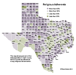
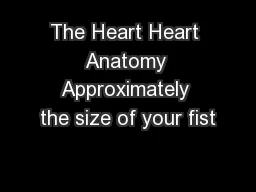
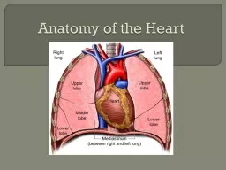
Located within bony thorax ribs and is flanked on each side by lungs Apex points toward your left hip and rests on your diaphragm Wall of the Heart The heart is enclosed by double sac of serous membrane called pericardium ID: 915290
Download Presentation The PPT/PDF document "Anatomy of the Heart Heart is approximat..." is the property of its rightful owner. Permission is granted to download and print the materials on this web site for personal, non-commercial use only, and to display it on your personal computer provided you do not modify the materials and that you retain all copyright notices contained in the materials. By downloading content from our website, you accept the terms of this agreement.
Slide1
Anatomy of the Heart
Slide2Heart is approximately the size of your fist and weighs less than a pound.
Located within bony thorax (ribs) and is flanked on each side by lungs
Apex points toward your left hip and rests on your diaphragm
Slide3Wall of the Heart
The heart is enclosed by double sac of serous membrane called pericardium
The heart walls are composed of 3 layers:
Epicardium
– visceral tissue that tightly hugs the heart
Myocardium – thick bundles of cardiac muscle; this is the layer that contracts
Endocardium
– thin, glistening sheet of endothelium that lines the heart chambers; continuous with linings of blood vessels
Slide4Coverings/wall of heart
Slide5Chambers of the Heart
The heart has four hollow chambers:
Two atria
Two ventricles
Each chamber is lined with
endocardium
to help blood flow smoothly through heart
Atria lie superior to ventricles and receive blood under low pressure from veins of body. Atria are separated by
interatrial
septum.
Ventricles lie inferior to atria and receive blood from atria. Ventricles contract to force blood into circulation. Ventricles are separated by
interventricular
septum
Slide6Slide7Valves of the Heart
The heart has 4 valves which allow blood to flow in only one direction
Slide8AV valves (
atrioventricular
) are located between each
atrial
and ventricular chambers on each side.
Bicuspid (mitral) valve – consists of two cusps of
endocardium
; located between left atrium and left ventricle
Tricuspid valve – consists of three cusps; located between right atrium and right ventricle
Chordae
tendinae
(“heart strings”) – tiny white chords which anchor wall of cusps of AV valves to ventricles. Anchor cusps of AV valves in a closed position when ventricles contract. This prevents blood from leaking back into atria.
Slide9Slide10Semilunar
Valves – Guard the bases of the two large arteries leaving the ventricles.
Pulmonary
Semilunar
Valve- guards the opening of the pulmonary trunk
Aortic
Seminlunar
Valve – guards opening of aorta
Each SL valve has 3 cusps which fit tightly together when closed and are forced open and flattened against
atrial
walls when ventricles contract to force blood out of heart
Slide11Valves
Slide12Mitral Valve
Prolapse
Slide13Circulation
The right side of heart functions as pulmonary circuit pump. It
receives
oxygen-poor blood from body through the Superior and Inferior
Venae
Cavae
and pumps this blood
out
through the Pulmonary Trunk
Slide14Pulmonary Circulation
The pulmonary trunk splits into right and left pulmonary arteries, which carry blood to lungs where it picks up Oxygen and unloads Carbon Dioxide.
Oxygen-rich blood drains returns from lungs to left side of heart through 4 pulmonary veins,
This path from right side of heart to lungs and back to left side of heart = Pulmonary Circulation
Slide15Systemic Circulation
Blood returning to left side of heart is deposited into the left atrium and then flows into the left ventricle. The left ventricle forces blood through the aorta to the systemic arties to supply oxygen to all body tissues and then back to right atrium
This path from left side of heart through body tissues and back to right side of heart = Systemic Circulation
Slide16Aorta
The largest artery of the bodySize of garden hose (diameter)
Springs from left ventricle (ascending aorta), arches to the left (aortic arch), and then plunges downward (descending aorta)
Slide17Slide18The Path of Circulation
Slide19The Path of Circulation through the Heart
Slide20Oxygen-poor blood returns from body through the inferior (5) and superior (1)
cavae
and empties into the right atrium(2). Blood then flows through the tricuspid valve into the right ventricle (4). Right ventricle contracts forcing blood against the pulmonary SL valve forcing it open. Blood exits the heart and enters the lungs via right and left
pulomonary
arteries (8).
Slide21Blood picks up Oxygen and deposits Carbon Dioxide in capillary beds of lungs (10). Oxygen-rich blood now returns to left atrium (7) via right and left pulmonary veins (3). As left atrium fills with blood, pressure is exerted on mitral valve (bicuspid) and it is forced open allowing blood to fill left ventricle (6). Left ventricle contracts forcing aortic SL valve open and allowing blood to leave heart through aorta (9).
Slide22Double Pump
Both sides of the heart are pumping at the same time.
Slide23In a healthy heart, the both atria contract simultaneously. At they begin to relax, contraction of the ventricles begins.
Systole = contraction of ventricles
Diastole = relaxation of ventricles
Slide24Cardiac circulation
Blood supply that nourishes the heart is accomplished by right and left coronary arteries which branch from base of aorta and encircle the heart.
Coronary arteries are compressed when ventricles are contracting and fill with blood when heart is relaxed
Myocardium is drained by several cardiac veins which empty into enlarge vessel on backside of heart called coronary sinus, which in turn empties into right atrium.
Slide25Coronary arteries
Slide26Coronary sinus
Slide27Slide28