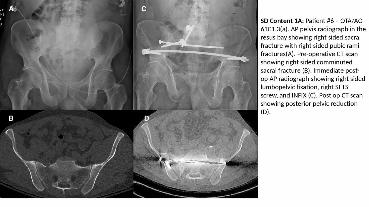
PPT-SD Content 1A: Patient #6 – OTA/AO 61C1.3(a). AP pelvis radiograph in the resus bay
Author : ariel | Published Date : 2023-07-28
A B C D A D B C SD Content 1B Example Case 2 right sided sacral fracture with extension into L5S1 facet Patient 7 OTAAO 61C31c AP pelvis xray in ED showing right
Presentation Embed Code
Download Presentation
Download Presentation The PPT/PDF document "SD Content 1A: Patient #6 – OTA/AO 61..." is the property of its rightful owner. Permission is granted to download and print the materials on this website for personal, non-commercial use only, and to display it on your personal computer provided you do not modify the materials and that you retain all copyright notices contained in the materials. By downloading content from our website, you accept the terms of this agreement.
SD Content 1A: Patient #6 – OTA/AO 61C1.3(a). AP pelvis radiograph in the resus bay: Transcript
A B C D A D B C SD Content 1B Example Case 2 right sided sacral fracture with extension into L5S1 facet Patient 7 OTAAO 61C31c AP pelvis xray in ED showing right sided sacral fracture with left sided pubic rami fractures A Coronal CT scan bone window showing right sided comminuted sacral ala fracture B Axial CT scan bone window showing extension of sacral fracture into right L5S1 facet C AP pelvis at three months follow up showing R sided triangular osteosynthesis and INFIX The posterior pelvic ring is well reduced and healed D. Basic to Intermediate Interpretation. Phillip Smith, BA, RRT. Relative Densities. The images seen on a chest radiograph result from the differences in densities of the materials in the body.. The . hierarchy of relative densities from least dense (dark on the radiograph) to most dense (light on the radiograph) include:. Skeletal anatomy of the hip/pelvis. 1. Ilium – upper, lateral section of pelvis. 2. Iliac crest – upper ridge of ilium. 3. ASIS (Anterior Superior Iliac Spine) – identifies anterior end point for iliac crest. Lecture One. Normal Anatomy of the Female Pelvis and Transvaginal Sonography. Holdorf. Outline. Skeletal. Ligaments. Musculature. True Pelvic Muscles. Pelvic Organs. Urinary Bladder. Vagina. Uterus. Cervix. Lecture 26. Normal Anatomy of the Female Pelvis and Transvaginal Sonography. Holdorf. Outline. Skeletal. Ligaments. Musculature. True Pelvic Muscles. Pelvic Organs. Urinary Bladder. Vagina. Uterus. Cervix. July/August 2007 Vol 78/No 6 RADIOLOGIC TECHNOLOGYhest radiography is the most common radiographic pro-cedure performed in medi-cal imaging departments and one of the most often repeated exams1-3 It i K W W S V G R L R U J 6 ; 3 X E O L V K H G R Q O L Q H E \ &