/
Abstract OBJECTIVE We report a minority of cases presented with abdo Abstract OBJECTIVE We report a minority of cases presented with abdo

Abstract OBJECTIVE We report a minority of cases presented with abdo - PDF document

bety
bety . @bety
Follow
342 views
Uploaded On 2022-08-16

Abstract OBJECTIVE We report a minority of cases presented with abdo - PPT Presentation

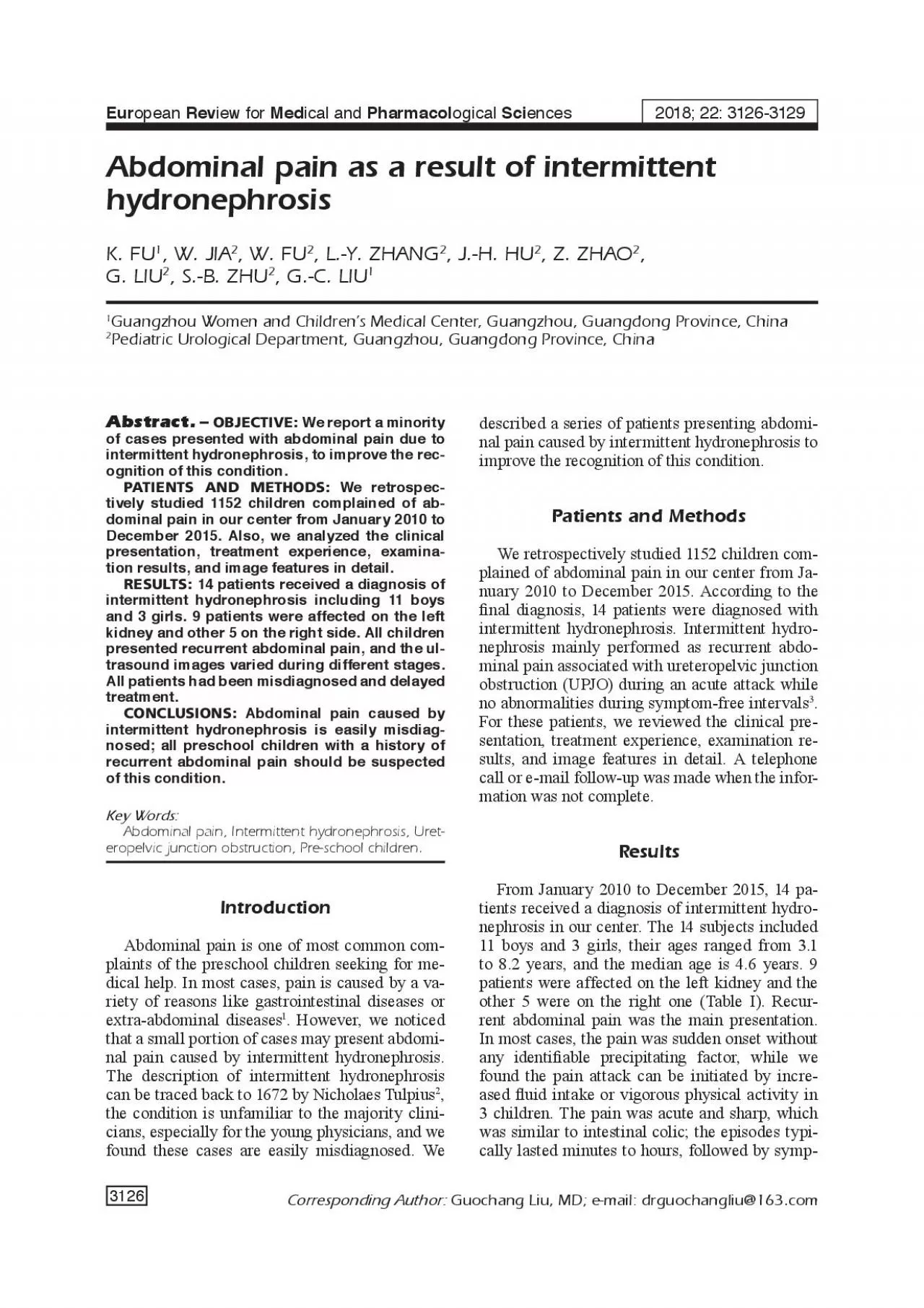
3126 European Review for Medical and Pharmacological Sciences2018 22 31263129 K FU1 W JIA2 W FU2 LY ZHANG2 JH HU2 Z ZHAO2 G LIU2 3127 tomfree intervals ranged from couple days to ID: 937550

pain hydronephrosis abdominal children hydronephrosis pain children abdominal intermittent patients x00660069 treatment clinical junction ureteropelvic renal obstruction urinary condition

Share:

Link:

Embed:

Download Presentation from below link

Download Pdf The PPT/PDF document "Abstract OBJECTIVE We report a minority ..." is the property of its rightful owner. Permission is granted to download and print the materials on this web site for personal, non-commercial use only, and to display it on your personal computer provided you do not modify the materials and that you retain all copyright notices contained in the materials. By downloading content from our website, you accept the terms of this agreement.


Presentation Transcript

3126 Abstract. OBJECTIVE: We report a minority of cases presented with abdominal pain due to intermittent hydronephrosis, to improve the recognition of this condition.PATIENTS AND METHODS: We retrospectively studied 1152 children complained of abdominal pain in our center from January 2010 to December 2015. Also, we analyzed the clinical presentation, treatment experience, examina European Review for Medical and Pharmacological Sciences2018; 22: 3126-3129 K. FU1, W. JIA2, W. FU2, L.-Y. ZHANG2, J.-H. HU2, Z. ZHAO2, G. LIU2 3127 tom-free intervals ranged from couple days to some months. Our patients hardly could tell us the sensation accurately; they predominantly located the pain in the renal area, upper abdominal, and periumbilical area. In addition to abdominal pain, 9 children had nausea and vomiting; other accompanied signs included palpable abdominal mass, gross hematuria, urinary frequency, urinary urgency, and hypertension can be found in half of the children. Laboratory tests were normal in almost all patients, while the image features of ultrasonography varied depending on the different period of the disease. During acute attack period, nearly all of the patients had either moderate or severe hydronephrosis on ultrasonography, while during symptom-free stage the patients had only mild hydronephrosis or pelvic dilation. Renal pelvic wall thickening can be detected in all patients during convalescence; it usually persisted for 6-9 days and then disappeared. 8 children conducted MR to show the renal image and to rule out extrinsic lower polar vessels. 3 patients also took a MAG3 scan to show the function of the affected kidney. In most cases, the abdominal pain can relieve spontaneously without any medical intervention. Only when the attack occurred many times or in severe conditions, the parents took the children to the hospital for medical help. Unfortunately, all of the 14 children were misdiagnosed at the �rst visit to hospital. In our group, 10 children (71.4%) were diagnosed with acute gastroenteritis or in�ammatory bowel disease, 3 children were diagnosed with urinary tract infection and the other one was diagnosed with urinary

stone. As a result of misdiagnose, most of our patients didn’t receive the correct treatment on time. Symptomatic treatment as analgesic and antibiotics have been most frequently used. The oral administration was effective in most cases, but in severe condition intravenous administration was necessary (Table II). In our work, the majority of patients were boys (78.6%) and the left kidney was affected more than the right one. Intermittent pain, ureteric colic with vomiting, and hematuria are the most common symptoms, also known as Dietl’s crisis. It was �rst described in patients with lower polar vessels causing intermittent hydronephrosis by Dietl in 1864. However, only a minority of patients possesses the diagnosis of intermittent hydronephrosis. A study of school-aged children showed that UPJO was the cause of abdominal pain in about 1 of 100. Besides, the pain of in Total cases42Median age (year)4.7M:F ratio38:4Affected sideLeft35 (83.3%)Right7 (16.7%)OperationOpen surgery30 (71.4%)Laparascopic12 (28.6%)Postoperative complications1 (2.63%) Clinical featuresNo. (%)Clinical featuresNo. (%)Quality of pain Duration of acute episode (d) Cramping 26 (61.9%)33 (78.6%) Dull 16 (38.1%) 1-26 (14.3%)Precipitating factors 2-33 (7.1%) None identi�ed24 (57.1%)Frequency of attacks Increased �uid intake 13 (31.0%) Every 1 week5 (11.9%) Vigorous exercise5 (11.9%) Every 2 weeks 7 (16.7%)Location of pain Every 1 month12 (28.6%) Flank/abdominal28 (66.7%) Every 2-3 months18 (42.8%) Periumbilical 8 (19.0%)Accompanied symptoms and signs Epigastric area6 (14.3%) Nausea and vomiting 28 (66.7%)Course of disease (month) Palpable abdominal mass5 (11.9%) 1M-6M19 (45.2%) Gross hematuria 4 (9.5%) 6M-12M14 (33.3%) Urinary frequency 2 (4.8%) >12M9 (21.5%)Hypertension3 (7.1%) 3128 termittent hydronephrosis has no speci�c nature, it can mimic almost any intra-abdominal pathology, so it is easy to miss the diagnosis unless a high index of suspicion. Compared with classic UPJO, intermittent hydronephrosis is a special condition that requires a different diagnostic strategy. A d

etailed history is the key to distinguish this condition; any pre-school children who had the history with recurrent abdominal pain should be suspected with the intermittent hydronephrosis. As the obstruction usually leads no loss of renal function, so the laboratory tests are unlikely helpful in the diagnosis. An immediate ultrasound is required when the next abdominal pain occurs, and another ultrasound during the symptom-free stage as a comparison. Further examinations like MAG3 scan, MR angiography, intravenous urography, retrograde pyelogram, and biopsy of the lesion, can all help in the diagnosis. The pain of intermittent hydronephrosis is due to an intermittent obstruction. The actual pathophysiology is not clear so far. The possible etiologies have been separated into extrinsic and intrinsic categories. Extrinsic etiology may be associated with improper rotation of kidney, or crossing lower pole renal vessels inhibiting the proper �ow of urine. Aberrant vessels crossing ureteropelvic junction are the most common extrinsic causes of UPJO. The incidence has been reported to range from 11% to 15% in children, but the rate may reach up to 58% in older children with symptomatic UPJOIntrinsic causes included congenital narrowing of the proximal ureter or abnormal peristalsis at the ureteropelvic junction. Patel et al11 had reported a child presented with recurrent attacks of Dietl’s crisis caused by upper ureteral polyp. Most of mild or moderate hydronephrosis may resolve spontaneously or improve with conservative treatment, but severe hydronephrosis can lead to glomerulosclerosis, medullary dysplasia or interstitial �brosis. Therefore, earlier diagnosis of intermittent hydronephrosis can prevent acute or chronic dysfunction. Once diagnosed, surgical is the best treatment.Abdominal pain caused by intermittent hydronephrosis is easily misdiagnosed; all preschool children with a history of recurrent abdominal pain should be suspect with this condition.Conflict of InterestThe Authors declare that they have no con�ict of interest.1) UEK SH . Recurrent abdominal pain in children: a clinical approach. Singapore Med J 2015; 56: 1

25-128. ERK M, SULYA B, POLOVITZER M, HEJJ, MOLNAR D, ZEVARYSS . [Intermittent hydronephrosis in childhood]. Orv Hetil 2013; 154: 940-946. AID, HUANG FIN CC, AIC, EE HC, SEUC, ANG P Intermittent hydronephrosis secondary to ureteropelvic junction obstruction: clinical and imaging features. Pediatrics 2006; 117: 139-146. LAGIRI M, POLEALLE S Dietl’s crisis: an under-recognized clinical entity in the pediatric population. Int Braz J Urol 2006; 32: 451-453. ANGF, S, CENH, ANGANG HM, ANG M . Expression and clinical signi�cance of aquaporin-1 and ET-1 in urine of children with congenital hydronephrosis. Eur Rev Med Pharmacol Sci 2017; 21: 4141-4146. ILLIAMS B, AREEN B, NI M . Pathophysiology and treatment of ureteropelvic junction obstruction. Curr Urol Rep 2007; 8: 111-117. CT scan showing the aberrant vessel crossing the K. Fu, W. Jia, W. Fu, L.-Y. Zhang, J.-H. Hu, Z. Zhao, G. Liu, S.-B. Zhu, G.-C. Liu 3129 CHINGOLO, MARESC, C S, ILI, PAOLUCC, BONOAGNOLI C, ENELI C. Post-natal ultrasound morpho-dynamic evaluation of mild fetal hydronephrosis: a new management. Eur Rev Med Pharmacol Sci 2013; 17: 2232-2239. 7) LONY B, PFNTEROLZNERHDER P, FMANNKJ, STRAAK, HERIG, PETEREN Accessory or additional renal arteries show no relevant effects on the width of the upper urinary tract: a 64-slice multidetector CT study in 1072 patients with 2132 kidneys. Br J Radiol 2011; 84: 145-152. INGRROVINARAJANKK, CANRAN H. Laparoscopic vascular relocation: alternative treatment for renovascular hydronephrosis in children. Pediatr Surg Int 2010; 26: 717-720. AIN MP, INKC, C, TIN PF, AEER M, ALEAJ Symptomatic ureteropelvic junction obstruction in children in the era of prenatal sonography – is there a higher incidence of crossing vessels? Urology 2001; 57: 338-341.10) IEEH, ON-WELCH S, BAKERLA, WILOX D Histologic differences between extrinsic and intrinsic ureteropelvic junction obstruction. Urology 2010; 76: 181-184.11) ATELRVALVAN, MSHTA Antenatal mild hydronephrosis with subsequent polyp of the upper ureter in a child presenting with recurrent Dietl’s crisis. BMJ Case Rep 2014; 2014: pii: r2013202967. Abdominal pain as a result of intermittent hydronephros