/
SEPTEMBEROCTOBER 2014 SEPTEMBEROCTOBER 2014

SEPTEMBEROCTOBER 2014 - PDF document

bitsy
bitsy . @bitsy
Follow
343 views
Uploaded On 2022-08-16

SEPTEMBEROCTOBER 2014 - PPT Presentation

CARDIAC INTERVENTIONS TODAY 29 A 
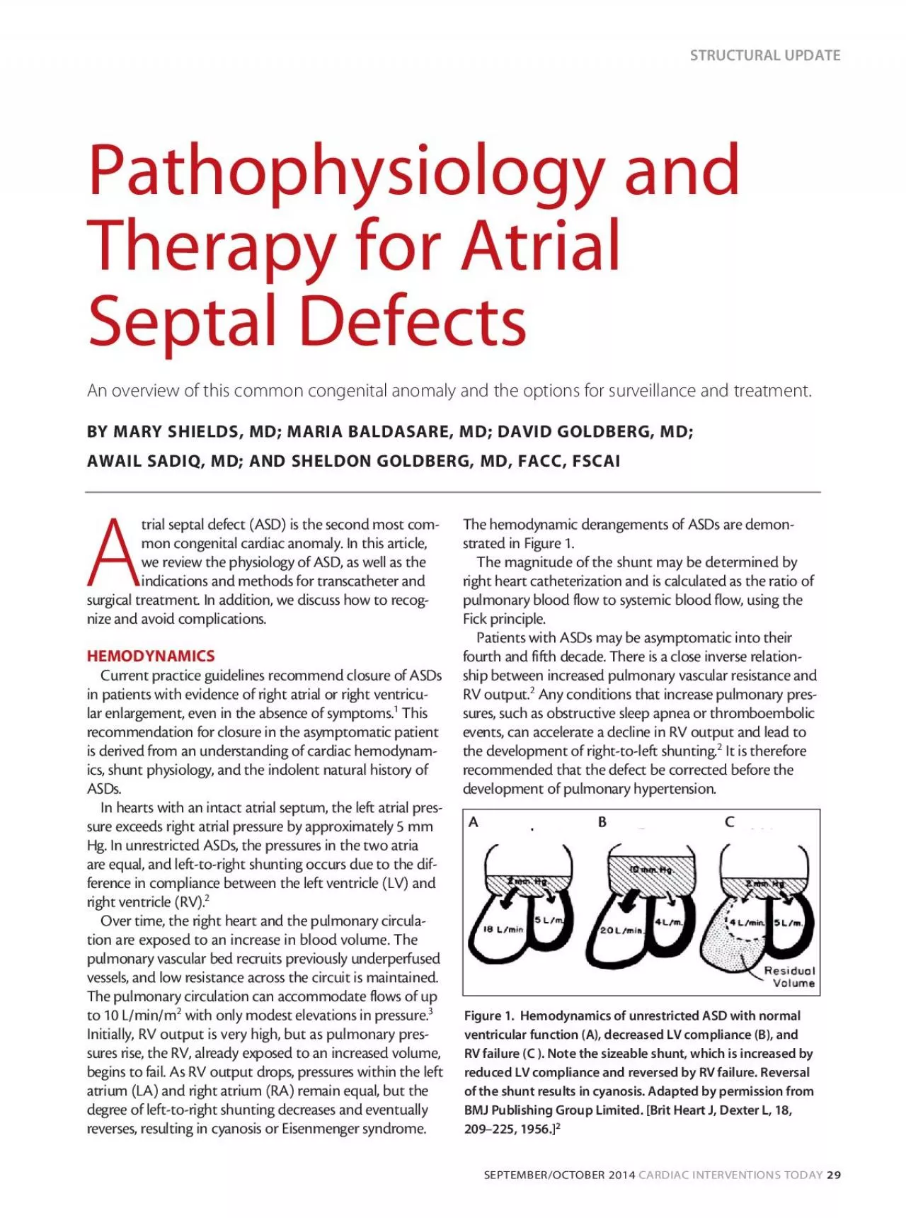
trial septal defect ASD is the second most com mon congenital cardiac anomaly In this article we review the physiology of ASD as well as the indications and m ID: 936873

atrial device septal closure device atrial closure septal asd defect patients heart pulmonary cardiac left rim defects erosion figure

Share:

Link:

Embed:

Download Presentation from below link

Download Pdf The PPT/PDF document "SEPTEMBEROCTOBER 2014" is the property of its rightful owner. Permission is granted to download and print the materials on this web site for personal, non-commercial use only, and to display it on your personal computer provided you do not modify the materials and that you retain all copyright notices contained in the materials. By downloading content from our website, you accept the terms of this agreement.


Presentation Transcript

SEPTEMBER/OCTOBER 2014 CARDIAC INTERVENTIONS TODAY 29 A trial septal defect (ASD) is the second most com - mon congenital cardiac anomaly. In this article, we review the physiology of ASD, as well as the indications and methods for transcatheter and surgical treatment. In addition, we discuss how to recog - nize and avoid complications. HEMODYNAMICS Current practice guidelines recommend closure of ASDs in patients with evidence of right atrial or right ventricu - lar enlargement, even in the absence of symptoms. 1 This recommendation for closure in the asymptomatic patient is derived from an understanding of cardiac hemodynam ics, shunt physiology, and the indolent natural history of ASDs. In hearts with an intact atrial septum, the left atrial pres - sure exceeds right atrial pressure by approximately 5 mm Hg. In unrestricted ASDs, the pressures in the two atria are equal, and left-to-right shunting occurs due to the dif - ference in compliance between the left ventricle (LV) and right ventricle (RV). 2 - tion are exposed to an increase in blood volume. The pulmonary vascular bed recruits previously underperfused vessels, and low resistance across the circuit is maintained. The pulmonary circulation can accommodate flows of up to 10 L/min/m 2 with only modest elevations in pressure. 3 Initially, RV output is very high, but as pulmonary pres sures rise, the RV, already exposed to an increased volume, begins to fail. As RV output drops, pressures within the left atrium (LA) and right atrium (RA) remain equal, but the degree of left-to-right shunting decreases and eventually reverses, resulting in cyanosis or Eisenmenger syndrome. The hemodynamic derangements of ASDs are demon - strated in Figure 1. The magnitude of the shunt may be determined by right heart catheterization and is calculated as the ratio of Fick principle. Patients with ASDs may be asymptomatic into their fourth and fifth decade. There is a close inverse relation - ship between increased pulmonary vascular resistance and RV output. 2 Any conditions that increase pulmonary pres - sures, such as obstructive sleep apnea or thromboembolic events, can accelerate a decline in RV output and lead to the development of right-to-left shunting. 2 It is therefore recommended that the defect be corrected before the development of pulmonary hypertension. Pathophysiology and Therapy for Atrial Septal Defects An overview of this common congenital anomaly and the options for surveillance and treatment. BY MARY SHIELDS, MD; MARIA BALDASARE, MD; DAVID GOLDBERG, MD; AWAIL SADIQ, MD; AND SHELDON GOLDBERG, MD, FACC, FSCAI STRUCTURAL UPDATE ventricular function (A), decreased LV compliance (B), and RV failure (C ). Note the sizeable shunt, which is increased by reduced LV compliance and reversed by RV failure. Reversal of the shunt results in cyanosis. Adapted by permission from BMJ Publishing Group Limited. [Brit Heart J, Dexter L, 18, 209–225, 1956.] 2 A B C 30 CARDIAC INTERVENTIONS TODAY SEPTEMBER/OCTOBER 2014 Electrocardiographic (ECG) findings are dependent on the type of defect. Patients with a secundum defect will have intraventricular conduction delay with incomplete right bundle branch block, RA enlargement, and normal or right axis deviation (Figure 2). This is a result of increased volume on the right side of the heart. Patients with ostium primum–type defects classically have first-degree atrio - ventricular (AV) block, incomplete right bundle branch block, and left axis deviation (Figure 3). The leftward axis is due to the proximity of the primum defect to the bundle of His, resulting in left anterior fascicular block. Finally, the ECG findings in patients with sinus venosus ASD wi

ll be similar to those of secundum defects (Figure 4). However, left axis deviation of the P wave may occur due to the proximity of the sinus venosus ASD to the sinus node, resulting in ectopic atrial pacing. 4 IMAGING Traditionally, a comprehensive preprocedure trans - esophageal echocardiogram (TEE) is used to determine the type and severity of the defect and to characterize the anatomic details of the atrial septum and associated rims. The rims are described as the superior vena cava rim (superior), aortic rim (anterosuperior), coronary sinus rim (anteroinferior), inferior vena cava rim (inferior), and pos - terior rim (posterior). 5 These rims are evaluated in three different planes. The transverse plane (0°) is the optimal view to assess the AV rim, as well as the posterior rim. Usually, the probe is moved at different levels to carefully evaluate the margins. From this position, the probe can be rotated to 30° to 40° to highlight the aortic rim. The bicaval, or 90° view, is best to evaluate the IVC and superior vena cava rim, and from this position, the aortic rim can again be assessed at 45°. Gastric views at 70° to 90° are helpful in assessing the IVC rim with the probe retroflexed. 6 Three-dimensional TEE can provide better visualization of the morphologic vari - ants of the defect and might be useful for complex cases. 7 Intracardiac echocardiography (ICE) may be used during percutaneous ASD closure. The ICE catheter is advanced through the IVC into the RA. Orthogonal views are used—mostly the transverse section of the aortic valve and the longitudinal section of the four-chamber view. The ICE images have higher resolution compared to TEE. ICE is especially useful for guiding placement of the sheath in the LA and deploying the left and right atrial discs. 5 Cardiac magnetic resonance (CMR) is a safe, nonin - vasive modality for imaging. CMR has demonstrated improved imaging for assessing anatomy, dimensions of surrounding rims, and device implantation. 8 In order to use CMR guidance, all devices and components should have no ferromagnetic components. 9 The Amplatzer septal occluder (ASO; St. Jude Medical, Inc.) meets the criteria for CMR-guided placement. 10 During ASD clo - sure, CMR can track the delivery system and device from the IVC to the RA to the LA at rates of 10 to 15 frames per second. 10 The same sequences are used to guide the device into position and confirm placement. 9,10 Multidetector CT with ECG-gated acquisition has high spatial and temporal resolution. 11 Although not consid - ered a first-line investigation, multidetector CT has both a high sensitivity (66%–100%) and specificity (86%–100%) for evaluating ASDs and quantifying the magnitude of the shunt. 11 ASD CLOSURE According to the 2008 American College of Cardiology/American Heart Association guidelines, it is Figure 2. TEE bicaval view demonstrating an ostium secundum defect (A). The ECG shows a normal axis and incomplete right bundle branch block (B). A B SEPTEMBER/OCTOBER 2014 CARDIAC INTERVENTIONS TODAY 31 reasonable to close a secundum ASD if there is evidence of RA and RV dilatation regardless of symptoms. 1 However, patients with defects measuring 5 mm without RV overload should be closely monitored. Paradoxical embolization is a class IIa indication for closure. Closure is contraindicated in patients with Eisenmenger physiol - ogy (pulmonary artery pressure more than two-thirds systemic pressure or pulmonary vascular resistance more than two-thirds systemic vascular resistance) (Table 1). Currently, percutaneous repair is considered first-line treatment for the majority of secundum ASDs. However, surgical repair may be indicated

for patients with defi - cient rims (5 mm), very large defects, multiple fenestra - tions, and mobile septae. 7 Surgery is required in patients with septum primum, sinus venosus, and coronary sinus defects. 7 Percutaneous Method In our practice, we perform closure of complex or larger defects with general anesthesia and TEE guidance. We most frequently use the ASO device. For smaller defects, an ICE catheter is placed via the left femoral vein into the RA. A second catheter (eg, multipurpose) is placed in the right femoral vein and passed over a soft guidewire (eg, Magic Torque [Boston Scientific Corporation]) into the LA and advanced to a left superior pulmonary vein. We then exchange for a stiff guidewire (eg, Amplatz Super Stiff [Boston Scientific Corporation]), over which a sizing balloon is placed across the defect and gently inflated to determine the stop-flow measurement (Figure 5). The balloon is removed, and a sheath (eg, Mullins or Hausdorf [Cook Medical]), large enough to accommodate the appropri - ate-sized device, is placed into the LA. Figure 3. TEE bicaval view of ostium primum (A) and corresponding ECG (B) demonstrating incomplete right bundle block and left axis deviation. TABLE 1. RECOMMENDATIONS FOR INTERVENTIONAL AND SURGICAL ASD SECUNDUM THERAPY* Class I Class IIa Class IIb Class III Level B RA/RV enlargement with or without symptoms Platypnea-orthodeoxia syndrome — Those with severe, irre - versible pulmonary artery hypertension without evidence of left-to-right shunting should not undergo ASD closure Level C — Paradoxical embolism Left-to-right shunting with pulmonary artery pressures - sure or responsive to pul - monary vasodilator therapy — *Adapted from Warnes CA, Williams RG, Bashore TM, et al. ACC/AHA 2008 Guidelines for the Management of Adults With Congenital Heart Disease: Executive Summary. Circulation. 2008;118:2395–2451. A B 32 CARDIAC INTERVENTIONS TODAY SEPTEMBER/OCTOBER 2014 We take special care to eliminate air from the system; after the dilator is removed, the sheath is withdrawn to the RA, and bleedback is allowed to occur. After the sheath is carefully flushed, it is gently passed to the mid- LA over the stiff guidewire already in place. Also, we take care to prepare the ASO under saline, eliminating entrapped air from the device. We then pass the ASO to the tip of the sheath and retract the sheath, exposing the LA disc. The entire assembly is withdrawn until the LA disc contacts the septum. We then expose the RA disc and use the sheath to flatten the discs (Figure 6). We check for residual shunts and proper device alignment (Figure 7) and gently perform a stability maneuver (push- pull). Only after we are satisfied with the device position and absence of residual leak and confirm stability, do we unscrew the delivery cable. It should be noted that up until the delivery cable is unscrewed, the device is com - pletely retrievable. The sheath can then be removed, and imaging is repeated. Transthoracic echocardiography is performed the next day to assess device position and exclude a pericardial effusion. Surgical Method Currently, minimally invasive surgery has replaced full thoracotomy in ASD repairs. The different surgical tech - niques include partial lower sternotomy, right antero - lateral small thoracotomy, and total endoscopic atrial septal repair. All surgical techniques use cardiopulmonary bypass and cardioplegia. The partial lower sternotomy technique involves a vertical skin incision at the lower sternal border fol - lowed by a partially inverted J-shaped sternotomy from the xiphoid to the right third intercostal space (ICS). Cardiopulmonary bypass is utilized with aortic c

ross- clamping. The ASD is then closed through a standard right atriotomy with direct or patch closure. 12,13 In contrast, the right anterolateral small thoracotomy technique involves a small anterolateral thoracotomy in the right fourth ICS. A 22- or 25-F venous cannula is placed in the femoral vein for port access; the femoral artery is also cannulated. The right lung is deflated, and the pericardium is opened 2 cm longitudinally along the phrenic nerve course. The ascending aorta is clamped through an incision in the third ICS. 12 The defect is then repaired with a standard right atriotomy. The total endoscopic atrial septal repair technique involves three incisions: one at the right anterior axillary line at the sixth ICS, the next at the right mid-axillary line at the third ICS, and the last at the parasternal line at the forth ICS. 13 These incisions are used for placement of the thoracoscopy, insertion of the aortic cross-clamping forceps and tissue forceps, and placement of the surgical instruments, respectively. Standard right atriotomy with patch closure is again utilized to repair the defect. COMPLICATIONS Death The mortality rate of an uncorrected ASD is 50% by 40 years of age and increases by 6% per year. 14,15 The periprocedural mortality rate of transcatheter closure is 0.01% and increases to 0.1% after 2 years. 16 Periprocedural and long-term complication rates are also relatively low, although not negligible, due to the high risk of mortality from these complications. The periprocedural major com - plication rate ranges from 0% to 9.4%, with pooled esti - mates of 1.6% (95% confidence interval, 1.4%–1.8%). 16 Device Embolization Device malpositioning and embolization is the most common acute complication, occurring in 1.1% of cases (range, 0.55%–1.7%). 16-19 The ASO has a lower incidence of embolization (0.5%) compared to the Helex septal occluder (Gore & Associates) (1.7%), which is most likely Figure 4. Inferior sinus venosus demonstrated on TEE bicaval view (A). The ECG shows right axis deviation with right ventricular strain and incomplete right bundle branch block (B). A B SEPTEMBER/OCTOBER 2014 CARDIAC INTERVENTIONS TODAY 33 due to the self-centering mechanism of the former. Ninety percent of embolizations occur within 24 hours; however, late embolization has been reported. 20 The most common risk factors are insufficient rims, large and eccentric defects, and device undersizing. Confirming delivery system to device lock, deployment of device parallel to the septum, and stable position with a push-pull maneuver reduces the incidence of this complication. Air Embolism Air embolism accounts for 0.7% to 0.8% of medical device–related adverse events. 21 Air in the right coronary artery may result in transient ST elevation. As previously noted in the ASD Closure section, there are several critical steps designed to eliminate the possibility of air emboliza - tion. Furthermore, prompt identification of air on fluoros - copy and administration of 100% oxygen may help lessen the consequences of air embolization. Erosion Device erosion is a rare but significant complication. Patients may present with pleuritic chest pain, dyspnea, syncope, hypotension, hemopericardium, tamponade, and/or sudden death. 22 The incidence of ASO device ero - sion has been estimated at 0.1% to 0.5%. Erosions have not been reported with the Helex septal occluder, but it should be noted that this device can only correct defects up to 18 mm in diameter. 23,24 The most common site of device erosion is at the roof of the RA or LA. A recent study reported that 50% occurred within 1 week, an addi - tional 11% up to 1 month, 26% from 1 mont

h to 1 year, and a disturbing 13% between 1 and 9 years. 25 The most common risk factors for device erosions are anterior-superior aortic rim deficiency, the use of over - sized devices, and a mobile septum. Aortic rim deficiency (5 mm) was noted among 89% of patients with device erosion. Device to unstretched ASD defect size was signifi - cantly larger in patients with device erosions, suggesting that oversized devices may contact the atrial walls with each cardiac cycle and lead to erosion and perforation. Certain sizes of the ASO device have increased stiffness, a factor which has been implicated in device erosion (eg, 36-mm device). 26 Patient selection with avoidance of aortic rim deficiency or use of an oversized ASD device (overlap of ASD device by only 2 mm) is suggested. Close follow-up of patients with pericardial effusion is mandatory. Urgent trans- thoracic echocardiography should be performed when pericardial effusion is suspected. TEE is able to visualize atrial and aortic erosion. CT angiography is an alternative to detect pericardial effusion, cardiac perforation, and device malposition. If erosion is detected, surgical device Figure 5. Fluoroscopic image of the sizing balloon with the waist in the center of the balloon. The diameter of the waist during cessation of shunting determined by TEE is measured, and a device with a central disc 2 to 4 mm larger than the stop-flow diameter is selected. Figure 6. Fluoroscopic image after exposure of both discs. The sheath has been used to flatten the RA disc against the septum. The delivery cable remains attached until optimal position is confirmed on TEE. 34 CARDIAC INTERVENTIONS TODAY SEPTEMBER/OCTOBER 2014 removal and repair (Figure 8) of the defect and the tear is recommended. 27 Device Fracture Device fracture is an uncommon complication, occurring in 6.5% of patients treated with the Helex device and in only one patient treated with the ASO device. 18,28,29 Larger devices have a higher incidence of wire frame fracture. Nonoverlapping regions of wire frame during the cardiac cycle may undergo differ - entially higher shear stress and may result in a higher incidence of fracture. Overall, the incidence of mor - bidity and mortality due to device frame fracture is minimal. Mitral valve perforation due to Helex frame fracture and traumatic LA thrombus due to ASO wire frame fracture have been reported. 29,30 Thrombosis and Thromboembolism Device thrombosis is the second most common long- term complication and is highly device-dependent. Thrombosis appears to be more frequent with the double-umbrella polyester implant but has recently been reported with the ASO device. 31 Risk factors for device thrombosis include atrial fibrillation, persistent atrial septal aneurysm, and hypercoagulable state. Initial treatment of acute device thrombosis is anticoagulation with heparin and warfarin. Aspirin and clopidogrel are recommended as prophylaxis for 6 months until endothelialization has occurred; thereafter, patients remain on aspirin. Surgery may be indicated for large left-sided mobile thrombus. 31 Arrhythmias Chronic volume overload leads to atrial dilation and remodeling, which predisposes to atrial tachyarrhyth - mias. 32 Atrial arrhythmias are common in patients with uncorrected ASDs, occurring in 10% of patients by age 40. 33 After closure, atrial tachyarrhythmias decline to 5% to 6%. 18,19,34-37 Patients younger than 40 and without a previous history of atrial arrhythmias have the lowest inci - dence of atrial arrhythmias after ASD closure. 38 Onset of new atrial fibrillation is not affected by the type or size of device used. The persistence of a residual shunt and onset of new A

V block are associated with a higher incidence of atrial fibrillation. 36 Stretching of the atrial septum and compression of Bachmann’s bundle by the device, as well as a local foreign body inflammatory reaction, are possible explanations for periprocedural atrial arrhythmias. Closure of the ASD leads to a decrease in RA stretch and reversal of chronic remod - eling and thus may explain the decrease in incidence of atrial tachyarrhythmia after ASD closure. Heart Block Conduction abnormalities, including first- and second- degree heart block, may occur after ASO placement (6.2%); however, complete heart block is relatively rare (0.4%; 95% confidence interval, 0.1–0.7). 16,33 AV conduc - tion abnormalities and complete heart block are attrib - uted to mechanical compression and ischemia of the AV node from an oversized device. 39 Most AV conduction abnormalities resolve without intervention, but complete AV block may require device removal or placement of a permanent pacemaker. 40 Residual Shunt Residual shunting after percutaneous ASD closure depends on the number, size, and location of the ASDs, type of device used, and timing of residual shunt assess - ment. The incidence of moderate to large residual shunts is 3.3% at 24 hours and decreases to 1.5% at 1 year. 19 LONG-TERM FOLLOW-UP Patients with unrepaired ASDs without signs of RV volume overload or pulmonary hypertension should be followed annually and monitored for arrhythmias, embolic events, pulmonary hypertension, and right-sided failure. A repeat echocardiogram should be obtained every 2 years to assess RV size, function, and for increase in pulmonary artery pressure. Patients who have had percutaneous ASD device clo - sure should have an echocardiogram performed at 24 hours to assess for device malposition, residual shunt, and pericardial effusion. Repeat echocardiography is Figure 7. TEE image showing flattened discs and lack of residual shunt prior to release of the delivery cable. SEPTEMBER/OCTOBER 2014 CARDIAC INTERVENTIONS TODAY 35 recommended at 3, 6, and 12 months. A routine clinical follow-up and echocardiogram should be done every 1 to 3 years thereafter. The complications with surgical closure usually occur within 1 month. In contrast, transcatheter closure patients, especially those with deficient rims, require long-term clinical and echocardiographic follow-up. Patients should be educated about the signs and symp - toms suggestive of device erosion and advised to seek urgent medical care if these events occur. Patients with percutaneous ASO repair should also be advised to avoid intense isometric exercises, as there is an isolated case report of cardiac perforation with such activity. 41 ENDOCARDITIS PROPHYLAXIS Periprocedural antibiotics are given for surgical and percutaneous ASD closure. Endocarditis prophylaxis is rec - ommended for the first 6 months after percutaneous ASD closure. Endocarditis prophylaxis is not required for an isolated ASD, after surgical ASD closure, and beyond 6 months after percutaneous ASD closure. SUMMARY AND FUTURE DIRECTIONS ASDs are the second most common cardiac congenital anomaly in adults. The diagnosis is dependent on the physi - cal examination, ECG, and TEE. Although transcatheter repair is the first-line therapy in the great majority of secun - dum ASDs, consideration for surgical repair should be given for patients with complex anatomy who may be at high risk for delayed complications, especially erosion. If transcath - eter therapy is chosen, life-long follow-up may be required. With advances in bioresorbable technology, long-term risks may be reduced. Bioresorbable septal occluders may act as temporary scaffolds for tissue

to grow and endothelialize the defect. One such device, the BioStar septal repair implant (Gore & Associates), has been tested in a clinical trial. The BioStar device consists of a double umbrella with stainless steel arms, a nitinol wire, and two discs of acellular porcine collagen. After insertion of the device, the device endothelializes and promotes regenera - tion of functional tissue with minimal scar formation. Although short-term complications are similar to current devices, the bioresorbable technology may decrease long-term complications, including late erosion. This approach allows for continued transseptal access and improved imaging with CT or mag - netic resonance imaging. Mary Shields, MD, is with the Department of Medicine, Pennsylvania Hospital in Philadelphia, Pennsylvania. She has stated that she has no financial interests related to this article. Maria Baldasare, MD, is with the Division of Cardiology, Drexel University College of Medicine, Philadelphia, Pennsylvania. She has stated that she has no financial inter - ests related to this article. David Goldberg, MD, is with the Department of Medicine, Pennsylvania Hospital in Philadelphia, Pennsylvania. He has stated that he has no financial interests related to this article. Awail Sadiq, MD, is with the Department of Medicine, Drexel University College of Medicine in Philadelphia Pennsylvania. He has stated that he has no financial inter - ests related to this article. Sheldon Goldberg, MD, FACC, FSCAI, is with the Department of Cardiology, Pennsylvania Hospital, University of Pennsylvania in Philadelphia, Pennsylvania. He has stated that he has no financial interests related to this article. Dr. Goldberg may be reached at sheldon.goldberg@uphs.upenn.edu. 1. Warnes CA, Williams RG, Bashore TM, et al. ACC/AHA 2008 guidelines for the management of adults with congenital heart disease: a report of the American College of Cardiology/American Heart Association Task Force on Practice Guidelines (Writing Committee to Develop Guidelines on the Management of Adults With Congenital Heart Disease). Developed in Collaboration With the American Society of Echocardiography, Heart Rhythm Society, International Society for Adult Congenital Heart Disease, Society for Cardiovascular Angiography and Interventions, and Society of Thoracic Surgeons. J Am Coll Cardiol. 2008;52:e143-263. 2. Dexter L. Atrial septal defect. Br Heart J. 1956;18:209-225. 3. Dexter L, Dow JW, Haynes FW, et al. Studies of the pulmonary circulation in man at rest; normal variations and the interrel - tions between increased pulmonary blood flow, elevated pulmonary arterial pressure, and high pulmonary capillary pressures. J Clin Invest. 1950;29:602-613. 4. Davia JE, Cheitlin MD, Bedynek JL. Sinus venosus atrial septal defect: analysis of fifty cases. Am Heart J. 1973;85:177-185 sc Diagn Ther. 2013;3:38-46. t of patent foramen ovale. Eur J Echocardiogr. 2010;11:i27-34. fect Figure 8. Example of late erosion occurring 6 years after device placement. The patient had a deficient aortic rim, mobile septum, and large defect. The right atrial disc (A) of the ASO device and atrial roof erosion (B). closure complications. Circ Cardiovasc Imaging. 2009;2:141-149. ardiac magnetic resonance imaging. Pediatr Cardiol. 2004;25:124-130. 9. Henk CB, Higgins CB, Saeed M. Endovascular interventional MRI. J Magn Reson Imaging. 2005;22:451-460. 10. Rickers C, Jerosch-Herold M, Hu X, et al. Magnetic resonance image-guided transcatheter closure of atrial septal defects. Circulation. 2003;107:132-138. 11. Hoey ET, Gopalan D, Ganesh V, et al. Atrial septal defects: magnetic resonance and computed tomography appearances. J Med Imaging Radiat Oncol. 20

09;53:261-270. 0-year single-institution experience. J Thorac Cardiovasc Surg. 2007;134:757-764. horac Surg. 1999;67:1648-1652. 14. Campbell M. Natural history of atrial septal defect. Br Heart J. 1970;32:820-826. 15. Adams CW. A reappraisal of life expectancy with atrial shunts of the secundum type. Dis Chest. 1965;48:357-375. 16. Abaci A, Unlu S, Alsancak Y, et al. Short and long term complications of device closure of atrial septal defect and patent foramen ovale: meta-analysis of 28,142 patients from 203 studies. Catheter Cardiovasc Interv. 2013;82:1123-1138. 17. Levi DS, Moore JW. Embolization and retrieval of the Amplatzer septal occluder. Catheter Cardiovasc Interv. 2004;61:543- 547. neous closure of secundum atrial septal defects. J Am Coll Cardiol. 2007;49:2215-2221. 19. Du ZD, Hijazi ZM, Kleinman CS, et al; for the Amplatzer Investigators. Comparison between transcatheter and surgical l. 2002;39:1836-1844. 20. Sahin DY, Koç M, Cakr H, et al. A silent and late embolization of atrial septal defect occluder device into the right pulmonary artery: a case report. Korean Circ J. 2012;42:781-783. 21. US Food and Drug Administration. FDA Executive Summary Memorandum: Transcatheter ASD Occluders: Clinical update and review of events. May 24, 2012. Available at http://www.fda.gov/downloads/AdvisoryCommittees/CommitteesMeet - ingMaterials/MedicalDevices/MedicalDevicesAdvisoryCommittee/CirculatorySystemDevicesPanel/UCM304924.pdf. Accessed May 1, 2014. zer septal occluder. J Am Coll Cardiol. 2005;45:1213-1218. ects: review of registry of complications and recommendations to minimize future risk. Catheter Cardiovasc Interv. 2004;63:496-502. he literature. Catheter Cardiovasc Interv. 2012;80:157-167. 25. Taggart NW, Dearani JA, Hagler DJ. Late erosion of an Amplatzer septal occluder device 6 years after placement. J Thorac Cardiovasc Surg. 2011;142:221-222. 26. Amin Z. Echocardiographic predictors of cardiac erosion after Amplatzer septal occluder placement. Catheter Cardiovasc Interv. 2014;83:84-92. 27. Sarris GE, Kirvassilis G, Zavaropoulos P, et al. Surgery for complications of trans-catheter closure of atrial septal defe institutional study from the European Congenital Heart Surgeons Association. Eur J Cardiothorac Surg. 2010;37:1285-1290. . Catheter Cardiovasc Interv. 2009;73:941-948. h atrial septal defect amplatzer occluder. J Card Surg. 2011;26:41-43. 30. Qureshi AM, Mumtaz MA, Latson LA. Partial prolapse of a Helex device associated with early frame fracture and mitral valve perforation. Catheter Cardiovasc Interv. 2009;74:777-782. diatr Cardiol. 2013;34:1269-1271. with an atrial septal defect. Circulation. 2003;107:1775-1782. eptal defect closure in adults. Int J Cardiol. 2009;134:47-51. . Heart Lung Circ. 2012;21:572-575. hmia in the short to medium term: a systematic review and meta-analysis. Heart. 2010;96:1789-1797. 36. Johnson JN, Marquardt ML, Ackerman MJ, et al. Electrocardiographic changes and arrhythmias following percutaneous atrial septal defect and patent foramen ovale device closure. Catheter Cardiovasc Interv. 2011;78:254-261. 37. Spies C, Khandelwal A, Timmermanns I, Schräder R. Incidence of atrial fibrillation following transcatheter closure of atrial septal defects in adults. Am J Cardiol. 2008;102:902-906. ptal defects in adults. Am J Cardiol. 2008;101:683-687. using the Amplatzer device. J Am Coll Cardiol. 2004;43:1677-1682. 40. Lin SM, Hwang HK, Chen MR. Amplatzer septal occluder-induced transient complete atrioventricular block. J Formos Med Assoc. 2007;106:1052-1056. tzer atrial septal defect closure: should isometric activity be limited in septal occluder holders? Ann Thorac Surg. 2012;93:671. STRUCTURAL UPDATE STRUCTURAL UPDA