/
Cranial Nerves Spinal Cord and Reflexes Cranial Nerves Spinal Cord and Reflexes

Cranial Nerves Spinal Cord and Reflexes - PDF document

blanko
blanko . @blanko
Follow
344 views
Uploaded On 2022-08-25

Cranial Nerves Spinal Cord and Reflexes - PPT Presentation

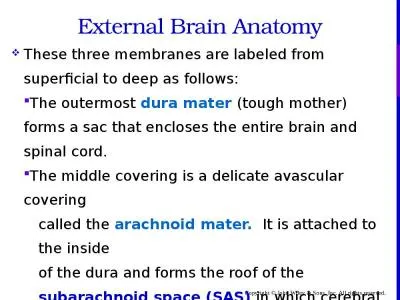
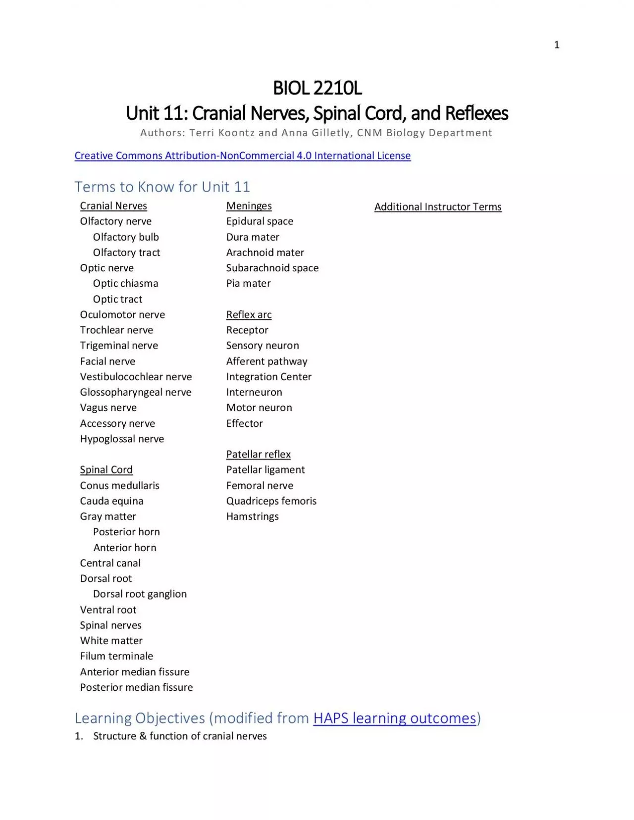
1 BIOL 2210L Unit 11 Authors Terri Koontz and Anna Gilletly CNM Biology Department Creative Commons Attribution NonCommercial 40 International License Terms to Know for Unit 1 1 Cranial Ner ID: 941660

spinal nerves cord cranial nerves spinal cranial cord nerve sensory motor reflex neurons function optic foramen structures patellar olfactory

Share:

Link:

Embed:

Download Presentation from below link

Download Pdf The PPT/PDF document "Cranial Nerves Spinal Cord and Reflexes" is the property of its rightful owner. Permission is granted to download and print the materials on this web site for personal, non-commercial use only, and to display it on your personal computer provided you do not modify the materials and that you retain all copyright notices contained in the materials. By downloading content from our website, you accept the terms of this agreement.


Presentation Transcript

1 BIOL 2210L Unit 11 : Cranial Nerves, Spinal Cord, and Reflexes Authors: Terri Koontz and Anna Gilletly, CNM Biology Department Creative Commons Attribution - NonCommercial 4.0 International License Terms to Know for Unit 1 1 Cranial Nerves Meninges Additional I nstructor T erms Olfactory nerve Epidural space Olfactory bulb Dura mater Olfactory tract Arachnoid mater Optic nerve Subarachnoid space Optic chiasma Pia mater Optic tract Oculomotor nerve Reflex arc Trochlear nerve Receptor Trigeminal nerve Sensory neuron Facial nerve Afferent pathway Vestibulocochlear nerve Integration Center Glossopharyngeal nerve Interneuron Vagus nerve Motor neuron Accessory nerve Effector Hypoglossal nerve Patellar reflex Spinal Cord Patellar ligament Conus medullaris Femoral nerve Cauda equina Quadriceps femoris Gray matter Hamstrings Posterior horn Anterior horn Central canal Dorsal root Dorsal root ganglion Ventral root Spinal nerves White matter Filum terminale Anterior median fissure Posterior median fissure Learning Objectives (modified from HAPS learning outcomes ) 1. Structure & function of cranial nerves 2 a. List and identify the cranial nerves by name and number. b. Describe the specific functions of each of the cranial nerves and classify each as sensory, motor or mixed. c. Identify the foramina that the cranial nerves pass through within the skull. 2. Anatomy of the sp inal cord a. Describe the gross anatomy of the spinal cord. b. Identify the anatomical features seen in a cross sectiona

l view of the spinal cord c. Identify the dorsal root ganglia, dorsal and ventral roots, and spinal nerves. 3. Reflexes & their roles in nervous system function a. Define the term reflex. b. Describe reflex responses in terms of the major structural and functional components of a reflex arc. c. Demonstrate a stretch reflex (e.g., patellar) d. Propose how specific reflexes would be used in clinical assessment of nervous system function. Explanation of Anatomy In this lab we begin with learn ing the twelve paired cranial nerves, where they are located and their functions. While learning the cranial nerves we’ll revisit the skull and identify wh ere eac h of the cranial nerves pass through the skull . Next, we’ll explore the major components of the spinal cord and describe what happens during a reflex arc. We’ll finish with applying knowledge of a reflex arc to what happens during the patellar reflex. Thin k about how nerve and spinal cord damage can cause dysfunction. One paired cranial nerve, the trigeminal nerve, causes severe pain when constant pressure is applied to it, but if removed on one side will cause that side of the face to feel numb. Damage to the spinal cord can cause paralysis of limbs. Think about how clinical application of our understanding of cranial nerves, the spinal cord, and reflexes, help determine the severity and location of damaged areas within our nervous system. With this informa tion , and as we continue to learn more about how our bod ies function, practitioners can provide better treatment. Cranial Nerves There are twelve paired cranial nerves (see Image 1) . Nine of the paired cranial nerves branch from the brainstem , two paired crania

l nerves branch from the forebrain (optic and olfactory), and one cranial nerve ( accessory ) emerges from the spinal cord but travels up to the brainstem and passes through the skull with the other cranial nerves . Unlike spi nal nerves, not all cranial nerves have both sensory and motor neurons within their structure. Some cranial nerves only have a sensory function like the optic nerves that are involved with vision where as other cranial nerves only have a motor function like the oculomotor nerves that are involved with eye movements. Two paired cranial nerves, accessory nerves and hypoglossal nerves , have some sensory function but are mainly motor. The accessory nerves control motor function for the sternocleidomastoid and tr apezius muscles and the hypoglossal nerves control tongue movement. There are four paired cranial nerves that are mixed nerves with a complex mix of sensory and motor function s : trigeminal, facial, glossopharyngeal and vagus nerve s . The trigeminal nerves ’ sensory function involves detecting skin sensations and its motor function involves activating muscles for mastication 3 Image 1: Cranial nerves Creative Commons Attribution 4.0 International Openstax URL: Cranial nerves ( chewing ) . The facial nerve s ’ sensory function allows us to taste food and the ir motor function allows us to make fac ial expressions . Glossopharyngeal nerves ’ sensory function m onitors carbon dioxide and oxygen blood levels to help regulate breathing and the motor function helps with swallowing. The last of the mixed nerves is the vagus nerve that monitor s and control s visce ra within the ventral body cavity using both sensory and motor neurons. Heart rate, breathi

ng, and digestive system processes are regulated by the vagus nerves. The olfactory and optic nerves have additional structures that you’ll learn in this lab. The o lfactory bulb is where the olfactory nerve neurons synapse with olfactory tract neurons . The olfactory bulb, along with the olfactory tract, are brain structures. The axons of the olfactory nerve are structures of the PNS. The olfactory tract is a bundle o f axons that originate within the olfactory bulb. Like the olfactory nerve and its “bulb”, the optic nerve travels from outside the cranial cavity and will create a notable structure, the optic chiasma. Axons of the optic nerve cross over forming an “X” like structure , which is the optic chiasma . After the optic chiasma , the name of the bundled axons will change to optic tracts as those fibers move toward the thalamus of the brain. Since the optic nerve and its structures are brain outgrowths, they ar e visible PNS structures within the cranial cavity . Listed in the following table are the twelve paired cranial nerves in the order that they branch from the forebrain, brain stem, and spinal cord. The pai red cranial nerves are listed using Roman numerals and by name. For example, the optic nerve is also called the second cranial nerve and can also be called “cranial nerve II.” In addition, next to each paired cranial nerve are their function s followed by where they pass through the skull. 4 Table 1: Cranial nerves, their functions, and skull features that they pass through Nerve # Name Neuron type Function Skull feature I Olfactory Sensory Sense of smell cribriform plate II Optic Sensory Sense of sight optic canal

III Oculomotor Motor Movement of most voluntary eye muscles and involuntary muscles of the iris that control the size of the pupil superior orbital fissure IV Trochlear Motor Controls a single extrinsic muscle of the eye that allows for eyeball movement superior orbital fissure V Trigeminal Sensory and Motor Touch perception for skin of the face and movement for muscles involved with mastication superior orbital fissure, foramen rotundum, and foramen ovale VI Abducens Motor Controls a single extrinsic muscle of the eye that allows for eyeball movement superior orbital fissure VII Facial Sensory and Motor Senses taste from taste buds, movement of muscles involved in facial expressions, and controls secretions of lacrimal and salivary glands internal acoustic meatus VIII Vestibulocochlear Sensory Sense of balance, equilibrium, and hearing internal acoustic meatus IX Glossopharyngeal Sensory and Motor Involved with chemoreceptors and pressure receptors of carotid artery and movement for pharyngeal muscles used in swallowing jugular foramen X Vagus Sensory and Motor both sensing and movement for visceral organs within thoracic and abdominopelvic body cavities jugular foramen XI Accessory Mainly motor Movement of sternocleidomastoid and trapezius muscles jugular foramen XII Hypoglossal Mainly motor Movement of tongue muscle hypoglossal canal 5 Mini Activity: Functions of some cranial nerves Match the cranial nerve with its function. 1. Trigeminal (V) A. Facial expressions and taste 2. Facial (VII) B. Tongue movement 3. Hypoglossal (X II) C. Blood gas regulation and swallowing 4. Glossopharynge

al (IX) D. Skin sensation and mastication 5. Vagus (X) E. Sternocleidomastoid movement 6. Olfactory (I) F. Vision 7. Optic (II) G. Moves the eyeball 8. Vestibulocochlear (VIII) H. Smelling 9. Oculomotor (III) I. Hearing 10. Accessory (XI) J. Controls visceral organs There are two cranial nerves that are not listed above, but are involved with “moving the eyeball”. What are the names of those two cranial nerves? a. b. You can come up with your own mnemonic to learn the order of the cranial nerves or you can use the one below: “ O, O, O, To, Touch, And, Feel, Very, Good, Velvet, A, H ” O lfactory, O ptic, O culomotor, T rochlear, T rigeminal, A bducen s, F acial, V estibulocochlear, G lossopharyngeal, V agus, A ccessory, H ypoglossal Spinal Cord The spinal cord travels through the vertebral foramina that make up the vertebral body cavity. The inferior portion of the spinal cord tapers forming a structure called the conus medullaris . An anchoring structure called the filum terminale (extension of t he pia mater) connects the conus medullaris to the coccyx, allowing the spinal cord to maintain its position during body movements. Also, the inferior end of the spinal cord has many spinal roots emerging that look like a horse’s tail and as a group are named the cauda equina . Cauda means tail and equina means horse. The reason for these long roots at the end of the spinal cord is because the vertebral column develops faster than the spinal cord, making it so th at spinal nerves chase from where the y e xit the dorsa l body cavity a s the body grows from a fetus to a young child . Because of th

is “chasing” and stretching, the cauda equina is the one place where spinal nerves are found within the vertebral body cavity; on the more superior portions of the spinal cord , the name doesn’t change to spinal nerve until the sensory and motor roots bundle together their axons as they emerge from the intervertebral foramen. Image 2 depicts inferior structures of the spinal cord. 6 Image 2: Spinal cord - 1. Conus medullaris, 2. Filum terminale, 3. Cauda equina Modified CC BY 2.5 by John A Beal, PhD URL: Spinal cord Image 3: Cross section of spinal cord Cropped Creative Commons Attribution 4.0 International Openstax U RL: Cross section of spinal cord A cross section of the spinal cord shows its functional areas (see Image 3) . The center of the cross section is the central canal that contains cerebrospinal fluid. Surrounding the central canal is gray matter in the shape of a butterfly. Gray matter in the CNS is made up of neuron al cell bodies and nonmyelinated neurons. The “wings” of the gray matter are called horns and it is within those horns that neurons communicate to one another. The posterior horns of the spinal cord allow for sensory neurons to send information to interneurons. The anterior horns of the spinal cord ’s gray matter allow for information to travel to motor neurons. Pay attention to those locations. Interneurons are commonly found in the space between the two horns. The white matter surrounding the gray matter are myelinated fiber tracts (see definition of tract above); the whiter color is due to the myelin sheath. Folds of the white matter are named based on their location and their depth. A deep fold is called a fissure an

d a shallow fold is called a sulcus. The deep anterior fold in the middle is called the anterior median fissure where as the shallow posterior fold in the middle is called the posterior median sulcus . As information is being sent to and away from the spinal cord, axons of neurons transition from being located within the PNS to t he CNS or within the CNS to the PNS. When the axons are within the PNS, they are called roots or nerves . Incoming information along the axons of sensory neurons travels 7 Image 4: Drawing of spinal cord CC BY 2.0 by Prof Alice Roberts URL: Drawing of spinal cord through the dorsal root where sensory neuronal axon terminals communicate with dendrite s and cell bodies of interneurons with in the posterior horn . The cell bodies of sensory neurons are in the dorsal root ganglion. The dorsal root ganglion is a noticeable bulge of PNS nervous tissue that can help orient what are posterior versus anterior sp inal cord structures. As information is being sent to motor neurons that originate in the anterior horn of the spinal cord, the axons of these motor neurons enter the PNS via a ventral root . The dorsal and ventral roots merge at the intervertebral foramen to form a spinal nerve that contains both sensory and motor neuronal axons. The ventral and dorsal roots are located inside the vertebral foramen and once they pass through the intervertebral foramina (mixing sensory and motor axons), the name of the roots changes to spinal nerves. To be clear , except for the cauda equina, no “nerves” are found inside the vertebral cavity. Mini Activity: Label r oots and s pinal n erves 1. In the drawing below, label the following: A . v entral r oot, B . d orsal r oot, C . s pinal n

erve 2. Now, look at a skeleton in the classroom and if available a vertebra. Find the following openings. Intervertebral foramen Vertebral foramen 3. What is the significance of those two structures , the intervertebral foramen and the vertebral foramen, regarding the spinal cord and its nerves? Meninges Meninges protect the CNS , both the brain and the spinal cord . There are three layers of meninges. Going from su perficial to deep , the meninges are the dura mater, arachnoid mater, and pia mater (see Image 5) . Dura - means tough and mater means mother, making the dura mater the toughest of all the meningeal layers. Arachnoid means spiderlike and th e arachnoid mater i s named because it contains 8 Image 5: Meninges of spinal cord public domain by Mysid who modified a Henry Vandyke Carter drawing URL: Meninge s of spinal cord Image 6: Epidural anesthesia within epidural space Creative Commons Attribution - Share Alike 4.0 Interna tional by BruceBlaus URL: Epidural anesthesia within epidural space connective tissue fibers that look like a spider web . These web - like fibers connect the arachnoid mater to the pia mater. The pia mater directly attaches to and cradles the nervous tissue of the CNS . The word pia means delicate or gentle; p ia mater is the gentle mother. Both the brain and spinal cord are surrounded by all three meninges. We will revisit the meninges again when we study brain anatomy. There are two spaces among the meninges that you need to know. The subarachnoid space is the space between the pia and arachnoid mater s where cerebrospinal fluid circulates around the outside of the brain and spinal cord (see Ima

ge 5) . The epidural space is superficial to the dura mater between the bone and dura mater membrane (see Image 6) . Adipose tissue is packed w ithin the epidural space to absorb shock from blunt trauma to the head and torso . The epidural space also contains blood vessels, nerves, and lymphatic vessels. As a result, it is a common location for bleeding and swelling , especially i n the head. It is also a convenient location to deliver anesthetic for some types of surgical procedures. This is commonly referred to as an “epidural.” Anesthetic is injected into that space because the medication can gently diffuse and numb sensory and/or motor nerve fibers without circulating through the cerebrospinal fluid. Epidural steroid injections are another type of treatment used to treat back pain. 9 Image 7: Reflex arc CC BY 3.0 by Ruth Lawson Otago Polytechnic URL: Reflex arc Reflex Arc You already know the basic components of a reflex arc. It is the same components that make up the control system used in maintain ing homeostasis. There are five components to a reflex arc. 1. Receptor – Detects a stimulus 2. Afferent pathway – Sensory neurons send information to the control center via a nerve 3. Control center – The integration center is the a rea in the CNS where interneurons receive information and sen d i nformation to motor neurons . 4. Efferent pathway – Motor neurons send information from the control ce nter to an effector via a nerve 5. Effector – Muscles or glands that receive information to provide a response (contraction or secretion) Mini Activity: “ Label ” c omponents of a r eflex a rc In Image 7

, some components of the r eflex a rc aren’t identified. Circle or highlight the anatomic structure that correctly corresponds t o the missing components . It is recommended that you use different colored pencils or pens to highlight the missing components: • Blue - Afferent Pathway, • Green - Control Center, • Pink - Efferent Pathway, and • Red - Effector. F or example, h ighlight or circle the sensory neuron in blue to indicate that it is part of the Afferent Pathway . 10 Image 8: Patellar Reflex Creative Commons Attribution - Share Alike 4.0 Interna tional by Amiya Sarkar URL: Patellar Reflex Patellar Reflex The patellar reflex is an example of a reflex arc that has its integration center within the spinal cord (see Image 8) . To initiate the patellar reflex the patellar ligament (patellar tendon) is tapped when a person is sitting dangling their legs from a table or chair. When the ligament is tapped it stretches , causing the quadriceps femor is muscles to lengthen or be stretched as well . This stretch of the quadriceps femoris muscle s are detected by special sensory receptors called muscle spindles. Muscle spindles a re proprio ceptors (a type of mechanoreceptor) within whole muscles that detect when a muscle is stretched , lengthened . These muscle spindles are associated with dendrites of sensory neurons that will send the message to the spinal cord. Motor neurons direc tly receive the message within the anterior horn of the spinal cord at the lumbar region. These motor neurons continue the reflex arc by sending information along their efferent axons within the femoral nerve to excite quadricep s femor al muscles to contract , causing knee extension

. In addition, sensory neurons are also communicating to interneurons in the posterior horn . The posterior horn interneurons inhibit different motor neurons that cause relax ation of three muscles commonly known as the hamstr ing muscles . This prevents the antagonizing movement of knee flexion during the patellar reflex. Click Here to watch a short demonstration of the patellar reflex . 11 Mini Activity: Q uadriceps f emoris and h amstring m uscles 1. Below list the four muscles that make up the Quadriceps Femoris. a. b. c. d. 2. Now list the three muscles that make up the Hamstrings. a. b. c. Activity 1: Identifying and Labeling Cranial Nerves Part 1 - Using the models set up in the lab , i dentify the following cranial nerves and structures : a. optic nerve b. oculomotor c. trochlear d. trigeminal e. abducens f. facial g. vestibulocochlear h. glossopharyngeal i. vagus j. accessory k. hypoglossal l. olfactory bulb m. olfactory tract n. optic chiasma o. optic tract It is recommended that you use painter’s tape provided in the lab to label the above nerves and structures on the model. If you use the provided letters , you’ll have a key to test your knowledge after labeling. U se the space below to ske tch out the cranial nerves and their structures. 12 Part 2 - Test your knowledge on cranial nerve function. 1. What cranial nerves are strictly sensory and what is the sensory function of each of those nerves ? 2. What cranial nerves are mixed nerve s and what is the sensory and motor functions of each of those nerves ? 3. List cranial nerves that move the skeletal muscle of the eyeball.

4. List the cranial nerve that controls the smooth muscle of the eye’s iris? Activity 2: Labeling Skull Features Where Cranial Nerves Pass Using the provided pipe cleaners in the classroom , l abel the following skull bone features . a. optic canal b. superior orbital fissure c. foramen rotundum d. foramen ovale e. jugular foramen f. hypoglossal canal M atch the foramen with the cranial nerves that travel through them. Next to the lettered bone feature , write the number given to the cranial nerves listed to the right. a. optic canal__________________________ 1. O ptic (II) b. superior orbital fissure_ ________________ 2. O culomotor (I) c. foramen rotundum____________________ 3. T rochlear (IV) d. foramen ovale________________________ 4. T rigeminal (V) e. jugular foramen_______________________ 5. A bducens (VI) f. hypoglossal canal______________________ 6. Facial (VII) 7. G lossopharyngeal ( IX) 8. V agus (X) 9. A ccessory (XI) 10. H ypoglossal (XII) Two cranial nerves and the ir associated skull structures are missing from the lists above. What are th ose cranial nerves and their associated skull struc tures? a. Name of c ranial nerves: b. The structures associated with those two cranial nerves: 13 Activity 3: Identifying Spinal Cord Structures Part 1 - Using the models set up in the lab, identify the following spinal cord structures. Use painter’s tape provided in the lab and label the listed structures below on the models. a. conus medullari s b. cauda equina c. posterior horn d. anterior horn e. dorsal root ganglion f. epidural space

g. subarachnoid space Part 2 - Test your knowledge on spinal cord structures by matching a description to its corresponding structure. a. conus medullaris ____ 1. Gray matter that contains cell bodies of motor neurons b. cauda equina ____ 2. Gray matter that contains cell bodies of interneurons c. posterior horn ____ 3. PNS structure that contains cell bodies of sensory neurons d. anterior ho rn ____ 4. Area where cerebrospinal fluid is located e. dorsal root ganglion ____ 5. Area where fatty tissue is located f. epidural space ____ 6. End of the spinal cord g. subarachnoid space ____ 7. Spinal nerves that are within the vertebral cavity Part 3 – Pick at least three structures above and describe their location and how they function within the nervous system. For example, if the filum terminale was listed above you could describe its function as stabilizing the spinal cord during movem ent, but you would describe it s location as attaching the spinal cord to the coccyx bone. 14 Activity 4: Testing Reflexes Use the medical hammer that is available in the lab and test a lab partner’s patellar reflex. 1. One partner sits w ith their feet dangling. 2. Once your partner is relaxed, gently tap on their leg where the patellar ligament is located. 3. Write below what you observe. 4. Why do doctor’s test the patellar reflex? 5. The patellar reflex is a stretch reflex. Now pick either a flexor (withdrawal) reflex or a cross - extensor reflex and complete a web search to learn more about it . From your web search explore the following : a. Outline, draw, or describe the components of the reflex. b. Why would a doctor test that refle