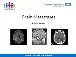
/
Brain Metastases Dr Saiqa Spensley Brain Metastases Dr Saiqa Spensley

Brain Metastases Dr Saiqa Spensley - PowerPoint Presentation

briana-ranney
briana-ranney . @briana-ranney
Follow
356 views
Uploaded On 2020-01-06

Brain Metastases Dr Saiqa Spensley - PPT Presentation

Brain Metastases Dr Saiqa Spensley Incidence of Brain Metastases 2040 Jain et al 2014 Most common primary tumours lung breast renal and melanoma CUP accounts for 1015 of patients with brain metastases ID: 772074

wbrt brain gpa months brain wbrt months gpa srs survival prognostic patients treatment neuro metastases kps mets qol disease

Share:

Link:

Embed:

Download Presentation from below link

Download Presentation The PPT/PDF document "Brain Metastases Dr Saiqa Spensley" is the property of its rightful owner. Permission is granted to download and print the materials on this web site for personal, non-commercial use only, and to display it on your personal computer provided you do not modify the materials and that you retain all copyright notices contained in the materials. By downloading content from our website, you accept the terms of this agreement.


Presentation Transcript

Brain Metastases Dr Saiqa Spensley

Incidence of Brain Metastases 20-40% (Jain et al 2014) Most common primary tumours- lung, breast, renal and melanomaCUP accounts for 10-15% of patients with brain metastasesSwedish observational study ( Smedby et al 2009):No. pts admitted to hospital because of brain mets doubled over a 20yr period Improvements in SACT Improvements in imaging techniques

Driving Legal obligation to inform DVLA Failure to inform is a criminal offenceCan be fined £1000Can have QOL implications for patients Always document you have informed patient and put in correspondence to GP

Treatment options Whole Brain Radiotherapy Surgical ResectionStereotactic RadiotherapyBest Supportive Care

WBRT

SRS

WBRT Median survival poor (2-8-4.5mths) Priestman et al 199612gy/2# vs 30gy/10# Headache control better in longer fractionationSignificant adverse effectsAlopecia, fatigue, nausea, vomiting, pain flare, constipation, reduced social functioning Effort of attending for treatment SRS Median Survival up to 8 mths Precise Single fraction Higher doses to sharply defined targets Surrounding tissue spared Outcomes comparable to neuro-surgery Rare AEs Treatment can be repeated Maximum tumour volume 20cc “Cost effective and safe but inequitable and variable access is a limiting factor” (NHS England 2015)

What do the RCTs tell us? SRS + WBRT or observation vs neurosurgery + observation or WBRT N= 95-359 (Patchell et al 1998, Sahgal et al 2015, Kocher et al 2011)No survival advantage with WBRT Sahgal’s data- + survival advantage for pts<50 treated with SRS alone Andrews et al (2004): WBRT + SRS boost vs WBRT alone for single brain met- survival advantage with SRS boost

What about QoL? Chang et al (2009)N=58 1-3 brain metsRCT: SRS vs SRS + WBRT QoL - primary outcome measure Neuro-cognition assessed at 4 months post treatment Considerable decline in learning and memory sustained at 6 months- WBRT arm Trial stopped Sofietti et al (2013) Data from the Kocher study Validated QoL questionnaire + brain cancer module used Assessed at baseline, 8 weeks and every 3 months Improved QoL scores: global health status + improved physical and cognitive functioning and less fatigue in SRS and neuro-surgery groups

Prognostic Index Models -recognition of need to tailor therapy to appropriate subgroups Recursive partitioning analysis (RPA) Validated tool but limitations GPA (Graded Prognostic Assessment) Multi-institutional 3940 patients Analysis of prognostic factors Site specific- Diagnosis specific GPA score (4.0 best and 0.0 worst prognosis) Accurate tool to estimate survival and select appropriate treatment If GPA 0-1.0 survival is 3 months irrespective of diagnosis and conservative management recommended ( Sperduto et al 2012)

GPA Overall Median survival 7.16 monthsHowever varied from 2.79 to 25.30 months dependent on diagnosis and GPANSCLC 7 months (3-14) SCLC 4.9 months (2.79-17.05)Breast 13.8 months (3.35-25.3)GI cancers 5.36 months (3.1-13.5)

Lung Cancer Prognostic Factor 0 0.5 1.0 Age >60 50-60 <50 KPS <70 70-80 >80 ECM Present - Absent No. BM >3 2-3 1 GPA Score 0-1 1.5-2.0 2.5-3.0 3.5-4.0 MS 3.0 5.5 9.4 14.8

Breast Cancer Prognostic Factor 0 0.5 1.0 1.5 2.0 Age >60 <60 KPS <50 60 70-80 90-100 - Subtype Triple negative - ER+, Her2- ER-, Her2+ ER+, Her2+ GPA Score 0-1 1.5-2.0 2.5-3.0 3.5-4.0 MS 3.4 7.7 15.1 25.3

Renal Cell Cancer GPA Score 0-1.0 1.5-2.0 2.5-3.0 3.5-4.0 MS 3.3 7.3 11.3 14.8 Prognostic Factor 0 1.0 2.0 KPS <70 70-80 90-100 No. BM >3 2-3 1

GI Cancers Prognostic Factor 0 1 2 3 4 KPS <70 70 80 90 100 MS 3.1 3.1 4.4 6.9 13.5

CUP Numbers too small to get big dataset for prognostic factors?extrapolate from lung data set In fit patient with limited disease refer to neuro-onc MDT In patients with poor PS or extensive extracranial disease, BSC may be more appropriate

CUP Limited dataRetrospective study (Bartelt et al 2003 Germany) 916 patients with brain mets treated with WBRT (total dose up to 50Gy)CUP n=47 16 had solitary met and31 multiple brain metsRPA class applied 0 in Class I, 23 Class II, 24 Class IIIMedian OS for CUP 4.8 months MS for solitary met 7.3 mths vs 3.9 mths for multiple MS for KPS>70 7.3 months vs 3.2 mths for KPS <70

NCCN Guidelines 1-3 metsDisseminated disease with poor systemic options – consider WBRT or BSC Limited systemic disease with available systemic options - consider resection or SRS4 or more mets Consider WBRT or SRS

NICE Guidelines Multiple metastases including brain involvement 1.4.2.1 Refer patients presenting with apparent brain metastases as the only sign of malignant disease after initial and special investigations to a neuro-oncology MDT for evaluation and treatment. 1.4.2.2 Do not offer chemotherapy to patients with brain metastases of unknown primary origin except as part of a controlled clinical trial. 1.4.2.3 Inform patients with brain metastases of unknown primary origin and their carers that there is no evidence that any treatment offers improved survival and there is limited evidence of improvement in neurological symptoms with surgery and/or whole brain radiotherapy

Management Symptoms suggestive of brain mets (headaches/seizures/confusion/nausea and vomiting)Contrast CT BrainRefer to Acute Oncology Bleep 3606 Dexamethasone 8mg bd with PPI cover If seizures prescribe antiepileptic If brain mets confirmed calculate GPA and chase inform Acute Oncology Advise NOT TO DRIVE

Management If GPA 0-1.0 probable best supportive care Get Oncology reviewConsider discharge planning with CHCFTReferral to palliative care teamTitrate dexamethasone

Management If GPA >1.0 refer to Bristol Neuro Oncology MDT (referral by 12 on Monday for discussion Wednesday) MRI Brain with contrast and staging CT CAPTitration of dexamethasoneEnsure patient has key worker for communication of MDT outcome

Any Questions?