/
DOG AND CAT SPAY/ DOG AND CAT SPAY/

DOG AND CAT SPAY/ - PDF document

briana-ranney
briana-ranney . @briana-ranney
Follow
444 views
Uploaded On 2016-07-01

DOG AND CAT SPAY/ - PPT Presentation

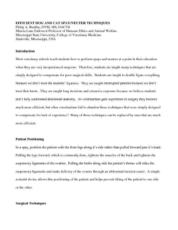
EFFICIENT NEUTER TECHNIQUES Philip A Bushby DVM MS DACVS Marcia Lane Endowed Professor of Humane Ethics and Animal Welfare Mississippi State University College of Veterinary Medicine Starkvil ID: 385074

Share:

Link:

Embed:

Download Presentation from below link

Download Pdf The PPT/PDF document "DOG AND CAT SPAY/" is the property of its rightful owner. Permission is granted to download and print the materials on this web site for personal, non-commercial use only, and to display it on your personal computer provided you do not modify the materials and that you retain all copyright notices contained in the materials. By downloading content from our website, you accept the terms of this agreement.


Presentation Transcript

EFFICIENT DOG AND CAT SPAY/ NEUTER TECHNIQUES Philip A. Bushby, DVM, MS, DACVS Marcia Lane Endowed Professor of Humane Ethics and Animal Welfare Mississippi State University , College of Veterinary Medicine Starkville, Mississippi, USA Introduction Most veterinary schools teach students how to perform spays and neuters at a point in their education when they are very inexperienced surgeons. Therefore, students are taught many techniques that are simply designed to compensate for poor surgical skills . Students are taught to double ligate everything because we don’t trust the students’ ligatures . They are taught interrupted patterns because we don’t trust their knots. They are taught long incisions and extensive exposure because we believe students don’t fully understand abdominal anatomy. As veterinarians gain experience in surgery they become much more efficient, but often veterinarians fail to abandon those techniques that were simp ly designed to compensate for lack of experience? Many of those techniques can be replaced by ones that are much more efficient. Patient Positioning In a spay, position the patient with the front legs along it’s side rather than pulled forward past it’s head. Pulling the legs forward, which is commonly done, tightens the muscles of the back and tightens the suspensory ligaments of the ovaries. Pulling the limbs along side the patient’s thorax will relax the suspensory ligaments and make delivery of the ovaries through an abdominal incision easier. A simple restraint devise allows this positioning of the patient and helps prevent tilting of the patient to one side or the other. Surgical Techniques Placement of incisions. One key to efficient ovariohysterectomies is making appropriately placed small incision s . Whil e most surgery instructors promote long incisions and maxi mum exposure; lengthy incisions are considerably more time consuming to close. Small incisions, obviously, can be closed much more rapidly than long incisions. The proper location of the incisio n varies with species and with age of the patient. In a cat spay the tissue that is more difficult to exteriorize is the uterine body. In the adult dog it is more difficult to exteriorize the ovaries. Puppie s are intermediate. Vary the location of your incisions accordingly. In the cat spay the skin incision should be located on the ventral abdominal midline at the midpoint between the umbilicus and the anterior brim of the pubis. In the adult dog, the skin incision is on the ventral abdominal midline just caudal to the umbilicus . In the puppy spay (6 months or younger) the skin incision is on the ventral abdominal midline a little cranial to the location of the cat spay incision and a little caudal to the location of the incision in an adult dog. I n the adult dog the right kidney and the right ovary are located further cranial in the abdomen than the left kidney and left ovary. It is, therefore, more difficult to exteriorize the right ovary than the left ovary. To equalize the difficulty of exteri orizing the two ovaries make the entry into the abdomen through a right paramedian incision. Incise the skin on the ventral abdominal midline, undermine only on the right side of the linea alba and, depending on the size of the dog, incise the rectus shea th 1 /2 to 2 cm to the right of the linea alba. To prevent hemorrhage incise only the fascia . Ent er the abdomen by bluntly separating the fibers of the rectus abdominis muscle and cutting the peritoneum. Castration incisions in the cat, the puppy and in the adult dog can be made through the scrotum. Ligation techniques. Most of you were probably taught to double ligate ovarian pedicles and uterine stumps and to ligate before transecting the tissue, but why . As stated above, y ou were, most likely, taug ht how to perform spays when you were very inexperienced a t surgery. Accordingly, at that stage of development it was not wise to trust your tissue handling and your ligations. Both of these techniques, however, can slow you down considerably . It is mu ch more efficient to transect the ovarian pedicles prior to ligation and to single ligate each pedicle. The most efficient technique is to place 3 hemostats, the first most proximal, the second several millimeters distal to the first, but still proximal t o the ovary, and the third between the ovary and the uterine horn. Close the first hemostat one click, the second two clicks and the third three clicks. The purpose of the 1, 2, 3 clicks is to avoid completely crushing the tissue at the most proximal cla mp. Complete crushing would predispose the pedicle to tearing. Before ligating, transect the ovarian pedicle just distal to the second hemostat. Ligate with a square, surgeon’s or Miller’s knot. If you are skilled at hand ties that, too, will improve your efficiency. Pedicle ties. The pedicle tie is a method of ligation in which the structure is tied to itself around a hemostat. The pedicle tie can be used in cat castrations, puppy castration s and in li gating the ovarian pedicles in cat spays. There are several variations of the pedicle tie in the cat spay. In the technique I use, deliver the ovary through the abdominal incision, cut the suspensory ligament and tear a hole in the broad ligame nt just caudal to the ovarian vessels. Hold the ovary in your no n - dominant hand and gently pull the ovary towards you . Using the dominant hand a curved hemostat is crossed over the ovarian vessels into the hole in the broad ligament and underneath and be hind the vessels. The hemostat should be held closed with the tip of t he hemostat facing away from you . The tip of the hemostat is then directed above the vessels as the hemostat is rotated counter - clockwise to end up facing you . The hemostat is opened a nd used to clamp the ovarian vessels. The vessels are cut or torn between the hemostat and the ovary and the knot is gently pushed off the tip of the hemostat. The knot should be pulled tigh t before releasing the hemostat . Miller ’ s knot. The Miller’s knot is a very secure, self - locking knot that can be placed either with an instrument or with a hand tie. T he Miller’s k not can be used on spermatic cords, on ovarian pedicles in dogs and uterine bodies of dogs and cats . To place a Miller’s knot pass the suture under the tissue to be ligated, bring the suture back over the tissue and under the tissue one more time. This creates a small loop of suture above the tissue to be ligated. Position the needle holder through that small loop , wrap the long strand once around the needle holder, grasp the short strand of suture with the needle holder and pull the needle holder towards you while pulling the long strand of suture away from you. Gentle upward tension while pulling this knot tight facilitates placement of the ligature. Complete the knot by place three or four more square knot throws. Abdominal closure . With a small incision the abdominal wall can be closed with one or two cruciate sutures. It the abdominal wound will take more than two cruciate sutures close it with a simple continuous pattern. In either case do not cut the ends of the suture at the second knot. Simply move directly into a continuous subcutaneous pattern and then into a continuous subcuticular pattern without cutting the suture. At completion of the subcuticular pattern tie the long strand of suture to the remaining short strand of suture from the abdominal closure. Scrotal Castrations in Adult Dogs . Scrotal castration are rarely ever taught in veterinary sc hool, in fact, for decades veterinary students have been taught to avoid making incisions in the scrotum of dogs. Scrotal castrations appear , however, to offer several advantages over the prescrotal approach including , smaller incisions, less surgical tim e, and less tendency for scrotal hematomas. The justification for avoiding scrotal castrations in dogs had been to prevent self - mutilation. As long as no external skin sutures are placed in the scrotum there appears, however, to be no greater risk of sel f - trauma in a scrotal castration than in a prescrotal castration. Position the patient in dorsal recumbency. Grasp one testicle and position it in a manner that elevates and exposes the median raphe. Make an incision through the skin and subcutaneous tissue along or near the median raphe over the displaced testicle. Continue the incision through the spermatic fascia to exteriorize the testicle. In the closed castration technique care is taken not to incise the parietal vaginal tunic and tunica albugin ea. Use gentle traction to exteriorize the testicle and reflect fat and fascia from the parietal tunic of the spermatic cord using a gauze sponge. Place three hemostats on the spermatic cord and transect the cord distal to the third hemostat. In smaller dogs (under 18 kg) a single ligature tied with a Miller’s knot and placed in the crushed area of the most proximal hemostat is sufficient for hemostasis. In larger dogs (18 kg and above) a transfixation ligature is placed in addition to and just distal t o the Miller’s knot. The second testicle is exteriorized through the same scrotal incision. A second incision in spermatic fascia is made over the second testicle to allow exteriorization , transection and ligation of the second spermatic cord is acc omplis hed in a manner identical to the first testicle. The technique for closure is the surgeon’s preference. Incisions can be left open to heal by second intention, can be partially closed with one buried subcutaneous suture of absorbable suture material, o r can be closed fully with skin glue. All three of these techniques are considered acceptable. Not all spays and neuters are “routine.” Conditions such as cryptorchidism , hermaphrodit ism, uterus unicornis, mammary hyperplasia and lactation may present surgical challenges, but approaches to each of these non - typical cases are actually quite simple. Flank Spay. Cats with mammary hyperplasia or lactating queens still nursing kittens are ideal candidates for flank spays. I also perform flank spays on fer al cats. Performing a flank spay will avoid any damage to mammary tissue, preventing abscesses due to leakage of milk into the tissues. A flank spay should be performed with the patient in left lateral recumbency. An incision is made paralleling the las t rib 2/3 the way back from the last rib and cranial to the wing of ilium and just ventral to the transverse spinous processes. Dissect through the subcutaneous tissue, separate fibers of the external abdominal oblique muscle and the internal abdominal ob lique muscle entering the abdomen. If the incision is positioned properly the right uterine horn and right ovary will be clearly visible. If not visible they can be retrieved using a spay hook. The spay is then performed the same as with a ventral midli ne approach. A three - layer closure is performed suturing internal abdominal oblique, external abdominal oblique and subcuticular tissue. Cryptorchidism Cryptorchidism is defined as the failure of one or both testicles to descend into the scrotum. The c ryptorchid testicle can be located anywhere along the path from the area of fetal development of the gonads (just caudal to the caudal pole of the kidney) to the subcutaneous tissue between the external inguinal ring and the scrotum. Thus a cryptorchid te sticle can be located in the abdominal cavity, in the inguinal canal, or in the subcutaneous tissue between the external inguinal ring and the scrotum. Testicles should be easily palpated in the scrotum of dogs and cats greater than 2 - 4 months of age. If one or both testicles are not located in the scrotum careful palpation will reveal which testicle(s) are involved and whether the testicle(s) are located in the subcutaneous tissue. Failure to palpate a testicle in the scrotum or the subcutaneous tissu e leads to a presumptive diagnosis of abdominal cryptorchidism. Palpation of the testicle in the subcutaneous tissue leads to a diagnosis of subcutaneous cryptorchidism. Subcutaneous Cryptorchidism . If the cryptorchid testicle is palpated in the subcu taneous tissue, incising directly over the testicle will allow exposure and removal of the testicle. Abdominal Cryptorchidism. Locating an abdominal testicle is generally very easy. The critical factor to remember is that both ductus deferens enter t he urethra at the prostate. If you trace the ductus deferens from the prostatic urethra cranially it is located dorsal to the bladder until it passes the junction of the ureter and the bladder. Cranial to the point where the respective ureter enters the bladder the ductus deferens turns laterally on its course to the inguinal canal and the testicle. This anatomical feature makes it extremely easy to find an abdominal testicle. In the dog the skin incision is made in the caudal abdominal skin just later al to the prepuce on the side of the cryptorchid testicle. Entry into the abdomen is either on the midline through the linea alba by undermining under the prepuce to the midline or by a paramedian incision incising the external rectus fascia and separati ng rectus abdominus muscle fibers. I first make a very small incision and pass a spay hook from medial to lateral, lateral to the bladder wall. Often that will catch the ductus deferens allowing exteriorization of the testicle. If that fails I extend th e incision exposing the urinary bladder. Caudal re flection of the urinary bladder exposes the dorsal surface of the bladder, allowing visualization of both ductus deferens. Gentle retraction of the ductus of the cryptorchid testicle will allow delivery o f the testicle into the surgical site, ligation of the testicular vessels and excision of the testicle. In the cat the skin incision is made in the caudal abdominal skin on the midline. Entry into the abdomen is on the midline through the linea alba an d allows exposure of the urinary bladder. Again, using a spay hook and sweeping laterally from the bladder wall will often catch the ductus deferens. If this fails, caudal reflection of the urinary bladder, exposing the dorsal surface of the bladder, wil l allow visualization of both ductus deferens. Gentle retraction of the ductus of the cryptorchid testicle will allow delivery of the testicle into the surgical site, ligation of the testicular vessels and excision of the testicle. On occasion cryptorchid testicles are trapped between the muscles layers in the inguinal canal. When this occurs gentle tension on the ductus deferens will allow visualization of the ductus deferens entering the inguinal canal. Gently teasing the musculature of the internal inguinal ring apart with a blunt instrument is often enough to allow delivery of the testicle back into the abdomen for removal. Frequently cryptorchid testicles are smaller than normal and it is possible that the cryptorchid testicle will be in the subcutaneous tissue but not be palpable. Entry into the abdomen, assuming abdominal cryptorchidism, would, therefore, fail to reveal the cryptorchid testicle. Gentle tension on the ductus deferens would confirm that the ductus deferens passes through the inguinal canal. The caudal abdominal skin incision is of value here, as from that incision you can undermine the skin between the incision and the external inguinal ring. Gentle traction on the abdominal ductus will allow you to locate the ductus de ferens as it exits the inguinal canal and will lead you to the cryptorchid testicle. Once the cryptorchid testicle is located, either in the abdomen or the subcutaneous tissue, it can be excised using any standard technique. For very small testicles with small vessels and a small ductus deferens I will use the cord tie or figure eight knot in the spermatic cord. For larger testicles, with larger spermatic cords I will clamp the spermatic cord with hemostats, transect distal to the most distal hemostat an d place a ligature using a Miller’s knot in the area of the spermatic cord crushed by the most proximal hemostat. In dogs weighing over 18 kg, I will clamp the spermatic cord with three hemostats, transect distal to the most distal hemostat, place a ligat ure using a Miller’s knot in the area of the spermatic cord crushed by the most proximal hemostat, and a transfixation ligature in the area of the spermatic cord crushed by the second hemostat. Ovariohysterectomy versus Ovariectomy In some countries ovari ectomy is the standard as opposed to ovariohysterectomy. Ovariectomy simply removes both ovaries leaving the uterine horns and uterine bodies intact. While some literature claims that ovariectomy is faster and can be done through a smaller incision, that may not be true for individuals who already perform spays through small incisions. I find that ovariectomy takes a little longer as it requires 3 structures to be ligated while ovariohysterectomy only requires three. Some people are of the belief that ov ariectomy would leave the patient subject to pyometra later in life. Since pyometra is related to ovarian hormones, pyometra would only occur if an ovarian remnant is present. Uterus unicornis Uterus unicornis is congenital absence of one horn of the ute rus, but both ovaries are always present. So when performing a spay and discovering that one uterine horn is absent you must search for the 2 nd ovary. It will be in the normal location and, if a broad ligament is present , is rather easy to find. If no b road ligament is present on the involved side use the biological retractors to help localize the ovary. Conclusions Becoming efficient at spays and neuters is a combination of many factors. One of which, of course, is the skill and comfort level of the surgeon. Adoption of specific techniques that are used commonly in high - volume spay neuter clinics is a key factor in improving efficiency. Being willing to question why you were taught specific manipulations in veterinary school and recognizing that it is acceptable to abandon some of them (such as always double ligating pedicles) will improve surgical efficiency greatly. While conditions such as cryptorchidism, hermaphroditism, uterus unicornis, mammary hyperplasia and lactation may present as chall enges to the veterinary surgeon, understanding the conditions, the anatomy involved and the surgical techniques that can be used will make spay neuter in these non - typical conditions relatively easy.