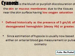
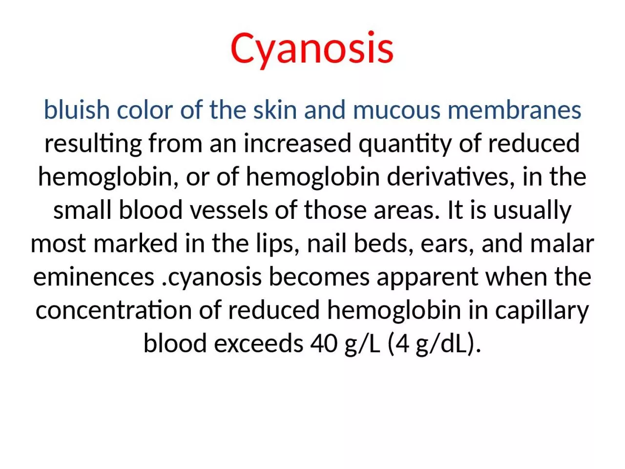
/
Cyanosis bluish color of the skin and mucous membranes Cyanosis bluish color of the skin and mucous membranes

Cyanosis bluish color of the skin and mucous membranes - PowerPoint Presentation

brianna
brianna . @brianna
Follow
68 views
Uploaded On 2023-05-20

Cyanosis bluish color of the skin and mucous membranes - PPT Presentation

resulting from an increased quantity of reduced hemoglobin or of hemoglobin derivatives in the small blood vessels of those areas It is usually most marked in the lips nail beds ears and malar ID: 998596

peripheral cyanosis blood central cyanosis peripheral central blood hemoglobin mucous reduced skin membranes pulmonary clinical disease flow oxygen increased

Share:

Link:

Embed:

Download Presentation from below link

Download Presentation The PPT/PDF document "Cyanosis bluish color of the skin and mu..." is the property of its rightful owner. Permission is granted to download and print the materials on this web site for personal, non-commercial use only, and to display it on your personal computer provided you do not modify the materials and that you retain all copyright notices contained in the materials. By downloading content from our website, you accept the terms of this agreement.


Presentation Transcript

1. Cyanosisbluish color of the skin and mucous membranes resulting from an increased quantity of reduced hemoglobin, or of hemoglobin derivatives, in the small blood vessels of those areas. It is usually most marked in the lips, nail beds, ears, and malar eminences .cyanosis becomes apparent when the concentration of reduced hemoglobin in capillary blood exceeds 40 g/L (4 g/dL).

2. central cyanosis can be detected reliably when the SaO2 has fallen to 85%; in others, particularly in dark-skinned persons, it may not be detected until it has declined to 75%. In the latter case, examination of the mucous membranes in the oral cavity and the conjunctivae rather than examination of the skin is more helpful in the detection of cyanosis.

3. Mechanism of CyanosisAbsolute increase of amount of reduced hemoglobin in blood, > 40g/L (4g/dl)(capillary)Nonfunctional hemoglobin such as methemoglobinor sulfhemoglobin) is present in blood.

4. Clinical Classification & EtiologyTrue Cyanosis (increased amount of reduced Hb) — Central Type — Peripheral Type — Mixed Type Cyanosis due to abnormal Hb derivatives — Methemoglobinemia — Sulfhemoglobinemia

5. 1-Central cyanosis is caused by decreased SaO2(increased amount of reduced Hb) and the mucous membranes and skin are both affected. only occurs when the oxygen saturation of arterial blood is less than 85%.

6. Peripheral cyanosis is due to a slowing of blood flow and abnormally great extraction of O2 from normally saturated arterial blood. It results from vasoconstriction and diminished peripheral blood flow, such as occurs in cold exposure, shock, congestive failure, and peripheral vascular disease. Often in these conditions, the mucous membranes of the oral cavity or those beneath the tongue may be spared.

7. Clinical differentiation between central and peripheral cyanosis may not always be simple, and in conditions such as cardiogenic shock with pulmonary edema there may be a mixture of both types

8. Causes of CyanosisCentral Cyanosis Decreased arterial oxygen saturation Decreased atmospheric pressure—high altitude Impaired pulmonary function  Alveolar hypoventilation  Impaired oxygen diffusion Anatomic shunts  Certain types of congenital heart disease  Pulmonary arteriovenous fistulas  Multiple small intrapulmonary shunts Hemoglobin with low affinity for oxygenHemoglobin abnormalities( Methemoglobinemia—hereditary, acquired Sulfhemoglobinema—acquired Carboxyhemoglobinemia (not true cyanosis)

9. Peripheral Cyanosis(Peripheral cyanosis is due to poor peripheral circulation and increased oxygen consumption in peripheral tissue.Reduced cardiac output Cold exposureRedistribution of blood flow from extremitiesArterial obstructionVenous obstruction

10. Possible clinical features include----peripheral cyanosisCool skin and mucous membranceSite (lower extremities,fingers)Diminish after massage

11. Possible clinical features include----central cyanosisthe warm mucous membranes are blue, for example the tongue, the inside of the lips central cyanosis increases immediately on exercise which is not the case for peripheral cyanosis there is polycythaemia with an abnormally high haemoglobin and haematocritclubbing is often seen in patients with central cyanosis

12. Differentiation of central as opposed to peripheralCyanosis Skin temp. Massage or warmingCentral Warm No change Peripheral Cool Cyanosis fades

13. Approach to the Patient: CyanosisCertain features are important in arriving at the cause of cyanosis:1. It is important to know the time of onset of cyanosis. Cyanosis present since birth or infancy is usually due to congenital heart disease. 2. Central and peripheral cyanosis must be differentiated. Evidence of disorders of the respiratory or cardiovascular systems are helpful. Massage or gentle warming of a cyanotic extremity will increase peripheral blood flow and abolish peripheral, but not central, cyanosis. 3. The presence or absence of clubbing of the digits (see below) should be ascertained. The combination of cyanosis and clubbing is frequent in patients with congenital heart disease and right-to-left shunting, and is seen occasionally in patients with pulmonary disease such as lung abscess or pulmonary arteriovenous fistulae. In contrast, peripheral cyanosis or acutely developing central cyanosis is not associated with clubbed digits. 4.,spectroscopic examination of the blood performed to look for abnormal types of hemoglobin (critical in the differential diagnosis of cyanosis