/
HISTOLOGY OF CIRCULATORY SYSTEM III HISTOLOGY OF CIRCULATORY SYSTEM III

HISTOLOGY OF CIRCULATORY SYSTEM III - PowerPoint Presentation

brooke
brooke . @brooke
Follow
64 views
Uploaded On 2024-01-29

HISTOLOGY OF CIRCULATORY SYSTEM III - PPT Presentation

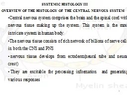
SMALL ARTERIES amp ARTERIOLES lt05mm lumen Thin subendothelial connective tissue Int elastic lamina absent in small arterioles T Media smooth muscle layer Adventitia is thin Hypertension ID: 1041430

basal muscles subendothelial layer muscles basal layer subendothelial smooth metabolic capillariesdiameter capillary edema cell continuous connective similar veins amp

Share:

Link:

Embed:

Download Presentation from below link

Download Presentation The PPT/PDF document "HISTOLOGY OF CIRCULATORY SYSTEM III" is the property of its rightful owner. Permission is granted to download and print the materials on this web site for personal, non-commercial use only, and to display it on your personal computer provided you do not modify the materials and that you retain all copyright notices contained in the materials. By downloading content from our website, you accept the terms of this agreement.


Presentation Transcript

1. HISTOLOGY OF CIRCULATORY SYSTEM III

2. SMALL ARTERIES & ARTERIOLES<0.5mm lumenThin subendothelial connective tissueInt. elastic lamina absent in small arteriolesT. Media: smooth muscle layerAdventitia is thinHypertension

3. CAPILLARIESDiameter : 5-7 µmSingle layer of endothelium; permit metabolic exchangeFunction in groups- Capillary BedsRichness conforms to metabolic activity of tissueStructure and pulsatile flow of blood through capillaries and thoroughfare channel.

4.

5. TYPESContinuousFenestratedSinusoidal

6. CONTINUOUS CAPILLARIESNo fenestrations, complete basal laminaPinocytic vesiclesLocation:Muscles, nervous tissue, exocrine glands

7. FENESTRATED CAPILLARIESTranscellular openings (80nm)Limited by cell membraneCovered by diaphragm ( thinner than cell membrane, proteoglycans)In endocrine glandsKidneys etc

8. SINUSOIDAL CAPILLARIESDiameter 30-40µmEndothelial cells discontinuous layerFenestrations without diaphragmIncomplete basal laminaMacrophagesFound in liver, hematopoietic organs

9. PERICYTESCells at periphery of continuous capillariesenclosed in own basal laminaMesenchymal originActin, myosin– contractile propertiesRole in repair processDiabetic Microangiopathy

10.

11. POST CAPILLARY VENULEDiameter 0.5-1mmEndothelium + thin subendothelial connective tissueMedia– pericytesMUSCULAR VEINSThin subendothelial layer, may be absentMedia: smooth muscles + reticular fibers valves

12. Inflammation & Edema

13.

14. LARGE VEINSWell developed intimaThin mediaVery thick adventitiaFurther three layersSmooth muscles

15.

16. LYMPH VASCULAR SYSTEMLYMPH CAPILLARIES:Single layer of endotheliumIncomplete basal laminaLYMPH VESSELS:Structure similar to veins, thinner wallsLARGE DUCTS:Similar to veins, smooth muscles

17.

18.

19. Medical ApplicationMetastasisLymph edema