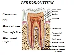
/
Cementum Cementum: Is a mineralized dental tissue that covering the anatomic roots of Cementum Cementum: Is a mineralized dental tissue that covering the anatomic roots of

Cementum Cementum: Is a mineralized dental tissue that covering the anatomic roots of - PowerPoint Presentation

cadie
cadie . @cadie
Follow
342 views
Uploaded On 2022-06-15

Cementum Cementum: Is a mineralized dental tissue that covering the anatomic roots of - PPT Presentation

Unlike bone however human cementum is avascular and noninnervated Cementum is thinnest at the cemento enamel junction and thickest toward the apex Like dentin cementum can form throughout the life of a tooth ID: 918461

teeth cementum dentin fibers cementum teeth fibers dentin bone tooth root cellular layer formed surface enamel fiber resorption cells

Share:

Link:

Embed:

Download Presentation from below link

Download Presentation The PPT/PDF document "Cementum Cementum: Is a mineralized dent..." is the property of its rightful owner. Permission is granted to download and print the materials on this web site for personal, non-commercial use only, and to display it on your personal computer provided you do not modify the materials and that you retain all copyright notices contained in the materials. By downloading content from our website, you accept the terms of this agreement.


Presentation Transcript

Slide1

Cementum

Slide2

Cementum:Is a mineralized dental tissue that covering the anatomic roots of human teeth. Cementum furnishes a medium for the attachment of collagen fibers that bind the tooth to surrounding structures.

Unlike bone, however, human cementum is

avascular

and

noninnervated

.

Cementum is thinnest at the cemento- enamel junction and thickest toward the apex.

Like dentin, cementum can form throughout the life of a tooth.

Slide3

PHYSICAL CHARACTERISTICS

1-The hardness of fully mineralized cementum is less than that of dentin.

2-Cementum is light yellow in color and can be distinguished from enamel by its lack of luster and its darker hue. Cementum is somewhat lighter in color than dentin.

3- Cementum has been shown to be permeable to a variety of materials.

Slide4

CHEMICAL COMPOSITION

On a dry weight basis, cementum from fully formed permanent teeth contains about 45% to 50% inorganic substances and 50% to 55% organic material and water. The inorganic portion consists mainly of calcium and phosphate in the form of

hydroxy

- apatite.


Slide5

Numerous trace elements are found in cementum in varying amounts. It is of interest that cementum has the

highest fluoride content

of all the mineralized tissues. The organic portion of cementum consists primarily of type

I collagen

and protein polysaccharides (

proteoglycans

) .

Slide6

CEMENTOGENESIS

Breaks occur in the epithelial root sheath allowing the newly formed dentin to come in direct contact with the cells of dental follicle.

Cells derived from his connective tissue are called

cementoblast

. They are responsible for the formation of cementum.

Slide7

Some sheath cells migrate away from the dentin toward the dental sac, whereas others remain near the developing tooth and ultimately are incorporated into the cementum.

Sheath cells that migrate toward the dental sac become the

epithelial rests of Malassez

found in the periodontal ligament of fully developed teeth.

Slide8

Slide9

Cementoblasts

Cementoblasts

synthesize collagen and protein polysaccharides, which make up the organic matrix of cementum.

At the superficial surface, the collagen fibrils produced by the

cementoblast

form a fibrous fringe perpendicular to periodontal space.

Slide10

Slide11

Slide12

Cementoid tissue

Under normal conditions ; growth of cementum is a rhythmic process, and as a new layer of

cementoid

is formed, the old one calcifies.

A thin layer of

cementoid

can usually be observed on the

cemental

surface. This

cementoid

tissue is lined by

cementoblasts

.

Connective tissue fibers from the periodontal ligament pass between the

cementoblasts

into the cementum.

Slide13

Slide14

These fibers are embedded in the cementum and serve to attach the tooth to surrounding bone. Their embedded portions are known as

Sharpey’s

fibers

.

Each

Sharpey’s

fiber is composed of numerous collagen fibrils that pass well into the cementum .

Cementoid

tissue is not observed in AEFC.

Slide15

Slide16

*

Light microscopic observations reveal two basic types of cementum, hence they are usually classified on the basis of presence of cementocytes (cellular cementum) or its absence (

acellular

cementum).

It can also be classified on the basis of the type of fibers (intrinsic/extrinsic fibers) presence or their absence (

afibrillar

cementum).

STRUCTURE

Slide17

*The

acellular

extrinsic fiber cementum is regarded as primary cementum, because it forms first.

*The cellular cementum are also known as

secondary cementum,

because it forms later than primary cementum

Slide18

Slide19

A-acellular cementum

1-Acellular

afibrillar

cementum (AAC):

is a mineralized ground substance containing no cells and is devoid of extrinsic and intrinsic collagen fibers. This type of cementum is seen chiefly as coronal cementum

Slide20

2-Acellular extrinsic fiber cementum:

extends from cervical margin to apical 1/3rd. It is the only type of cementum seen in single rooted teeth. The extrinsic fibers are seen perpendicular to surface of cementum and they are known as

Sharpey’s

fibers

. When cementum remains relatively thin,

Sharpey’s

fibers cross the entire thickness of the cementum. With further apposition of cementum, a larger part of the fibers is incorporated in the cementum. The attachment proper is confined to the most superficial or recently formed layer of cementum .The main function of this type of cementum is anchorage especially in single rooted teeth.

Slide21

Slide22

Slide23

B-Cellular cementum:

is also known as secondary cementum as this is formed later than the AEFC.

The cellular cementum found in the

apical third

is mainly of two types

1-

the cellular mixed fiber cementum which forms the bulk of secondary cementum and occupies the

apical and

interradicular

regions

and the

2-

CIFC:

which

is present in the

middle and apical third

. These types are mainly involved in the

adaptation and repair

of

cementum

Since the secondary cementum is formed rapidly the incremental lines are placed further apart than in AEFC.

Slide24

Slide25

Slide26

Slide27

  1-Cellular intrinsic fiber cementum (CIFC):

This cementum contains cells but has no extrinsic fibers. The fibers present are intrinsic fibers which are secreted by the

cementoblasts

. It is formed on the root surface and in

cases of repair

.

Slide28

Slide29

2-Cellular mixed fiber cementum (CMFC):

The cellular mixed fiber cementum is a variant formed at a faster rate with less mineralized fibers. The collagen fibers of CMFC are derived from the periodontal ligament fibroblasts and the

cementoblasts

. These intrinsic and extrinsic fibers form an intricate pattern running between each other at almost right angles and different orientations, though the number of intrinsic fibers is comparatively less than the extrinsic fibers. The fiber bundles originating from the fibroblast are ovoid or round and the fibers originating from the

cementoblasts

are much more delicate and smaller measuring

Slide30

Slide31

3-Cellular mixed stratified cementum (CMSC):

In this type of cementum the cellular intrinsic fiber cementum alternates with

acellular

extrinsic fiber cementum. It is formed by

cementoblasts

and fibroblasts. It appears primarily on

apical third of the root and

furcation

areas

Slide32

Slide33

Cementocytes :The cells incorporated into cellular cementum, are similar to

osteocytes

. They lie in spaces designated as lacunae. A typical

cementocyte

has numerous cell processes, or canaliculi, radiating from its cell body. These processes may branch, and they frequently

anastomose

with those of a neigh- boring cell.

Slide34

Most of the processes are directed toward the periodontal surface of the cementum. They are best viewed in mineralized ground sections.

These characteristics indicate that cementocytes are either degenerating or are marginally active cells and lacunae in the deeper layers of cementum appear to be empty, suggesting complete degeneration of cementocytes located in these areas.

Slide35

Slide36

Slide37

Differences between cementocytes and osteocytes

Though the cementocytes resemble the

osteocytes

, there are a few important differences. The lacunae of cementocytes varies from being ovoid or tubular, but the

osteocytic

lacunae is invariably oval.

The canaliculi are less complicated and sparse, with the majority of them facing the periodontal ligament when compared to

osteocytes

, whose canaliculi are radiating, more dense and arranged in a complex network. However in both, the

cytoplasmic

processes are connected with the cells lining the surface

Slide38

CEMENTODENTINAL JUNCTION(CDJ):

The dentin surface upon which cementum is deposited is relatively smooth in permanent teeth. The

cementodentinal

junction in deciduous teeth, however, is sometimes scalloped.

The attachment of cementum to dentin in either case is quite firm. increased water content which contributes to the stiffness.

This reduction in its mechanical property, helps it to redistribute occlusal loads to the alveolar bone.

Slide39

Slide40

Sometimes dentin is separated from cementum by a zone known as the intermediate cementum layer

, which does not exhibit characteristic features of either dentin or cementum .As it appears hyaline (

structureless

), it is also known as Hyaline layer.

Slide41

This layer is predominately seen in the apical two thirds

of roots of molars and premolars and is only rarely observed in incisors or deciduous teeth.

It is believed that this layer represents areas where cells of

Hertwig’s

epithelial sheath become trapped in a rapidly deposited dentin or cementum matrix.

The intermediate cementum layer is considered to be of dentinal origin.

Slide42

It contains no tubules but wide spaces which are thought to be enlarged terminals of dentinal tubules. It appears very similar to

aprismatic

enamel, as it is an amorphous layer of

noncollagenous

material devoid of odontoblasts and

cementoblasts

.

it is ether a continuous layer or in isolated areas. The probable function might be to seal the sensitive root dentin.

Slide43

CEMENTOENAMEL JUNCTION

1-In about 10% of the teeth, enamel and cementum do not meet. this occurs when enamel epithelium in the cervical portion of the root is delayed in its separation from dentin. Instead, a zone of the root is devoid of cementum and is, for a time, covered by reduced enamel epithelium

Slide44

Slide45

2- In approximately 60% of the teeth, cementum overlaps the cervical end of enamel for a short distance .This occurs when the enamel epithelium degenerates at its cervical termination.

Slide46

3-In approximately 30% of all teeth enamel and cementum meeting edge to edge. CEJ may exhibit all of these patterns in teeth of an individual.

Slide47

FUNCTIONS:

1-Anchorage

The primary function of cementum is to furnish a medium for the attachment of collagen fibers that bind the tooth to alveolar bone. Since collagen fibers of the periodontal ligament cannot be incorporated into dentin, a connective tissue attachment to the tooth is impossible without cementum. This is dramatically demonstrated in some cases of

hypophosphatasia

, a rare hereditary disease in which loosening and premature loss of anterior deciduous teeth occurs. The exfoliated teeth are characterized by an almost total absence of cementum.

Slide48

Slide49

2-Adaptation

Cementum may has functional adaptation of teeth possible. For example, deposition of cementum in an apical area can compensate for loss of tooth substance from occlusal wear. The continuous deposition of cementum is of considerable functional importance. In contrast to the alternating resorption and new formation of bone, cementum is not resorbed under normal conditions. As the most superficial layer of cementum ages, a new layer of cementum must be deposited to keep the attachment apparatus intact.


Slide50

Slide51

3-Repair:

Cementum serves as the major reparative tissue for root surfaces. Damage to roots such as fractures and resorption can be repaired by the deposition of new cementum. Cementum formed during repair resembles cellular cementum because it forms faster but it has a wider

cementoid

zone and the apatite crystals are smaller. If the repair takes place slowly, it cannot be differentiated from primary cementum.

Slide52

Slide53

HYPERCEMENTOSIS

Hypercementosis is an abnormal thickening of cementum

.

This condition frequently is found in teeth that are exposed to great stress.

It may be diffuse or circumscribed

.

It may affect all teeth of the dentition

, or

be confined to a single tooth, or even affect only parts of one tooth

.

In localized hypertrophy a spur or prong like extension of cementum may be

formed which

provide a larger surface area for the attaching fibers; thus a firmer anchorage of the tooth to the surrounding alveolar bone is assured.

Slide54

Slide55

Excementoses:

knob like projections or Localized hypercementosis may sometimes be observed in areas in which enamel drops have developed on the dentin.

The

hyperplastic

cementum covering the enamel drops occasionally is irregular and sometimes contains round bodies that may be calcified epithelial rests.

The

same type of embedded calcified round bodies frequently are found in localized areas of

hyperplastic

cementum. They too develop around degenerated epithelial rests.

Extensive

deposition of cementum is occasionally associated with

chronic periapical inflammation

. The excessive deposition is circumscribed and surrounds the root like a cuff.

Slide56

A thickening of cementum is often observed on teeth that are not in function. It may extend around the entire root of the nonfunctioning teeth or may be localized in small areas teeth is characterized by a reduction in the number of

Sharpey’s

fibers embedded in the root

.

The cementum is thicker around the apex of all teeth and in the

furcation

of

multirooted

teeth than on other areas of the root.

Slide57

This thickening is found in embedded and in newly erupted teeth. Hyper-

cementosis

is associated with a large number of

neoplastic

and non

neoplastic

diseases. Generalized thickening is seen in Paget’s disease.

Slide58

Localized

forms can be seen in benign

cementoblastoma

, florid cemento-osseous dysplasia,

acromegaly

,

calcinosis

and some forms of arthritis Hypoplasia or

aplasia

of cementum is of rare occurrence. It is associated with

hypophosphatasia

.

Slide59

The extraction of such teeth may necessitate the removal of bone. They can anchor the tooth so tightly to the socket that the jaw or parts of it may be fractured in an attempt to extract the tooth. This possibility indicates the necessity for taking radiographs before any extraction. Small fragments of roots left in the jaw after

extraction

of teeth may be surrounded by cementum and remain in the jaw without causing any disturbance.

Slide60

CLINICAL CONSIDERATIONS

Cementum is more resistant to resorption than bone, and it is for this reason that orthodontic tooth movement is possible. When a tooth is moved by means of an orthodontic appliance, bone is resorbed on the side of the pressure, and new bone is formed on the side of tension.

On

the side toward which the tooth is moved, pressure is equal on the surfaces of bone and cementum.

Slide61

Resorption of bone as well as of cementum may be anticipated. so careful orthodontic treatment, cementum resorption is minimal or absent, but bone resorption leads to tooth migration. The difference in the resistance of bone and cementum to pressure may be caused by the fact that bone is richly

vascularized

,

Slide62

Cementum resorption can occur after trauma or excessive occlusal forces.

In

severe cases cementum resorption may continue into the dentin

.

After resorption has ceased, the damage usually is repaired, either by formation of

acellular

or cellular cementum or by alternate formation of both

.

In

most cases of repair there is a tendency to reestablish the former outline of the root surface. This is called

anatomic repair.

Slide63

However, if only a thin layer of cementum is deposited on the surface of a deep resorption, the root outline is not reconstructed, and a bay like recess remains.

In

such areas sometimes the periodontal space is restored to its normal width by formation of a bony projection so that a proper functional relationship will result.

The

outline of the alveolar bone in these cases follows that of the root surface. In contrast to anatomic repair, this change is called

functional repair.

If

teeth are subjected to a severe blow, fragments of cementum may be severed from the dentin. The tear occurs frequently at the

cementodentinal

junction, but it may also be in the cementum or dentin.


Slide64

Slide65

Transverse fractures of the root may occur after trauma, and these may heal by formation of new cementum. This also applies to extensive

excementosis

, as

.

In periodontal pockets, plaque and its by-products can cause numerous alterations in the physical, chemical, and structural characteristics of cementum. The surface of

pathologically

exposed cementum becomes

hypermineralized

because of the incorporation of calcium, phosphorus, and fluoride from the oral environment.