/
D RAW ME A PICTURE OF . . . D RAW ME A PICTURE OF . . .

D RAW ME A PICTURE OF . . . - PowerPoint Presentation

calandra-battersby
calandra-battersby . @calandra-battersby
Follow
343 views
Uploaded On 2020-01-14

D RAW ME A PICTURE OF . . . - PPT Presentation

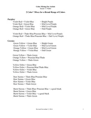
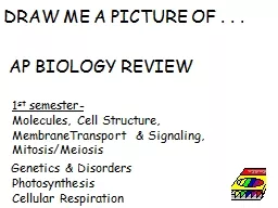
D RAW ME A PICTURE OF AP BIOLOGY REVIEW 1 st semester Molecules Cell Structure MembraneTransport amp Signaling MitosisMeiosis Genetics amp Disorders Photosynthesis Cellular Respiration ID: 772761

cell draw picture diagram draw cell diagram picture label showing show reaction energy blood reactants person fatty change polar

Share:

Link:

Embed:

Download Presentation from below link

Download Presentation The PPT/PDF document "D RAW ME A PICTURE OF . . ." is the property of its rightful owner. Permission is granted to download and print the materials on this web site for personal, non-commercial use only, and to display it on your personal computer provided you do not modify the materials and that you retain all copyright notices contained in the materials. By downloading content from our website, you accept the terms of this agreement.


Presentation Transcript

DRAW ME A PICTURE OF . . . AP BIOLOGY REVIEW1st semester- Molecules, Cell Structure, MembraneTransport & Signaling, Mitosis/Meiosis Genetics & DisordersPhotosynthesisCellular Respiration

Draw a diagram of a typical biological membrane including the lipid bilayer and both integral and peripheral proteins. Label your diagram and GIVE AN EXAMPLE of an integral and a peripheral protein you learned about.

Peripheral proteins- G protein Last ETC protein Integral proteins: transport proteins (ion channels, carriers, aquaporins, ATP synthase . . . .).

Draw a picture of an amino acidShow how 2 amino acids could be joined together.Identify this process and the type of bond formed

DEHYDRATION SYNTHESIS (CONDENSATION) REACTIONRemove H2OMAKES A PEPTIDE BOND

Draw a picture of the subunit used to make nucleic acids.Circle the parts of this subunit that make the backbone of a DNA moleculeWhich nitrogen bases could be found in the nitrogen base spot if this were used to makeRNA?

AdenineGuanineCytosineUracilNO THYMINE IN RNANUCLEOTIDE

Draw a chloroplast Label all the places & spaces. Mark the locations of ETC and Calvin Cycle. Show where H + ions build up during ETC.

Draw a chloroplast

Draw a picture of a human red blood cell placed in distilled water. Use arrows to show how the water will move. What vocab word(s) describe what will happen to this cell?

Draw a picture of a human red blood cell placed in distilled water. Use arrows to show how the water will move. What vocab word describes what will happen to this cell? More water will enterthan leave. Animal cell will swell and possibly burst = cytolysis

Draw a PUNNETT SQUARE that shows theoffspring of a cross between a father withcolorblindness and a non-colorblind mother whose father had normal vision.Tell the possible outcomes of this cross.

XC XC XcyXCXc X CXcXCy XCy50% normal female (carriers)50% normal males

Diagram the differences between cells that are haploid, diploid, triploid, trisomic, and monosomic USE 2n=6

Diagram the differences between cells that are haploid, diploid, triploid, trisomic, and monosomic USE 2n=6 DIPLOID 2n TRISOMY 2n+1 TRIPLOID 3n MONOSOMY 2n-1

Draw a diagram showing the glycoproteins on the surface of a blood cell from a person with A positive blood.WHAT KIND OF ANTIBODIES WOULD THIS PERSON MAKE?

Draw a diagram showing the glycoproteins on the surface of a blood cell from a person with A positive blood.WHAT KIND OF ANTIBODIES WOULD THIS PERSON MAKE? ANTI-B antibodies

DRAW A DIAGRAM showing the molecules in the thylakoid membrane involved in the light reactions and label them.Add H+ ions to show where H+ ions build up during the light reaction.Tell 2 things that happen to create the H+ gradient.

DRAW A DIAGRAM showing the molecules in the thylakoid membrane involved in the light reactions and label them.H+ accumulate in the thylkoid space due to :1. Splitting water to replace electrons in chlorophyll 2. ETC (proton pumps) move H+ from stroma into thylakoid spaceH+ H+ H+ H+ H+ H+ H+ H+ H+ H+

Draw a diagram of a diploid cell with 4 chromosomes in Metaphase I of meiosis.Compare this to the same cell in Metaphase of mitosis

Metaphase I of meiosis Metaphase of mitosis Metaphase of mitosis

Draw the 2 kinds of shapes found in the secondary structure of proteins.What kinds of groups are involved in holding this shape in position?

Beta pleated sheetAlpha helix Hydrogen bonds between C=O on one amino acid and the N-H on another amino acid

Show how alpha and beta glucose are different.Give examples of polysaccharides made with each of these.Which of these polysaccharide are humans and other animals unable to digest?

Alpha (α)glucose Beta (β) glucose glycogen & starch cellulose & chitin Humans and other animals are unable to break polysaccharides with β linkages

Draw a picture showing the components used to make a FAT molecule. What kind of reaction joins the “pieces”?How is a fat different than a phospholipid?How does adding unsaturated fatty acid tails change the shape of the phospholipid?How does this affect its behavior in cell membranes?

FAT = 1 glycerol + 3 fatty acid tailsJoined by dehydration synthesisPhospholipid = 1 glycerol + 2 fatty acid tails + 1 phosphate group

GLYCEROL3 FATTY ACIDS MAKE A FAT

PHOSPHOLIPID

STRUCTURE/FUNCTION!UNSATURATED FA’s affectstructure.Can’t pack astightly.Makes membranes MORE fluid

Draw a diagram showing the glycoproteins on the surface of a blood cell from a person with IB i genotype.WHAT KIND OF ANTIBODIES WOULD THIS PERSON MAKE?

Draw a diagram showing the glycoproteins on the surface of a blood cell from a person with IB i genotype.

Draw a mitochondrion. Label all the places & spaces. Mark the locations of glycolysis, Krebs Cycle and ETC. Show where H + ions build up during ETC. Add some mitochondrial DNA.

Draw the shape of a phospholipid and identify the parts that are polar and non-polar. Identify the parts that are hydrophobic and hydrophilic.How does adding unsaturated fatty acids change the shape of the molecule and impact membrane fluidity?

Unsaturated fatty acids put bends in the tails; They pack less tightly together in membrane;Increase fluidityHEAD = polar/hydrophilicTAILS = non-polar/hydrophobic

Draw a diagram showing the action of a non-polar hormone like cholesterol in acell signaling pathway.

Draw a diagram showing the action of a non-polar hormone like cholesterol in acell signaling pathway.

Draw a bacterial cell engulfed by phagocytosis and label 3 parts that are similar to mitochondria and chloroplastsand provide evidence for Lynn Margulis’sEndosymbiotic theory

DNA is one circular loopRibosomes are smaller than cellular ribosomes like prokaryotesInner mitochondrial/chloroplast membranes have bacterial phospholipids Outer mitochondrial/chloroplast membrane like cell membranesInner membranes have enzymes for respiration/photosynthesis like bacterial outer membranes

Draw a pathway that shows the movement of a protein like insulin from where it is made in a cell to where it is secreted into the blood stream.

Made on ribosomes.Travels through Rough ERSent via vesicle to Golgi Sent via vesicle to plasma membraneExocytosis to exit

Draw a gene map based on the following recombination frequencies:A-B 8%A-C 28%A-D 25%B-C 20%B-D 33%

Draw a gene map based on the following recombination frequencies:A-B 8%A-C 28%A-D 25%B-C 20%B-D 33%

Draw a picture of a nerve axon and show how the two kinds of Na+/K+ transporters work together to polarize and depolarize a nerve cell. Which of these transporters is active/passive?

POLARIZATIONNa + - K+ pumpsSet potential on membraneMore + outside than inACTIVE TRANSPORTrequires energyPASSIVE TRANSPORTNo energy neededMove down gradient from [high] → [low] DEPOLARIZATIONIon channels allow Na + and K+ to where they started

Draw a picture of a chromosome in G1 of interphase. Show what it looks like in G2

Draw a picture of a chromosome in G1 of interphase. Show what it looks like in G2

Draw a diagram to show 2 ways (ALLOSTERIC) NON-COMPETITIVE INHIBITORS work

NON-COMPETITVE (ALLOSTERIC) INHIBITION

Draw a picture of a chromosome in prophase of interphase. Label its parts.

Draw a picture of a chromosome in prophase of interphase. Label its parts.chromatidchromatid centromeretelomeres

Draw a graph of FREE ENERGY of PRODUCTS and REACTANTS over time in a NEGATIVE ∆ G chemical reaction. Label ACTIVATION ENERGY and ∆GHow does adding an enzyme change the graph?How does it change the ∆G of this reaction?

Draw a graph of FREE ENERGY of PRODUCTS and REACTANTS over time in a NEGATIVE ∆ G chemical reaction. Label ACTIVATION ENERGY and ∆GHow does adding an enzyme change the graph?How does it change the ∆G of this reaction?

Draw a picture to explain why NADH makes more ATP than FADH2 when electrons are passed to the ETC during cellular respiration.Image from: http://study.com/cimages/multimages/16/Electron_Transport_Mitochondrion.png

NADH drops its electrons at beginning of ETC so as electrons pass down ETC 3 proton pumps move H+ ions into the intermembrane space = 3 ATP when they return through ATP synthase.FADH2 drops its electrons farther down ETC skipping the 1st proton pump so less H+ moved = 2 ATP Image from: http://study.com/cimages/multimages/16/Electron_Transport_Mitochondrion.png

Draw a graph of FREE ENERGY versus time showing the PRODUCTS and REACTANTS for a NEGATIVE ∆ G and a POSITIVE ∆G REACTION. How is the ∆ G in these reactions different? Which of these is spontaneous?Which is endergonic/exergonic?

Draw a graph of FREE ENERGY versus time showing the PRODUCTS and REACTANTS for a NEGATIVE ∆ G and a POSITIVE ∆G REACTION. How does the energy of reactants and products compare in these two kinds of reactions? Label these as: spontaneous or not?endergonic/exergonic?

EXERGONIC REACTION ∆G < 0Reaction is spontaneousEnergy of reactants is greater than energy of productsEXERGONIC REACTION ∆G > 0Reaction is NOT spontaneousEnergy of products is greater than energy of reactants

asdfasdf

asdfasdf

asdfasdf