/
Patient information factsheet Patient information factsheet

Patient information factsheet - PDF document

callie
callie . @callie
Follow
344 views
Uploaded On 2022-08-16

Patient information factsheet - PPT Presentation

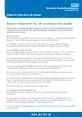
wwwuhsnhsuk Tachyarrythmias fast heart rhythms This factsheet has been written to help you understand more about heart rhythm problems If there is anything you do not understand or you would l ID: 936435

electrical heart www factsheet heart electrical factsheet www pathway uhs nhs node rhythm tachycardia ventricular arrhythmia ventricles arrhythmias x00660069

Share:

Link:

Embed:

Download Presentation from below link

Download Pdf The PPT/PDF document "Patient information factsheet" is the property of its rightful owner. Permission is granted to download and print the materials on this web site for personal, non-commercial use only, and to display it on your personal computer provided you do not modify the materials and that you retain all copyright notices contained in the materials. By downloading content from our website, you accept the terms of this agreement.


Presentation Transcript

Patient information factsheet www.uhs.nhs.uk Tachyarrythmias (fast heart rhythms) This factsheet has been written to help you understand more about heart rhythm problems. If there is anything you do not understand, or you would like more information, please ask a member of your healthcare team, who will be happy to explain further. To help you understand this factsheet please refer to our “how the heart works” factsheet �rst. This is available on: www.uhs.nhs.uk What is an arrhythmia? Sometimes if the heart’s conduction pathway is damaged, blocked, or an extra pathway exists • too quickly (tachycardia) • too slowly (bradycardia) • irregularly. This may a�ect the heart’s ability to pump blood around the body. Arrhythmias can occur in the heart’s upper chambers (atria) or lower chambers (ventricles). Causes of an arrhythmia Any interruption in the heart’s electrical system can cause an arrhythmia. such as myocardial infarction (heart attack) or congenital heart disease you may be at risk of Diagnosing your arrhythmia Electrocardiogram (ECG) Your heart’s www.uhs.nhs.uk Patient information factsheet Holter monitor A Holter monitor shows changes in your heart rhythm over the course of a 24-hour period that may not be detected during a resting or exercise ECG. You will be asked to go about your daily activities as usual (except for showering or bathing) while you wear a small, portable recorder that connects to electrode stickers on your chest. You will then come back to the Cardiac event monitor have a cardiac event monitor. This type o

f recording device is used if your arrhythmias are infrequent. This device is about the size of a large pager, and can be clipped to your belt or recorder against your chest and press a button. The device then records up to 70 seconds of Internal loop recorder (ILR) Your doctor may feel an ILR is appropriate to investigate your symptoms. This is a long-term activator, when you have symptoms you place the activator over the ILR and it will store your device will transmit this data automatically to the hospital. The ILR is inserted under local Types of arrhythmia Arrhythmias that occur in the atria are either ‘atrial’ (in the heart’s upper chambers) or ‘supraventricular’ (above the ventricles) in origin, whereas ventricular arrhythmias start in Supraventricular tachycardia (SVT) as ‘re-entry tachycardia’. This is because the electrical impulse does not fade out as with the is due to an extra electrical pathway that can form a short circuit within the heart’s conduction system. SVT is usually a rapid, regular rhythm. The two most common types of SVT are: • AV-nodal re-entry tachycardia • AV re-entry tachycardia (AVRT), most commonly known as Wol�-Parkinson-White AV nodal re-entry tachycardia (AVNRT) This type of arrhythmia occurs when a problem arises in the way the electrical impulses pass through the AV node. Normally, the AV node acts as a gateway, slowing and regulating AVNRT there are two AV node. This type of arrhythmia usually starts following an early beat (ectopic). An electrical short circuit then occurs where the

electrical impulse rotates around the circuit and with www.uhs.nhs.uk Patient information factsheet AV re-entry tachycardia (AVRT) or Wol�-Parkinson-White syndrome (WPW) In AVRT an extra electrical pathway exists that bypasses the normal conduction system. The pathway directly connects the atria to the ventricles. This extra pathway is known as an accessory pathway. The electrical impulses travel along the accessory pathway, bypassing the AV node. The tissue in the pathway does not slow the impulse down, as in the AV node. Therefore the electrical impulses reach the ventricles before the normal electrical impulse shows the existence of an extra electrical pathway. Very fast heart rates may occur as the The electrical impulse normally originates at the SA node. However, in atrial �brillation, many the ventricles. This can cause blood clots to form. Therefore, to prevent you being at an Atrial �utter also occurs in the atria. The electrical impulses �re rapidly but the resulting rhythm is regular and organised. The rhythm is due to a re-entry circuit within the atria, where Ventricular tachycardia (VT) VT occurs when the electrical impulses arise in the ventricles. The ventricles start beating at work as e�ciently, causing symptoms of weakness, dizziness, chest pain, shortness of breath or even collapse. There are several di�erent types of VT and the seriousness of the condition can vary. VT can be a potentially life-threatening heart rhythm as it can progress to ventricular There are a number of reasons that people

may develop VT. For example, in people who have had a previous myocardial infarction (heart attack) the area of the heart muscle damaged by the heart attack forms scar tissue. This can make the heart susceptible to abnormal heart rhythms. Other people who may experience VT are patients with cardiomyopathy, previous corrective congenital heart surgery or inherited arrhythmias. There is also a small group of people who have VT with a structurally normal heart. Ventricular �brillation Ventricular �brillation occurs in the ventricles. In ventricular �brillation, the electrical impulses heart to quiver rather than to beat and pump blood. Ventricular �brillation is a life-threatening Treatments The results of the tests you have had will determine the type and seriousness of your arrhythmia. Your doctor will then discuss your treatment options with you. Many patients with www.uhs.nhs.uk Patient information factsheet Medicines medicines that change the electrical signals in your heart and help prevent irregular or rapid Permanent pacemaker node ablation. A pacemaker is a small device used to treat slow heart rhythms. It is implanted beneath the skin below the collarbone and connected to a pacing wire placed inside the heart. The pacemaker delivers a small electrical impulse to stimulate the heart to beat when it is going too slowly. An AV node ablation and permanent pacemaker insertion will regulate the heart rate and Radiofrequency (heat) energy / cryo (cold) energy catheter ablation have a catheter ablation. A catheter ablation creates scar tissue

that blocks the area of extra electrical activity causing the arrhythmias. This provides relief for patients who may not have Internal cardioversion Two catheters are inserted into a vein in your groin and a small electrode pad applied to your sleepy. unsuccessful in returning the heart’s rhythm back to a normal sinus rhythm. and stop fast ventricular arrhythmias by using extra paced beats known as ‘anti-tachycardia pacing (ATP)’ or by delivering an electric shock to the heart. It is also capable of pacing the heart to stop it from going too slowly. Cancellations always try to explain the reason. We fully appreciate that this is a stressful time for you and your family and we will do our best to provide you with a new date that is convenient for you Who will perform my procedure? Your procedure will be performed by a specially trained doctor with appropriate experience (although we aren’t able to guarantee that you will be treated by a particular member of www.uhs.nhs.uk Patient information factsheet Contact us factsheet please call: 023 8120 8436 clinical nurse specialist. You can also email: uhs.crmnurses@nhs.net management coordinator on: 023 8120 8772 Useful links The following websites also provide useful information: www.bhf.org.uk www.heartrhythmcharity.org.uk An online version of this factsheet is available on our website www.uhs.nhs.uk mat such as easy read, large print, Braille or audio, please telephone 023 8120 4688. accessing the hospital, please visit www.uhs.nhs.uk/additionalneeds Version 5. Published December 2020. Due for review December 2023. 727