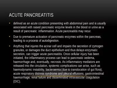
/
Role of CT in  acute pancreatitis Role of CT in  acute pancreatitis

Role of CT in acute pancreatitis - PowerPoint Presentation

callie
callie . @callie
Follow
342 views
Uploaded On 2022-02-10

Role of CT in acute pancreatitis - PPT Presentation

MBBCh MS FRCR Consultant radiologist Riyadh Military Hospital Dr Ahmed Refaey Normal CT anatomy of the upper abdomen Anterior pararenal space Normal Anatomy by CT Pancreas is located in the anterior ID: 908125

acute pancreatitis pancreatic necrosis pancreatitis acute necrosis pancreatic collections necrotizing early severity mortality infection sign severe mild tail peripancreatic

Share:

Link:

Embed:

Download Presentation from below link

Download Presentation The PPT/PDF document "Role of CT in acute pancreatitis" is the property of its rightful owner. Permission is granted to download and print the materials on this web site for personal, non-commercial use only, and to display it on your personal computer provided you do not modify the materials and that you retain all copyright notices contained in the materials. By downloading content from our website, you accept the terms of this agreement.


Presentation Transcript

Slide1

Role of CT in acute pancreatitis

MBBCh, MS, FRCR

Consultant radiologist Riyadh Military Hospital

Dr. Ahmed

Refaey

Slide2

Normal CT anatomy of the upper abdomen

Slide3

Slide4

Slide5

Anterior pararenal space

Slide6

Slide7

Normal Anatomy by CTPancreas is located in the anterior pararenal space

Head adjacent to duodenumTail extending toward spleenSplenic vein posterior to body and tail

Slide8

Normal Morphology by CTNo capsuleAP dimensions

Head 2-2.5 cm Body and tail 1-2 cmPancreatic duct Maximal diameter 3 mm in adults (5 mm in elderly)

Slide9

Evaluation of Acute PancreatitisContrast-enhanced CT is imaging modality of choiceOral and IV contrast differentiate pancreatic tissue from adjacent blood vessels and duodenum

Slide10

There is no additional value of an early CT (within 72 hours) in patients with acute pancreatitis. The diagnosis is usually made on clinical and laboratory findings.An early CT may be misleading concerning the severity of the pancreatitis, since it can underestimate the presence and amount of necrosis.

Slide11

Etiology

Slide12

PathophysiologyActivated pancreatic enzymes escaping the

ductal system and auto digesting the pancreas and adjacent structures ( mainly amylase, lipase & trypsin ).Lack of capsule facilitates spread

Slide13

Acute pancreatitisMild acute pancreatitis ----- 80 %

* edematous ( interstitial ) * exudativeSevere acute pancreatitis ----20 %

Slide14

Mild acute pancreatitis - run a mild course without development of multiple organ failure - improvement within 3 days following conservative therapy with gradual decrease of elevated enzymes.

- has a mortality rate of < 1%

Slide15

Severe acute pancreatitis “ necrotizing “ - run a serious clinical course with pancreatic necrosis and the development of multiple organ failure

- of these, 60% of pancreatic necrosis remain sterile , while 40% becomes infected - this last category ( infected necrosis ) , has the highest mortality rate ( 25-70%)

Slide16

Clinical outcome

Slide17

CT Imaging of acute pancreatitis

Slide18

Acute edematous “interstitial” pancreatitisAcute exudative pancreatitis

Mild acute pancreatitis

Slide19

Acute edematous “interstitial” pancreatitis Edematous pancreas

with/without peripancreatic fat stranding. No collections or necrosis

Slide20

Slide21

Slide22

Slide23

Slide24

Slide25

Acute exudative pancreatitis

an intermediate form of pancreatitis without pancreatic necrosis with an intermediate clinical course.This is called extrapancreatic necrosis (EXPN)

Slide26

Slide27

Slide28

Slide29

Avoid early drainage of collections and avoid introducing infection! 50% of these collections show spontaneous regression The other 50% either remain stable (

pseudocyt ) or develop infection ( abscess ) .

Slide30

Peripancreatic fluid50% spontaneous regression 50% stable

sterile ( pseudocyst) infection ( asbscess )

Slide31

Spontaneous regression of peripancreatic collection

Slide32

Severe pancreatitis “ necrotizing pancreatitis”

occurs in 20% of patients. * partial necrotizing pancreatitis * total necrotizing pancreatitis

Slide33

Partial necrotizing Total necrotizingDelayed or no response to conservative therapy

Delayed or no normalization of enzymesMortality : 30 – 75 %Deterioration under conservative therapyMortality : 100 % - 40% by 2nd day - 75% by 5th day - 100% by 10th day

Slide34

Slide35

Slide36

Slide37

Slide38

Slide39

Slide40

Central gland necrosis

Slide41

Central gland necrosis

Subtype of necrotizing pancreatitis.Necrosis between the pancreatic head and tail and is nearly always associated with disruption of the pancreatic duct.This leads to persistent collections as the viable pancreatic tail continues to secrete pancreatic juices.These collections react poorly to endoscopic or percutaneous drainage.Definitive treatment often requires distal pancreatectomy

.

Slide42

Slide43

An early CT may be misleading concerning the severity of the pancreatitis, since it can underestimate the presence and amount of necrosis.

Slide44

Slide45

Slide46

Mortality Early mortality in acute pancreatitis is the result of the systemic inflammatory response with multiple organ failure.

Late mortality is the result of infection of pancreatic necrosis and peripancreatic fluid collections which results in sepsis and is seen in more than 50% of deaths.

Slide47

CT Severity IndexIt is critical to identify patients who are at high risk for severe disease, since they require close monitoring and possible intervention

Slide48

Balthazar et al constructed a CT severity index (CTSI) for acute pancreatitis that combines the grade of pancreatitis (A-E) with the extent of pancreatic necrosis.

Slide49

Slide50

CT Severity Index1- 3 …………….. Mild 4-6 …………….. Moderate

7-10 ……………. Severe

Slide51

Complications of pancreatits

Slide52

ComplicationsPancreatic PseudocystsAbscess

Hemorrhagic PancreatitisSplenic Artery Pseudoaneurysm

Slide53

Pancreatic PseudocystFluid collection surrounded by fibrous capsule

Time of onset : > 4 weeks from the onsetAmylase rich-fluidPrognosis : spontaneous resolution in 44%

Slide54

Slide55

Slide56

Slide57

AbscessWell demarcated fluid collection of pus Suspected clinically with fever and septicemia

Pathognomonic finding → presence of gasTime of onset : 2- 4 weeks after onset

Slide58

Slide59

Slide60

Slide61

Hemorrhagic pancreatitis Type of severe pancreatitisPeripancreatic fat necrosis and hemorrhage due to erosion of small vessels

Falling hematocrit Cullen sign and Grey-Turner sign

Slide62

(Grey-Turner sign): flank ecchymosis

(Cullen sign) : periumbilical ecchymosisassociated with a 37% mortality rate

Slide63

Grey Turner signCullen’s sign

Slide64

Slide65

Slide66

Splenic Artery PseudoaneurysmPresents similarly to hemorrhagic pancreatitis with a

↓ in hematocrit

Slide67

Slide68

Slide69

Take home messages

Slide70

Severity of acute pancreatitis and pancreatic necrosis can only be reliably assessed by imaging after 72 hours. CT can not reliably differentiate between collections that consist of fluid and those that contain solid debris.In these cases MRI can be of additional value.

Slide71

Central gland necrosis is a subtype of necrotizing pancreatitis with important implications. Avoid early drainage of collections and avoid introducing infection!

Slide72

Thank you