PDF-How CT scans of the chest work: What You Need to Know
Author : carebox | Published Date : 2024-04-13
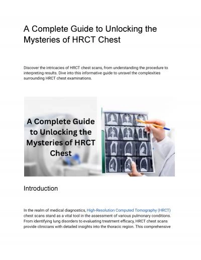
A CT Computed Tomography scan of the chest is a diagnostic imaging procedure that provides detailed crosssectional images of the chest area It is commonly used to
Presentation Embed Code
Download Presentation
Download Presentation The PPT/PDF document "How CT scans of the chest work: What You..." is the property of its rightful owner. Permission is granted to download and print the materials on this website for personal, non-commercial use only, and to display it on your personal computer provided you do not modify the materials and that you retain all copyright notices contained in the materials. By downloading content from our website, you accept the terms of this agreement.
How CT scans of the chest work: What You Need to Know: Transcript
Download Rules Of Document
"How CT scans of the chest work: What You Need to Know"The content belongs to its owner. You may download and print it for personal use, without modification, and keep all copyright notices. By downloading, you agree to these terms.
Related Documents