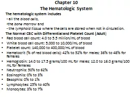
The hematologic system includes all the blood cells the bone marrow and the lymphoid tissue where the cells are stored when not in circulation The Normal CBC with Differential and Platelet Count Adult ID: 909767
Download Presentation The PPT/PDF document "Chapter 10 The Hematologic System" is the property of its rightful owner. Permission is granted to download and print the materials on this web site for personal, non-commercial use only, and to display it on your personal computer provided you do not modify the materials and that you retain all copyright notices contained in the materials. By downloading content from our website, you accept the terms of this agreement.
Slide1
Chapter 10
The Hematologic System
The hematologic system includes
- all the
blood cells
,
-the
bone marrow
and
- the
lymphoid tissue
where the cells are stored when not in circulation.
The Normal CBC with Differential and Platelet Count (Adult
)
Red blood cell count: 4.0 to 5.5 million/
mL
of blood
White blood cell count: 5,000 to 10,000/
mL
of blood
Platelet count: 140,000 to 400,000/
mL
of blood
Hematocrit
(% of red blood cells): 42% to 52% for males; 36% to 48% for females
Hemoglobin: 14.0 to 17.5 grams/100
mL
for males; 12.0 to 16.0 grams/100
mL
for females
Neutrophils
: 50% to 62%
Eosinophils
: 0% to 3%
Basophils
: 0% to 1%
Lymphocytes: 25% to 40%
Monocytes
: 3% to 7%
Slide2Tests of Red Blood Cell Size and Hemoglobin (Adult)
- MCV: 82 to 98
fL
/red cells
- MCHC: 32 to 36 g/
dL
- Sedimentation Rate(SED) Rate: 0 to 20 mm/hour
Bleeding Time
-Bleeding time refers to the length of time bleeding occurs after a standardized puncture wound to the skin.
- It indicates the effectiveness of the platelet plug.
-
Bleeding time should not exceed 15 minutes (normal: 3.0 to 9.5 minutes) .
Slide3Partial
Thromboplastin
Time/
Prothrombin
Time
Partial
thromboplastin
time (PTT) and
prothrombin
time (PT) detect deficiencies in the activity of various clotting factors.
Both tests evaluate clotting in a
venous blood
sample.
PTT should not exceed 90 seconds (normal: 30 to 45 seconds). This test is important in determining the effectiveness and safety of
heparin
therapy.
PT should not exceed 40 seconds .PT demonstrates the effectiveness of the
vitamin K dependent coagulation factors
.
PT is used to determine the effectiveness of
warfarin
(Coumadin) therapy.
Slide4Pathophysiologic
Concepts
Anemia
Is decrease in oxygen carrying ability of
blood.
1-
Anemia Caused by a Disorder in Red Cell
Production.
Occur if there is:
- inadequate
iron
,
folic acid
, or
vitamin B
12
.
- bone marrow disease, as would occur in
leukemia
,
after radiation
exposure
and
a deficiency in erythropoietin
(renal failure ).
Anemias
due to disorders in RBC production may result in a red cell that is too small (
microcytic
) or too large (
macrocytic
), and low hemoglobin (
hypochromic
).
2-Anemia Caused by Sudden or Chronic Hemorrhage or
Lysis
Result in a decrease in the total number of circulating red cells.
This type of anemia may be associated with an increased percentage of circulating immature red cells (
reticulocytes
).
-red
blood cells live approximately 120 days.
-
cell
destruction or loss occurring before 100 days is abnormal.
Slide6Polcythemia
Is an increase in the number of red blood cells.
-
Primary
polycythemia
(
polycythemia
vera
) is characterized by an increase in platelets and granulocytes as well as red blood cells, and is believed to be the result of a precursor cell abnormality.
-
S
econdary
p
olycythemia
may occur
after
chronic hypoxia. Chronic hypoxia causes increased release of the renal hormone erythropoietin, which stimulates the production of red blood cells.
Individuals
who live at
high altitude
or suffer from
chronic lung disease
frequently experience
secondary
polycythemia
.
Slide7Leukopenia
Leukopenia
is a decrease in the number of white blood cells.
Leukopenia
may be caused by
prolonged stress, viral infection, bone marrow disease or destruction,
(
radiation
, or
chemotherapy ).
Leukocytosis
Leukocytosis
is an increase in the number of circulating white blood cells.
Leukocytosis
is a
normal
response to infection or inflammation.
Thrombocytopenia
Is a decrease in the number of circulating platelets. It is associated with increased risk of severe bleeding. Thrombocytopenia is characterized by small spots of subcutaneous bleeding, called
petechiae
, or larger areas of subcutaneous bleeding, called
purpura
.
Slide8Thrombocythemia
-
Is an
increase
in the number of circulating platelets.
-It is associated with increased risk of thrombosis.
-Depending on the site of clot formation or trapping,
stroke, myocardial infarct, or respiratory distress
may develop
.
Lymphadenopathy
-
Is the enlargement of the lymph nodes in response to a proliferation of B or T lymphocytes.
- It typically occurs after infection by a microorganism.
- Regional
lymphadenopathy
indicates a localized infection.
- Generalized
lymphadenopathy
usually indicates a systemic infection such as
AIDS
or an
autoimmune
disorder such as rheumatoid arthritis or systemic lupus
erythematosus
(SLE).
-Occasionally,
lymphadenopathy
may indicate a malignancy.
Slide9Conditions of Disease or Injury
Anemia
Classic systemic signs of anemia are common to all of the
anemias
and include the following:
- Increased heart rate.
- Increased respiratory rate .
- Dizziness caused by decreased brain
oxygenation.
- Skin pallor .
- Nausea caused by decreased gastrointestinal and central nervous system
oxygenation.
-
Fatigue.
Slide101-Aplastic Anemia
- caused by dysfunction of the bone marrow such that dying blood cells are not replaced.
- is usually associated with a deficiency in red blood cells, white blood cells, and platelets.
Causes
:
- cancers of the bone marrow,
- vitamin deficiency,
- ingestion of many different drugs or chemicals, and
- high-dose
radiation or chemotherapy.
- various
viral infections, including mononucleosis, hepatitis, and AIDS.
- Frequently, the cause is unknown.
Slide11Clinical Manifestations
- signs of
anemia(
normocytic
,
normochromic
)
- if platelets and white
cells
are involved, additional symptoms include
:
-
Bleeding
from the gums and teeth; easy bruising, including
petechia
and
purpura
.
-
Recurrent
infection .
Poor
healing of skin and mucosal sores.
Diagnostic Tools
- CBC with differential and platelet count, MCV, and MCHC will diagnose anemia.
- Bone marrow biopsy will determine involved cells.
Complications
- Heart failure and death as a result of cardiac overload .
- Death from infection and hemorrhage
.
Treatment
- Treat underlying disorder .
- Transfusions to reduce
symptomatology
.
- Bone marrow transplant.
Slide122-Hemolytic Anemia
Hemolytic anemia is a decrease in red blood cell number caused by excessive destruction of red cells. Remaining red cells are
normocytic
and
normochromic
.
Red blood cell production in the bone marrow will increase to replace destroyed cells, and the advancement into the blood of immature red cells, or
reticulocytes
, will be accelerated.
Specific causes of hemolytic anemia include:
Slide13a-
thalassemia
It is an anemia characterized by
microcytic
,
hypochromic
, and short-lived red blood cells caused by deficient synthesis of hemoglobin polypeptide chains.
All types are transmitted by
autosomal
-recessive genes, and people of Mediterranean origin are more often affected.
The anemia is classified according to the chain involved:
-
α-
thalassemia
that caused by diminished synthesis of alpha chains of hemoglobin. The
homozygous
form is
incompatible with life
, the stillborn infant displaying severe
hydrops
fetalis
. The
heterozygous
form may be asymptomatic or marked by mild anemia
Slide14-
β-
thalassemia
that caused by diminished synthesis of beta chains of hemoglobin.
The homozygous form is called
thalassemia
major and the heterozygous form is called
thalassemia
minor.
thalassemia
major
(
Cooley,s
anemia)
the homozygous form in which beta chain is completely absent; it appears in the newborn period and is marked by hemolytic,
hypochromic
,
microcytic
- anemia,
hepatosplenomegaly
, skeletal deformation, mongoloid
facies
, and cardiac enlargement.
thalassemia
minor
the heterozygous form , usually asymptomatic, although there is sometimes mild
anemia.
Slide15Clinical manifestation
thalassemia
major
(
Cooley,s
anemia)
- Fatigue
- Pale skin
- Jaundice
- Poor bone growth
- Protruding abdomen
- Enlarged liver or spleen
Diagnostic tool
- complete blood count
- hemoglobin electrophoresis
- serum iron levels
Slide16Treatment
regular blood transfusions, usually on a monthly basis. This helps prevent severe anemia and allows for more normal growth and development.
desferoxamine
is administered, usually five nights per week over a period of several hours, using an automatic pump that can be used during sleep or taken anywhere the person goes. This medication is able to bind to the excess iron, which can then be eliminated through
urine.
bone marrow transplantation
in
which the bone marrow of an affected individual is replaced with the bone marrow of an unaffected donor.
Slide17b-Hemolytic Disease of the Newborn
Is a
normocytic
,
normochromic
anemia seen in an Rh-positive fetus or infant born to an Rh-negative mother who has previously developed antibodies to the
Rh
antigen.
Clinical Manifestations
Mild hemolytic disease may be relatively asymptomatic, with slight
hepatomegaly
and minimally elevated
bilirubin
.
Moderate and severe disease manifest with pronounced signs of anemia.
Hyperbilirubinemia
, resulting from excessive red cell
lysis
, may occur, leading to jaundice.
Complications
-
Kernicterus
:
is
a form of brain damage caused by excessive jaundice
.
- Severe anemia may cause heart failure.
-
Hydrops
fetalis
. Affected fetuses often abort spontaneously at approximately 17 weeks' gestation.
Slide18Treatment
- If, after birth, the
infant is
Rh
positive and the woman is still
Rh
negative, she is again given
RhoGAM
within 72 hours.
- The fetus is observed by serial amniocentesis to determine
bilirubin
level.
-Mildly affected fetuses are delivered at term;
- Moderately affected fetuses may be delivered before term.
- Severely affected fetuses may receive an intrauterine transfusion and be delivered before term.
-In the newborn with hemolytic disease, exchange blood transfusions may be required.
- In mild cases, phototherapy to reduce the levels of
unconjugated
bilirubin
may be sufficient.
Slide19c-Transfusion Reaction
Is destruction of incompatible red blood cells received in a blood transfusion.
Clinical Manifestations
- Immediate flushing of the face.
- A feeling of warmth in the vein receiving the blood.
- Fever and chills.
- Chest, flank, or low back pain.
- Abdominal pain with nausea and vomiting.
- Decreased blood pressure with increased heart rate.
-
Dyspnea
(a sensation of breathing difficulty).
Slide20Complications
Renal failure may result from red blood cell casts and hemoglobin obstruction of the
nephrons
.
Treatment
- The transfusion must be stopped immediately.
- Fluids may be given to reduce the risk of renal damage.
- Anaphylactic responses are treated by anti-inflammatory drugs, including antihistamines and steroids.
Slide213
-
Posthemorrhagic
Anemia
Is a
normocytic
,
normochromic
anemia that results from sudden loss of blood. The hemorrhage may be obvious or hidden.
Clinical Manifestations
- Increased heart rate and respiratory rate, with a decrease in blood pressure.
- Consciousness may be impaired.
Diagnostic Tools
Reduction in red cell count,
hematocrit
, and hemoglobin .
Complications
Hypovolemic
shock with the possibility of renal failure, respiratory failure, or death.
Treatment
Restore blood volume with intravenous infusion of plasma or type-matched whole blood (or O negative). Saline or albumin may also be infused.
Slide224-Pernicious Anemia
Is a
megaloblastic
anemia characterized by abnormally large red cells.
Caused by a deficiency of vitamin B
12
which
is essential for
- DNA synthesis in red blood cells
-
neuronal functioning.
Vitamin B
12
is provided in the diet and absorbed across the stomach by a gastric hormone,
intrinsic factor
.
Most cases of pernicious anemia result from :
-
intrinsic factor deficiency
- dietary deficiency
of vitamin
B
12
.
-
Surgical removal
of all or part of the stomach .
Slide23Clinical Manifestations
- Systemic signs of anemia.
- Dementia related to neurologic deterioration.
- Ataxia (poor muscle coordination) and sensory loss resulting from myelin degeneration.
Diagnostic Tools
- large cells with immature (
blastic
) nuclei
- Elevated MCV > 103, normal MCHC.
- A decrease in serum B12 will confirm the disease.
Complications
Severe anemia may cause heart failure, especially in the elderly.
Treatment
- diet rich in b12 as
liver
(best source);
beef;
chicken;
fish
; whole
egg ,milk
; cheese;
yoghurt.
-
Lifelong
intramuscular
injections of vitamin
B12 if
intrensic
factor is deficient.
Slide245-Folate-Deficiency Anemia
Is a
megaloblastic
anemia characterized by enlarged red cells with immature nuclei.
- Folic acid is essential for red blood cell production and maturation.
- It is also important for DNA and RNA synthesis .
- Folic acid is provided in the diet, absorption occurs across the small intestine and does not require intrinsic factor.
Clinical Manifestations
Systemic signs of anemia are present.
Slide25Diagnostic Tools
- Elevated MCV > 98, normal MCHC.
- Typically, the MCV will be elevated less than in pernicious anemia, and there will be no vitamin B deficiency.
Complications
- Maternal deficiencies in folic acid are associated with an increased risk of fetal malformations, especially neural tube defects.
Treatment
- Administration of oral
folate
.
Meat
,Legumes ,Starches
Fruit and Vegetables
.
Fruit
and vegetables should be eaten raw whenever possible as cooking destroys Folic
Acid.
- Women intending to become pregnant should begin vitamin supplementation at least 3 months before conception.
- Blood transfusions may be required in severe cases.
Slide266-Iron-Deficiency Anemia
Iron-deficiency
anemia is a
microcytic-hypochromic
anemia that results from :
- a diet deficient in iron,
- or from the slow, chronic loss of blood.
Clinical Manifestations
-Systemic signs of anemia .Individuals usually do not seek treatment for symptoms until hemoglobin decreases to 8 g/100
mL
or below.
- Pale palms, pale conjunctivae, and pale earlobes may also be present
.
Slide27Diagnostic Tools
-
Microcytic
cells (MCV < 87) and decreased serum iron.
- Iron-binding capacity in the blood is high .
- Stool test for occult blood may be positive.
Complications
A hemoglobin value of less than 5 g/100
mL
can lead to heart failure and death.
Treatment
- An iron-rich diet containing red meat and dark green vegetables, such as spinach.
- Oral iron supplementation.
- Treat the cause .
Slide28Leukemia
Is a cancer of one class of white blood cells in the bone marrow, which results in the proliferation of that cell type preventing other blood cells from developing normally. This reduces blood levels of all
nonleukemic
cells.
Types of Leukemia
Leukemia is described as
acute or chronic
, depending on the suddenness of appearance and how well differentiated the cancerous cells are.
The cells of
acute leukemia
are poorly differentiated.
The of
chronic leukemia
are usually well differentiated.
Leukemia is also described based on the proliferating
cell type
.
-
Acute lymphoblastic
leukemia, the most common childhood leukemia, describes a cancer of lymphocyte cell line.
-
Granulocytic
leukemias
are
leukemias
of the
eosinophils
,
neutrophils
, or
basophils
.
Leukemia in adults is usually chronic lymphocytic or acute
myeloblastic
.
Slide29Clinical Manifestations
- Acute leukemia has marked clinical manifestations.
- Chronic leukemia progresses slowly and may have few symptoms until advanced.
- Pallor and fatigue from anemia.
- Frequent infections caused by a decrease in white blood cells.
- Bleeding and bruising caused by thrombocytopenia and coagulation disorders.
- Bone pain caused by accumulation of cells in the marrow.
- Weight loss caused by poor appetite and increased caloric consumption
-
Lymphadenopathy
,
splenomegaly
, and
hepatomegaly
caused by leukemic cell infiltration of these lymphoid organs may develop.
Slide30Diagnostic Tools
- Alterations in specific blood cell counts.
- Bone marrow tests demonstrate
clonal
proliferation and blood cell accumulation.
Treatment
- Multiple drug chemotherapy.
- Antibiotics to prevent infection.
- Transfusions of red blood cells and platelets to reverse anemia and prevent bleeding.
- Bone marrow transplant may successfully treat the disease.
Slide31Hodgkin Lymphoma
(Hodgkin's Disease)
Is a cancer of the lymphoid tissue, usually the lymph nodes and spleen. It is one of the most common cancers in young adults, especially young males.
The
abnormal cell population appears to be derived from a B cell or, less frequently, a T cell or
monocyte
.
Staging of Hodgkin lymphoma is important because it
-guides treatment
- and strongly influences outcome.
The early stages of the disease, stages I and II, are usually curable. Cure rates for stages III and IV are approximately 75% and 60%, respectively.
The cause of Hodgkin lymphoma is unknown.
Slide32Diagnostic Tools
Lymph node biopsy can diagnose Hodgkin lymphoma.
Complications
Secondary malignancies and
cardiotoxicity
may develop after aggressive treatment. Because of these and other treatment complications, Hodgkin lymphoma patients have a higher chance of dying from acute and late treatment toxicities than from the disease itself.
Treatment
- Multidrug chemotherapy.
- Radiation therapy.
- Bone marrow transplant.
Slide33Hemophilia A
Hemophilia A, also called classic hemophilia, is an X-linked recessive disease resulting from deficient coagulation factor VIII.
It is the most common inherited coagulation disorder. It is seen in boys who inherit the defective gene on the X chromosome from their mother. The mother is usually heterozygous for the disorder and shows no symptoms.
Without factor VIII, the intrinsic coagulation pathway is interrupted and extensive bleeding from small wounds or
microvascular
tears occurs.
Bleeding is frequently into the joints and can cause significant pain and disability.
Slide34Other Types of Hemophilia
These
hemophilias
result from the absence of different coagulation factors.
Hemophilia B is caused by a lack of factor IX.
Hemophilia C is caused by a lack of factor XI.
Von
Willebrand
disease results from an abnormality of von
Willebrand
factor (
vWF
). This factor is released from endothelial cells and platelets and is essential for the formation of the platelet plug. .
Clinical Manifestations of Classic Hemophilia
-
Spontaneous or excessive bleeding after a minor wound.
-
Joint swelling, pain, and degenerative changes
Slide35Diagnostic Tools
-
Laboratory studies show a normal bleeding time, but prolonged PTT.
-
Measurement of factor VIII is reduced.
-
Prenatal testing for the gene is possible.
Complications
-
Intracranial hemorrhage may occur.
-
Infection with HIV was common before artificial production of factor VIII reduced the need for transfusions.
Treatment
Factor VIII replacement.
Slide36Liver Disease and Vitamin K Deficiency
-The liver is the site of synthesis for many coagulation factors, several of which are vitamin K dependent.
-Disease of the liver or inadequate plasma levels of vitamin K will interrupt the coagulation pathways.
- Vitamin K is a fat-soluble vitamin absorbed in the diet by means of bile. Because bile is produced in the liver, a healthy liver and a clear bile duct are required for successful coagulation.
-Vitamin K also is synthesized by bacteria in the gut.
Slide37Newborns are vitamin K deficient because of
a lack of vitamin K producing bacteria in the intestine
and immature liver function.
Clinical Manifestations
Bleeding characterized by
petechia
(small hemorrhage spots on the skin) and
purpura
(purplish discoloration of the skin).
Treatment
Vitamin K is administered intramuscularly to the neonate and orally in children or adults
.
Slide38Disseminated Intravascular Coagulation
(DIC)
Is a unique condition characterized by the formation of multiple blood clots throughout the microvasculature.
Eventually, the components of the blood clotting cascade and the platelets are
used up
, and hemorrhages begin to occur at all bodily orifices, at sites of injury or venous puncture, and throughout many organ systems.
DIC is
never a primary condition
. Instead, it occurs as a complication of major clinical incidents or trauma such as shock, widespread infection, major burn, myocardial infarct, or obstetric complication.
Multiple endothelial cell injuries initiate extensive activation of the platelets and the intrinsic coagulation pathway, leading to
microthrombi
throughout the vascular system.
Slide39Clinical Manifestations
Hemorrhage from puncture sites, wounds, and mucous membranes in a patient with shock, obstetric complications, sepsis (widespread infection), or cancer.
Altered consciousness indicates a cerebral thrombus.
Abdominal distention indicates a GI bleed.
Cyanosis and
tachypnea
(increased respiratory rate) caused by poor tissue perfusion and oxygenation are common.
Hematuria
(blood in urine) caused by hemorrhage or
oliguria
(decreased urine output) caused by poor renal perfusion.
Slide40Diagnostic Tools
- Blood tests demonstrate accelerated clotting and decreased platelets.
- Platelets and plasma fibrinogen levels are reduced.
Complications
Obstruction to blood flow in all organs of the body.
Widespread organ failure may occur.
Mortality is greater than 50%.
Treatment
- Removal of the precipitating event.
- Heparin therapy may be initiated .Heparin is not suggested when central nervous system bleeding occurs.
- Fluid replacement is important to maintain organ perfusion as high as possible.
- Plasma containing factor VIII, red cells, and platelets may be administered.