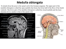
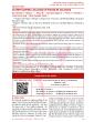
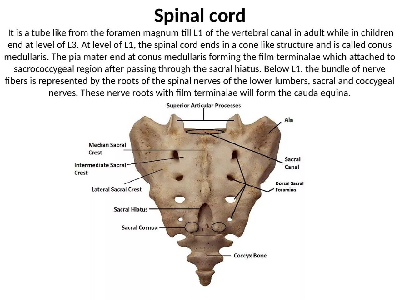
/
Spinal cord  It is a tube like from the foramen magnum till L1 of the vertebral canal Spinal cord  It is a tube like from the foramen magnum till L1 of the vertebral canal

Spinal cord It is a tube like from the foramen magnum till L1 of the vertebral canal - PowerPoint Presentation

catherine
catherine . @catherine
Follow
342 views
Uploaded On 2022-06-15

Spinal cord It is a tube like from the foramen magnum till L1 of the vertebral canal - PPT Presentation

terminalae which attached to sacrococcygeal region after passing through the sacral hiatus Below L1 the bundle of nerve fibers is represented by the roots of the spinal nerves of the lower lumbers sacral and coccygeal nerves These nerve roots with film ID: 919223

region spinal sacral cord spinal region cord sacral mater motor nerves neurons medullaris conus level vertebra segments plexus anterior

Share:

Link:

Embed:

Download Presentation from below link

Download Presentation The PPT/PDF document "Spinal cord It is a tube like from the ..." is the property of its rightful owner. Permission is granted to download and print the materials on this web site for personal, non-commercial use only, and to display it on your personal computer provided you do not modify the materials and that you retain all copyright notices contained in the materials. By downloading content from our website, you accept the terms of this agreement.


Presentation Transcript

Slide1

Spinal cord It is a tube like from the foramen magnum till L1 of the vertebral canal in adult while in children end at level of L3. At level of L1, the spinal cord ends in a cone like structure and is called conus medullaris. The pia mater end at conus medullaris forming the film terminalae which attached to sacrococcygeal region after passing through the sacral hiatus. Below L1, the bundle of nerve fibers is represented by the roots of the spinal nerves of the lower lumbers, sacral and coccygeal nerves. These nerve roots with film terminalae will form the cauda equina.

Slide2

■ Lumber puncture is performed below L1. The dura and arachnoid mater end at the level of second sacral vertebra. The subarachnoid space caudal to the conus medullaris is known as lumbar cistern.

Slide3

Slide4

The spinal cord is formed of thirty-one segments, eight segments in cervical region, twelve segments in the thoracic region, five in lumber region, five in sacral region and one in coccygeal region. Above C8

spinal segment, the spinal nerves emerge from above the corresponding vertebra. While below C

8

spinal segment, the spinal nerves emerge from below the corresponding vertebra.

The spinal cord has cervical enlargement (from C4-T1) for brachial plexus, other swelling at (L2-S3) for lumbosacral plexus.

Slide5

Slide6

The pia mater forms an extension to dura mater to form the denticulate ligaments.

Slide7

The gray matter of the spinal cord is similar to butter fly, has two dorsal or posterior horns, intermediate horn and two anterior or ventral horns.

Anterior horn:

It has a motor function and it is composed of two groups of cell bodies or neurons:

Alpha motor neurons (the highest number).

Gamma motor neurons.

■ Polio virus that cause poliomyelitis affect alpha motor neurons causing paralysis.