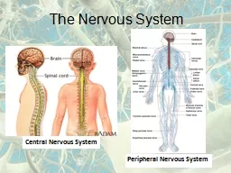
/
Chapter 44 Care of Patients with Problems of the Central Nervous System: The Brain Chapter 44 Care of Patients with Problems of the Central Nervous System: The Brain

Chapter 44 Care of Patients with Problems of the Central Nervous System: The Brain - PowerPoint Presentation

celsa-spraggs
celsa-spraggs . @celsa-spraggs
Follow
377 views
Uploaded On 2018-11-21

Chapter 44 Care of Patients with Problems of the Central Nervous System: The Brain - PPT Presentation

Headaches Migraine headache chronic episodic disorder with multiple subtypes Stages Prodrome Aura phase Headache phase Interventions Recognize migraine symptoms Respond and see health care provider ID: 731570

seizures disease drug seizure disease seizures seizure drug management therapy patient brain meningitis interventions patient

Share:

Link:

Embed:

Download Presentation from below link

Download Presentation The PPT/PDF document "Chapter 44 Care of Patients with Problem..." is the property of its rightful owner. Permission is granted to download and print the materials on this web site for personal, non-commercial use only, and to display it on your personal computer provided you do not modify the materials and that you retain all copyright notices contained in the materials. By downloading content from our website, you accept the terms of this agreement.


Presentation Transcript

Slide1

Chapter 44

Care of Patients with Problems of the Central Nervous System: The BrainSlide2

Headaches

Migraine headache

chronic, episodic disorder with multiple subtypes

Stages:

Prodrome

Aura phase

Headache phaseSlide3

Interventions

Recognize migraine symptoms

Respond and see health care provider

Relieve pain and associated symptoms Slide4

Drug Therapy

Abortive therapy

alleviating pain during the early aura phase or soon after the headache has started

Preventive therapySlide5

Complementary and Alternative Therapies

Yoga, meditation, massage, exercise, biofeedback, relaxation techniques

Acupuncture

Use of herbs and nutritional therapies with approval

Avoidance of trigger events that may result in migraine episodes, such as tension and stressSlide6

Cluster Headache

Histamine cephalalgia

Cause unknown; attributed to vasoreactivity and oxyhemoglobin desaturation

Unilateral, radiating to forehead, temple, or cheek

Ipsilateral tearing of the eye, rhinorrhea, ptosis, and miosisSlide7

Therapy

Same types of drugs used for migraines

Patient to wear sunglasses and avoid sunlight

Oxygen via mask

Avoidance of precipitating factors, such as anger, excitement

Surgical managementSlide8

Tension Headache

Neck and shoulder muscle tenderness and bilateral pain at the base of the skull and in the forehead

Head pain without associated symptoms

Treatment

non-opioid analgesics, muscle relaxants, occasional opioids

Ibuprofen plus caffeine

Prophylactic treatment similar to that used in treating migraine headachesSlide9

Seizures and Epilepsy

Seizure

abnormal, sudden, excessive, uncontrolled electrical discharge of neurons within the brain; may result in alteration in consciousness, motor or sensory ability, and/or behavior

Epilepsy

—t

wo or more seizures experienced by a person; chronic disorder with recurrent, unprovoked seizure activity, may be caused by abnormality in electrical neuronal activity and/or imbalance of neurotransmitters (e.g., GABA)Slide10

Types of Seizures

Generalized seizures

Partial seizures

Unclassified seizuresSlide11

Types

Primary or idiopathic epilepsy

not associated with any identifiable brain lesion

Secondary seizures

result from an underlying brain lesion, most commonly a tumor or traumaSlide12

Seizures Risks

Seizures may result from:

Metabolic disorders

Acute alcohol withdrawal

Electrolyte disturbances

Heart disease

High fever

Stroke

Substance abuseSlide13

Nonsurgical Management

Antiepileptic drugs (AEDs)

Importance of compliance

Health teachingSlide14

Seizure Precautions

Oxygen

Suction equipment

Airway

IV access

Siderails up

No tongue bladesSlide15

Seizure Management

Will depend on the type of seizure

Observation and documentation

Patient safety

Side-lying position

No restraints Slide16

Acute Seizure Management

Lorazepam

Diazepam

Diastat

IV phenytoin or fosphenytoinSlide17

Status Epilepticus

Medical emergency

Prolonged seizures lasting more than 5 minutes or repeated seizures over the course of 30 minutes

Establish an airway

ABGs

IV push lorazepam, diazepam; rectal diazepam

Loading dose IV phenytoinSlide18

Drug Therapy

Evaluate most current blood level of medication, if appropriate.

Be aware of drug-drug and drug-food interactions.

Maintain therapeutic blood levels for maximal effectiveness.Slide19

Drug Therapy (Cont’d)

Do not administer warfarin with phenytoin.

Document and report side and adverse effects.Slide20

Patient and Family Education

Antiepileptic drugs (AEDs) may not be stopped, even if seizures stop.

Refer limited-income patients to social services.

All states prohibit discrimination against people who have epilepsy.Slide21

Patient and Family Education (Cont’d)

Alternative employment may be needed.

Vocational rehabilitation may be subsidized.Slide22

Seizure Precautions

Oxygen and suctioning equipment should be readily available.

Saline lock may be necessary.

Siderails should be

up

at all times.

Padded siderail use is controversial.

Place bed in lowest position.

Never insert padded tongue blades into

the patient’s mouth during a seizure.Slide23

Seizure Management

If simple partial seizure, observe the patient and document the seizure.

Turn the patient on the side during a generalized tonic-clonic seizure; if possible, turn the patient’s head to prevent aspiration.

Cyanosis usually is self-limiting.

Do not restrain.Slide24

Surgical Management

Vagal nerve stimulation (VNS)

Conventional surgical procedures

Anterior temporal lobe resection

Partial corpus callosotomy Slide25

Meningitis

Meningitis

inflammation of the meninges that surround the brain and spinal cord

Viral meningitis

usually self-limiting and the patient has a complete recovery

Bacterial meningitis

potentially life-threateningSlide26

Physical Assessment and Clinical Manifestations

Signs and symptoms of meningitis

headache, nausea, vomiting, and fever

Photophobia and indications of increased intracranial pressure

Nuchal rigidity and positive Kernig’s and Brudzinski’s signs

Seizure, decreased mental status, focal neurologic deficitsSlide27

Laboratory Assessment of Meningitis

Cerebrospinal fluid analysis

Computed tomography scan

Blood cultures

Counterimmunoelectrophoresis

Polymerase chain reaction

Complete blood count

X-ray study to determine presence of infectionSlide28

Drug Therapy

Broad-spectrum antibiotic

Hyperosmolar agents

Anticonvulsants

Steroids (controversial)

Prophylaxis treatment for those who have been in close contact with the meningitis-infected patientSlide29

Encephalitis

Inflammation of the brain tissue and surrounding meninges

Caused by viral agents, bacteria, fungi, or parasites

Degeneration of neurons of the cortex

Hemorrhage, edema, necrosis, small lacunae develop in cerebral hemispheresSlide30

Hemorrhagic EncephalitisSlide31

Interventions

Prompt recognition and treatment of signs of cerebral edema, hemorrhage, and necrosis of brain tissue

Establishment of patent airway

Assessment of vital signs

Continuous supportive care and assessmentSlide32

Parkinson Disease

Progressive neurodegenerative disease that is the third most common neurologic disorder of older adults

Tremor, rigidity, bradykinesia, or akinesia

DopamineSlide33

Parkinson Disease (Cont’d)Slide34

Assessment

Fatigue, slight tremor, problems with manual dexterity

Rigidity, changes in facial expression, uncontrolled drooling, dementia, changes in voluntary movement, excessive perspiration, orthostatic hypotension

No specific diagnostic testsSlide35

Drug Therapy in Parkinson Disease

Dopamine agonists

Catechol

O

-methyltransferases (COMTs)

Monoamine oxidase type B (MAO-B) inhibitors

Dopamine receptor antagonistsSlide36

Drug Toxicity

Long-term drug therapy regimens often cause delirium, cognitive impairment, decreased effectiveness of the drug, or hallucinations.

Reduce medication dose.

Change medications or frequency of administration.

Take “drug holiday,” especially in the use of levodopa therapy.Slide37

Management of Parkinson Disease

Exercise and ambulation

Self-management

Injury prevention

Nutrition

Communication

Psychosocial supportSlide38

Management of Parkinson Disease

(Cont’d)

Surgical management includes:

Stereotactic pallidotomy/thalamotomy

Deep brain stimulation

Fetal tissue transplantationSlide39

Alzheimer’s Disease

Chronic, progressive, degenerative disease that accounts for 60% of dementias occurring in people older than 65 years

Loss of memory, judgment, and visuospatial perception and change in personality

Increasing cognitive impairment, severe physical deterioration, death from complications of immobilitySlide40

Structural Changes in the Brain

Alzheimer’s disease creates changes that include:

Neurofibrillary tangles

Neuritic plaques

Vascular degeneration

Changes in neurotransmitters

Increased amounts of an abnormal protein, beta amyloid

Slide41

Manifestations

Changes in cognition

Alterations in communication and language abilities

Changes in behavior, personality, and judgment

Changes in self-care skills

Psychosocial assessment, especially patient’s reaction to changes in routineSlide42

Interventions in Alzheimer’s Disease

Answer patient’s questions truthfully.

Assess and treat other medical problems.

Provide cognitive stimulation and memory training.

Structure the environment to increase patient’s ability to function.

Prevent overstimulation.Slide43

Interventions

Orientation and validation therapy.

Promote self-management.

Promote bowel and bladder continence.

Assist with facial recognition.

Promote communication.Slide44

Drug Therapy

Donepezil, galantamine, rivastigmine

Memantine

Antidepressants

Psychotropic drugsSlide45

Risk for Injury

Interventions for the patient with Alzheimer’s disease include:

Coping with restlessness and wandering; ensuring patient wears identification bracelet; registering patient in Safe Return Program; providing frequent walks and structured activities

Slide46

Risk for Injury (Cont’d)

Ensuring safety by removing all potentially dangerous objects, particularly in case seizures occur

Minimizing agitation by talking calmly and softly; displaying positive affect; making calm movements; offering diversionSlide47

Compromised Family Coping

Interventions for the caregiver role:

Encourage family to seek legal counsel regarding patient’s competency, need to obtain guardianship, or durable medical power of attorney, when necessary.

Make caregivers and family aware of their own health and stress resulting from new responsibilities for care.Slide48

Disturbed Sleep Pattern

Difficulty sleeping at night with frequent naps in the day

Interventions for establishing sleep pattern:

Re-establish the usual day-night pattern by providing activity and exercise during the day.

Establish before-bedtime ritual.

Slide49

Disturbed Sleep Pattern (Cont’d)

Adjust treatment and medication schedule to provide for uninterrupted sleep.

Give mild antianxiety agent or hypnotic.Slide50

Huntington Disease

Hereditary disorder transmitted as an autosomal dominant trait at the time of conception

Movement disorder characterized by both neurologic and behavioral symptoms

Gradual clinical onset of progressive mental status changes, leading to dementia and choreiform movements in the limbs, trunk, and facial muscles

Three stages, each lasting about 5 years over an average 15 years of the diseaseSlide51

Management of Huntington Disease

No known cure or treatment

Genetic counseling

Antipsychotic agents or monoamine-depleting agents used to manage movement abnormalities that are disabling or interfere with ADLs

Medications to treat depression, anxiety, and obsessive-compulsive behaviors