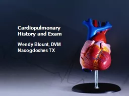
BY DRVIDHU MITTAL JUNIOR RESIDENT DEPTT OF CHEST AND TB Anterior lung surface markings REMEMBER 246810 Lungs Each lung extends 3cm above the clavicle apex Anterior borders of lungs are closest at the sternal angle 2 ID: 363344
Download Presentation The PPT/PDF document "PERCUSSION AND AUSCULTATION" is the property of its rightful owner. Permission is granted to download and print the materials on this web site for personal, non-commercial use only, and to display it on your personal computer provided you do not modify the materials and that you retain all copyright notices contained in the materials. By downloading content from our website, you accept the terms of this agreement.
Slide1
PERCUSSION AND AUSCULTATION
BY DR.VIDHU MITTAL
JUNIOR RESIDENT
DEPTT. OF CHEST AND TBSlide2
Anterior lung surface markings
REMEMBER: 2,4,6,8,10 Lungs
Each lung extends 3cm above the clavicle (apex)
Anterior borders of lungs are closest at the sternal angle – 2nd costal cartilage (cc) Both reach to 4thccLeft:Moves away from the midline at the 4th ccRight: Moves away from the midline at the 6th ccBoth cross the midclavicular line at the 8th ccBoth cross the midaxillary line at the 10th cc
Note about pleura: They have the same surface markings as the lungs but reach further down to the 12
th
cc
REMEMBER: 2,4,6,8,10,12 PleuraSlide3Slide4Slide5
Percussion
Historical note
Amongst the notable advances of 18th century, one is the introduction of a new method of clinical investigation i.e. percussion by “Joseph Leopold Auen brugger in 1761. It was the simple expedient of tapping the barrels of wine in his father’s cellar to determine their contents that gave Auen brugger the idea of percussion of the thorax as an aid to diagnosis, a discovery first announced in 1761, in his Inventum Novum.Slide6
Definition:
Percussion is a method of examination which depends on the interpretation of sounds heard and the sense of resistance encountered on subjecting the chest to a series of strokes or taps with the fingers .
Main purposes of respiratory percussion :
Diagnostic Percussion.Topographical Percussion.Slide7
Methods of Percussion
Direct (immediate)
Where the strokes are aimed directly at the chest wall.
Percussion over bony structure.Indirect (Intermediate)
Where the stroke are aimed at some intermediate object (e.g. a finger) applied to surface of chest wall.Slide8Slide9
The other special types of percussion are:
Light percussion :
on the clavicles is useful to determine the character of the lung substance at the apices.Heavy percussion : by using two fingers instead of one or using several fingers without any intermediate finger.(c) Flicking percussion: it is useful for eliciting metallic resonance in case of pneumothorax
.(d) Direct or indirect palpatory percussion: it may be used for detecting the presence of fluid or consolidation within the chest.Slide10
TECHNIQUE OF PERCUSSION:
Position of Patient
: The patient may be
percussed in the sitting, standing or recumbent position. Recumbent position: best avoided The standing position : tiring both to patient and examine.The position of choice is sitting up posture.Slide11
IDEAL POSTURES AUAUSCULTATION OF RESPECTIVE AREAS
Area to be
percussed
Posturea) Front wall of chestPatients sits on a stool opposite the examiner with the body bolt upright, completely relaxed and with sides symmetrical b) Back
Patients bends slightly forwards with the head flexed on the chest, the shoulders sagging and the arms resting, either crossed or uncrossed, on the thighs. c) Interscapular / scapular regionPatient is directed to place his hands over the shoulders after crossing the arms in front of the chest.d)
Axillae
Patient
is instructed to put his hands over the head. Slide12
AREAS OF EXAMINATIONSlide13
Direction of percussion:
It is customary to start percussion to at the apices of the lungs comparing identical or corresponding area on 2 sides and slowly proceeding downwards.
Cardinal Rules of Technique
:The pleximeter, the middle phalanx of the 3rd finger of the examiner’s left hand must be firmly applied to the chest wall, so that no air pockets are interposed between the finger and chest wall.Slide14Slide15
The ‘
plesser
’ which is the third finger of the examiner’s right hand, is kept flexed at a right angle and must hit the middle phalanx of the
pleximeter finger perpendicularly, with the pad and not the tip of the finger. The percussion stroke must be sudden, the plessor finger being withdrawn immediately after the stroke, to prevent a damping of the note.
The movement of percussion must originate at the wrist and not at the elbow or finger. The force of the stroke must be varied according to the purpose of the percussion the tissue or organ being percussed , thickness of the chest wall or area of the chest wall.Slide16
Percussion should proceed from resonant to dull area or from more resonant to less resonant areas whenever possible the auditory appreciation of any change of note being better in this direction.
Whenever delineating the border of an organ, such as the heart or the liver, the long axis of the pleximeter finger must be kept parallel to the expected position of that border. The area percussed must be more or less equidistant from the two ears of the examiner, in order to prevent wrong interpretation of sounds; the examiner must therefore directly face the centre of the patient chest, whenever possible.Slide17Slide18Slide19
Types of percussion notes:
Normal lung resonance (Vesicular resonance)
:
The normal percussion note of the chest is due to an underlying lung tissue, containing a normal amount of air in the air vesicles, air sacs and air passages which has a distinctive and clear character with a low pitch.Slide20
Abnormal types of percussion notes
Quantitatively different
Tympany SubtympanyHyperresonance
Impairment of note dullness stony dullness
Qualitative different
Cracked pot resonance
Amphoric
resonance
Bell
tympanySlide21
Tympany
:
Drum like note.
When such a note in heard once a region of the chest mall, the possibility of a superficial cavity in the lung or pneumothorax should be considered.Subtympany (Skodaic
resonance) : Hyperresonant note with boxy quality.heard just above the level of a pleural effusion or pneumonic consolidation .Slide22
Hyperresonance
:
A note intermediate in pitch between normal lung resonance and tympany which can be elicited normally by percussing in full inspiration. Pathologically it can be encountered either B/L in case of emphysema. U/L in pneumothorax, large
bullae or compensatory emphysema.Impaired note : When part of the lung becomes comparatively airless, it fails to vibrate sufficiently to the percussion stroke and gives rise to an impairment of note or loss of resonance. Slide23
Flatness (absolute dullness)
: A percussion note completely devoid of resonance a displaying absolute or extreme dullness in referred to as flat dull notes.
Stony dullness: A type of absolutely dull percussion note associated with pain when percussing in the examiner’s pleximeter finger as one would experience when
percussing oves stone. Slide24Slide25
TYPE
OF PERCUSSION NOTE
SEEN
IN TympaniticGas containing hollow viscera.Subtympany Above pleural effusion or consolidation HyperresonantPneumothoraxResonant
Normal aerated lungImpairedPulmonary fibrosis, sometimes consolidation or collapse.DullThickened pleura, consolidation, collapse
Flat
Pleural
effusion
Stony dull
Massive growth
in lung or pleuraSlide26
OTHER ABNORMAL PERCUSSION NOTES
Cracked pot resonance
: tympanic resonance which is due to the sudden expulsion of air from the cavity into the bronchus through the narrow opening or communication.
Amphoric resonance: A low pitched and hollow note that can be elicited in pneumothorax or large cavity in the lung.
Bell tympany Slide27
Topographic Percussion of Lungs
Percussion of the chest to determine the boundaries or extent of lungs is referred to as topographic percussion . It can be :-
APICAL percussion
BASAL percussionTIDAL percussionSlide28
Apical percussion
: Can be carried out in the
supraclavicular
fossae anteriorly by determining the upper borders of lung resonance on the two sides. decreased/absent supraclavicular
zone of resonance on one orBoth side. increased extent of resonance
bilateral suggests.
good evidence of pulmonary tuberculosis.
emphysemaSlide29
An alternative method
:
Kronig’s
isthmus :- which is a band of 5-7cm in width of resonance connecting the large zones of lung resonance over the anterior and posterior aspects of each side. .
Decreased / absent kronig’s istmusSuggestive of apical tuberculosisIncrease in width Suggestive of emphysemaSlide30
Basal Percussion
:
Percussion of lower border of the lung necessitates light percussion
anteriorly and heavy percussion at the back (because of thicker musculature)A change of note from vesicular resonance to dullness (posteriorly, on both sides and anteriorly, on the right side). From vesicular resonance to
tympany when percussing downwards over the chest, serves to delineate the lower or basal limit of lung resonanceSlide31
Applied :
lower border of the lung resonance tends to be depressed in case of emphysema or
pneumothorax
raised in case of lung fibrosis, collapsed lung, consolidation, ascites, massive abdominal tumor or pleural effusion.Tidal percussion: Percussion of the lower border of lung resonance, on each side, at the height of deep inspiration and expiration serves to determine the extent of diaphragmatic excursion.decreased or restricted movement suggests some underlying lung disease such as pulmonary fibrosis.Slide32
Areas of abnormal percussion note in health
:
Area of cardiac dullness.
Area of liver dullness. Area of splenic dullness
Traube’s area. Ewart’s signSlide33Slide34
Special
Percussional
findings in disease
: Reduction of both cardiac and liver dullness. Shifting dullness. Obliteration of Traube’s area.
William’s tracheal resonance. Wintrich’s sign
Gerhardts
’ sign
Friedreich’s
sign
Lines of Demarcation
Myotatic
irritability. Slide35
AUSCULTATIONSlide36
HISTORICAL NOTE
A discovery of the greatest importance in the early part of 19
th
century was that of auscultation with the aid of stethoscope by the French physician LAENNEC . Remembering a well known acoustic fact that “ if the ear be applied to one end of a plank , it is easy to hear a pin’s scratching on the other end .”Slide37
UNAIDED EAR
TYPE
OF SOUND
FOUND IN 1- STERTOROUS breathing coma and sleep( snoring) 2- RATTLING breathing ineffective cough due to suppression of cough reflex 3- GASPING , GRUNTING and SIGHING physical and emotional stimuli- exercise , pain, cold fear , grief 4-HISSING ( KUSSMAULS ) breathing
signifies hyperventilation without dyspnoea. sign of severe acidosis as in diabetic keto acidosis 5-WHEEZING asthma 6- STRIDOR
narrowing
of
extrathoracic
airway Slide38
OBJECTIVES OF AUSCULTATION
To determine whether the breath sounds are equal on both sides .
To ascertain the character of the breath sounds .
To detect any added sounds and decide their nature and whether they are intra or extrapulmonary.To compare the voice sounds on different parts of lungs.Slide39
METHOD OF BREATHING
Patient is asked for forced or deep breathing , through the mouth as this increases the tidal volume sufficiently without producing additional upper airway noise or provoking
hypocapnia
.Slide40
DEFECTIVE AUSCULTATION OF RESPIRATORY SOUNDS
Auscultation may be defective if the patient is
Breathing through the nose , especially in the presence of nasal obstruction.
Breathing noisily or too forcibly giving added sounds .Shallow breathing due to pain on breathing .Hair on the chest produces crackling sounds which may be mistaken for lung sounds .Slide41
POSITION OF THE PATIENT
Ideal posture - upright , either sitting or standing
For examination of the back – patient may lean slightly forward , with the head flexed arms crossed in front or resting on the thighs .
Examination in the recumbent position although undesirable but may be required in seriously ill patient . Slide42
FEATURES TO NOTE DURING AUSCULTATION OF BREATH SOUNDS
Intensity or loudness.
Quality or character whether rustling or wheezy .
Comparison of inspiratory and expiratory elements in terms of intensity , duration or length and pitch. Presence or absence of intermediatory pause between them .Characteristics such as prolongation or jerky or interrupted nature .Presence of other sounds or accompanimentsSlide43
FACTORS DETERMINING TRANSMISSION OF BREATH SOUNDS
The intensity of the breath sounds heard through the chest wall depends on :-
rate of airflow into the territory of lung under the stethoscope .
The acoustic properties of the two media namely the lung and the chest wall .Slide44
Therefore..
Transmission is almost complete between the two well matched media like consolidated lung and the chest wall ,hence the similarity between the bronchial breathing and the breath sounds heard over the trachea .
Sound is reflected at the interface between the lung and air or fluid in the pleural cavity so that in
pneumothorax and in pleural effusion no breath sounds reaches the chest wall Slide45
MODE OF PRODUCTION OF NORMAL BREATH SOUNDS
Normal breath sounds are generated by turbulent airflow in the upper airways i.e. in the pharynx and larger airways of the lugs ( frequency range of 200 to 2000 Hz or cycles / sec per sound )
As this sound is transmitted through the lungs it is dampened ; the higher frequencies are lost and a softer , lower pitched sound ( 200 to 400 Hz ) is heard , which are the normal vesicular breath sounds. In the smaller airways airflow is slow and laminar , turbulence cannot be developed hence smaller airways acts as filter and not a source of lung sounds .Slide46
TYPES OF NORMAL BREATH SOUNDS
TYPE OF SOUND
AREA WHERE FOUND
VESICULAROver most areas of the chest BRONCHIALLarynx , trachea BRONCHOVESICULAROver
and around the upper part of the sternum Slide47
VESICULAR BREATH SOUNDS
It is
characterised
by active inspiration due to inflow of air into bronchi and alveoli followed without a pause by passive expiration from elastic recoil of the alveoli. QUALITY :- rustling noise louder and more prolonged in inspiration than expiration. MODE OF PRODUCTION :- It is attributed to distention and separation of the alveolar walls by the in-rushing current of air .Slide48
BRONCHIAL BREATHING
The less the filtering of the breath sounds the more closely will the sound approximate to its source in the trachea and bronchi i.e. is bronchial breathing .
QUALITY :- higher pitched and clearer than vesicular breathing .
Inspiration and expiration are of equal length or expiration is somewhat longer than inspiration and they have a distinct gap between them .MODE OF PRODUCTION :- Due to the in and out movement of the air through the narrow aperture of the glottis . The lower pitch of the sound during inspiration is due to the glottic aperture being wider during inspiration because of a wider separation of vocal cords .Slide49
BRONCHOVESICULAR BREATHING
Its a type of breathing intermediate in character in between vesicular and bronchial breathing . Its recognition usually depends on the nature of the expiratory sound which is louder , longer and higher in pitch than the
inspiratory
sound .MODE OF PRODUCTION :- it usually arises when the normal air containing lung tissue is interposed between a large bronchus and the chest wall , thus combining the characteristics of both vesicular and bronchial types of breathing .Slide50Slide51
ABNORMAL BREATH SOUNDS
Breath sounds may be heard abnormal due to two main reasons :-
ABNORMAL
GENERATIONABNORMAL CONDUCTION Narrowed airways intensify the linear velocity of breath sounds and thus increasing their turbulence making the breath sounds louder . Abnormal lung will conduct the centrally generating breath sounds abnormally for e.g. consolidated lung and the overinflated lung of emphysema.Slide52
ABNORMAL TYPES OF VESICULAR BREATHING
ABNORMALITY
PHYSIOLOGICALLY SEEN IN
PATHOLOGICALLY IN 1- Exaggerated or loud breath sounds childrenThin chested individualsIn women
Bilaterally in states of dyspnoea or bronchitis unilaterally in pulm. Tb or compensatory emphysema
2-Diminished or feeble
habitual shallow breathers
During
quiet breathing
Thick chest wall
obesity
Defective production of respiratory sounds
-defective conduction of respiratory sounds .
3-Absent breath sounds
Massive pleural
effusion ,
pneumothorax
, area of lung collapse , secondary occlusion of a bronchus Slide53
ABNORMAL TYPS OF BRONCHIAL BREATHING
TYPE OF BREATHING
QUALITY
/CHARACHTERCAN BE IMMITATED BYSUGGESTIVE OFCavernous Low pitched with hollow characterAuscultating at occipital region of skullUnderlying cavity in the lung Open pneumothoraxPulled trachea syndrome
Tubular High pitched with tubular or aspirate quality PneumoniaPulm infarctionAtelectasis or collapse of lungAmphoric
High pitched with metallic or ECHO like quality
Blowing intensely across the mouth of the bottle
Large cavity in the lungs with smooth walls
Pneumothorax
communicating with the bronchusSlide54
ABNORMAL BRONCHOVESICULAR BREATH SOUNDS
METAMORPHOSING breath sounds :- occasionally , the type of breathing may change suddenly in type, character or intensity during one and the same breath . It is usually due to dislodgement of a mucus plug that is partially occluding a bronchus , by the incoming rush of air during inspiration .Slide55
ADVENTITIOUS (ADDED) SOUNDS
Chest diseases can give rise to 3 types of added sounds , namely :-
Wheezes
CracklesPleural friction Slide56
WHEEZES
These are continuous musical sounds caused by flow through narrowed airways .
MODE OF PRODUCTION :- as air is forced past a point at which opposing airway walls are just touching ; these vibrate , generating the wheeze .
TYPES OF WHEEZES:- 1- Fixed monophonic wheeze – this is a single note of constant pitch , timing and site . It results from air passing at high velocity through a localized narrowing of one airway . Bronchial carcinoma is the commonest cause Slide57
2 Random monophonic wheeze :- these are random single notes which may be scattered and overlapping throughout inspiration and expiration and are of varying duration , timing and pitch . They signify widespread airflow obstruction as in asthma or bronchitis .
POLYPHONIC WHEEZES
Expiratory polyphonic wheeze :-it results from the oscillation of several large bronchi simultaneously brought to the point of closure by congestion of the mucus lining , contraction of smooth muscle and thickening of layer of mucus . E.g. in asthma and COPD.Slide58
SEQUENTIAL INSPIRATORY WHEEZES
( SQUAWKS ):- A series of sequential but not overlapping
inspiratory
sounds or occasionally a single sound , resulting from opening of airways which had become abnormally apposed during previous expiration . They tend to occur in deflated areas of lung and hence are heard in lung fibrosis , especially fibrosing alveolitis . Slide59
Fixed monophonic wheeze
Random monophonic wheeze
Expiratory polyphonic wheeze
Sequential inspiratory wheeze(squakes)Slide60
CRACKLES
Crackles result from the explosive equalization of gas pressure between two airway compartments when a closed section between them suddenly opens . Expiratory closure of airways is gravity dependent , so that crackles are mainly basal in site . Slide61
Classification of cracklesSlide62
EARLY INSPIRATORY CRACKLES :-Are coarser
Come from larger airways so pattern is same over different parts of the lung .
are scanty , audible at the mouth and not posture dependent.Slide63
LATE INSIPIRATORY CRACKLES
Are due to restrictive conditions of the lung resulting in expiratory closure of small peripheral airways with re-opening at the end of inspiration .
Come from smaller airways so have fine pattern and varies over small areas of the lung .
Dependent on the gravitational forces on the lung so best heard at lung bases where the small airways close on expiration.Slide64
EXPIRATORY CRACKLES
They arise by re-opening of the airways , temporarily closed by the trapping mechanism as air is redistributed distal to larger and more proximal airways narrowed by the trapping mechanism during expiration .
They are characteristic of severe airway obstruction .Slide65
COVENTIONAL CLASSIFICATION OF RALES
COARSE CRACKLES
:- they originate within large bronchial tubes and are heard equally in inspiration and expiration.
. Are often altered with coughing and can be heard over segments and lobes affected with bronchiectasis. . May also be heard at the mouth without stethoscope and are caused by air bubbling through collections of mucus or pus in areas of bronchiectasis.Slide66
FINE CRACKLES
Are due to sudden separation of sticky alveolar walls , at the end of inspiration by the inrushing of air .
These lack the bubbling quality of coarse crackles and have “crackling” quality .
These may be artificially imitated by rubbing a lock of hairs between the finger and the thumb.Slide67
PLEURAL RUB ( PLEAURAL FRICTION )
Definition :- oscillations arising from the frictional resistance between two layers of
inflammed
or roughened pleura produce a creaking sound ; the pleural friction rub . SITE OF AUSCULTATION :- commonest site is lower part of axilla as movement of two layers of pleura is maximum in this area . Slide68
CHARACTERISTICS OF PLEURAL RUB
Rubbing or creaky in quality
Interrupted or jerky in nature
Loud and superficial Audible during both phases of respiration Unaltered after bouts of coughUsually confined to small or localized area on chest.Usually associated with pain and tenderness.Slide69
PLEURAL RUB AND CRACKLES : COMPARISION
PLEURAL RUB
Superficial and loud .
Continuous sound.Heard over a localized area.Remains unaffected by coughing.Pressure of chest piece of stethoscope intensifies the sound.Associated with pain or local tenderness.CRACKLESNot so superficial and loudInterrupted soundHeard over a wide area Intensified or abolished by coughingPressure of chest piece produces no effectNo pain or local tendernessSlide70
MISCELLANEOUS SOUNDS AND SIGN
Succussion
splash
:- Splashing sound heard when the chest of the patient is shaken suddenly by the examiner . It can be seen in 2. Clicking sounds :- A small left sided pneumothorax may at times give rise to clicking sounds in tune with the heart beat.Herniation of stomach or colon into the thoracic cavity Hydro or pyopneumothoraxSlide71
3
.
Mediastinal
crunch :- it may result from sudden movement of air by the systolic contraction of the heart or sudden contact and separation of the two pleural layers . Seen in . mediastinal emphysema bullous emphysema of lingula4. Forced expiratory time :- normally it takes less than 4 seconds . A value of 6 seconds or more indicates airway obstruction.Slide72
VOICE SOUNDS ( VOCAL RESONANCE)
It is the
auscultatory
equivalent of the palpatory phenomenon of tactile vocal fremitus . MODE OF PRODUCTION :- vibrations initiated by the spoken voice or whispered voice are transmitted along the air passages and through the lung parenchyma to the chest wall , resulting in certain auditory or auscultatory sound phenomenon refered to as either spoken voice sounds or whispered voice sounds.Slide73
SPOKEN VOICE SOUNDS ( CONDUCTED)
TECHNIQUE :- while
auscultating
the patient is made to repeat over and over again , in a slow, loud, uniform and deep voice some phrase such as ‘ninety-nine’ or ‘ one-one-one’ or ‘one-two-three’. Normal – sounds heard are weak , muffled and indistinct with blurred and indistinguishable individual syllable.Slide74
Physiological variations :- depends on factors like
1. age
2. sex
3. state of nutrition Regional variations :- . Louder on right than left . Louder anteriorly than posteriorly Slide75
SPOKEN VOICE SOUNDS IN DISEASE
INCREASED VR
Consolidation
Infilteration of lung tissue BronchiectasisCompensatory emphysemaDECREASED VRPartial laryngeal stenosisThickened pleuraSmall pleural effusion Edema of chest wall Slide76
ABSENCE OF VR
ABSENCE OF PRODUCTION
Deaf-
mutismVocal cord paralysisABSENCE OF CONDUCTION Large pleural effusion Severe emphysema PneumothoraxAcute pulmonary edemaSlide77
BRONCHOPHONY
When spoken voice sounds appear unduly loud , clear with individual words or syllables however remaining indistinguishable.. Its presence indicates:-
. Lung consolidation
. Compressed lung tissue as in pleural effusion. . Tuberculous or bronchiectatic lung cavitySlide78
SPOKEN PECTORILOQUY
When spoken voice sounds appear unduly loud , clear and syllabic individual words being clearly distinguishable . It may be seen in :-
. Large cavity in the lung communicating with the bronchus
. Obstruction of a large bronchus by the tumor Slide79
AEGOPHONY
When the spoken voice sounds during auscultation display a peculiar quivering , nasal quality like the “ bleating of goat “, the condition is referred as
aegophony
.It can be initiated by saying ‘ ninety – nine ‘ while holding one’s nose. It may be seen in . Pleural effusion . Cavity half filled with secretion .Slide80
AMPHORIC VOICE SOUNDS( ECHOING RESONANCE)
When spoken voice sounds besides being intense and clear , display a characteristic metallic , echoing sound , they are referred as
amphoric
voice sounds . They may be seen in :- . Large cavity with tense or rigid walls communicating with bronchus . Over open pneumothoraxSlide81
THE SCRATCH SIGN
It is useful in diagnosis of
pneumothorax
It is elicited by placing the stethoscope at some midline position on the chest and equidistant from it on both sides the skin is scratched with finger or blunt object and the sound from the two sides is compared. Positive sign – louder and harsher sound on the side of pneumothorax .Slide82
WHISPERED VOICE SOUNDS
Sounds heard over the chest wall during the act of whispering by the patient.
MODE OF PRODUCTION :- here the phenomenon of articulation takes over the entire function of sound production . Also there is no vibratory tremor and sympathetic vibration on the part of the thoracic walls .
METHOD OF ELICITING :- patient is asked to whisper phrases like ‘one-one-one’, ‘one –two-three’.Slide83
WHISPERED SOUNDS
IN HEALTH
Usually inaudible over normal lung parenchyma
IN DISEASEConsolidationBronchopneumoniaTuberculous infilterationSlide84
WHISPERED PECTORILOQUY
It is when the whispered voice is transmitted to the chest wall with sufficient clarity and individual syllables or words being clearly distinguishable .
It may be indicative of :-
. Fairly large cavity in the lung communicating with a bronchus . Massive or diffuse consolidation of lung tissue overlying or adjacent to bronchus . Obstruction of a large bronchus by a tumor.Slide85
SUMMARY
DISEASES
PERCUSSION NOTE
BREATH SOUNDSVOICE SOUNDSADDED SOUNDSCONSOLIDATIONDULLBRONCHIALBRONCHOPHONYWHISPERING PECTORILOQUYFINE INSPIRATORY CRACKLES
COLLAPSEDULLABSENT OR BRONCHIALDIMINISHED OR BRONCHOPHONYNONE
FIBROSIS
DULL
DIMINSHED OR BRONCHIAL
DIMISHED OR BRONCHOPHONY
COARSE CRACKLES( IF BRONCHIECTATIC)Slide86
DISEASES
PERCUSSION NOTE
BREATH SOUNDS
VOICE SOUNDS ADDED SOUNDSEMPHYSEMAHYPERRESONANCE,, LOSS OF LIVER AND HEART DULLNESSDIMINISHEDDIMINISHEDEXPIRATORY WHEEZE
EFFUSIONFLAT OR STONY DIMINISHEDDIMINISHEDFRICTION RUB(IN EARLY STAGES)
PNEUMOTHORAX
TYMPANY
DIMINISHED OR AMPHORIC
DIMINISHED
METALLIC CRACKLESSlide87